Abstract
Oligomeric forms of Aβ peptides are implicated in Alzheimer's disease (AD) and disrupt membrane integrity, leading to cytosolic calcium (Ca(2+)) elevation. Proposed mechanisms by which Aβ mediates its effects include lipid destabilization, activation of native membrane channels, and aggregation of Aβ into Ca(2+)-permeable pores. We distinguished between these using total internal reflection fluorescence (TIRF) microscopy to image Ca(2+) influx in Xenopus laevis oocytes. Aβ1-42 oligomers evoked single-channel Ca(2+) fluorescence transients (SCCaFTs), which resembled those from classical ion channels but which were not attributable to endogenous oocyte channels. SCCaFTs displayed widely variable open probabilities (P(o)) and stepwise transitions among multiple amplitude levels reminiscent of subconductance levels of ion channels. The proportion of high P(o), large amplitude SCCaFTs grew with time, suggesting that continued oligomer aggregation results in the formation of highly toxic pores. We conclude that formation of intrinsic Ca(2+)-permeable membrane pores is a major pathological mechanism in AD and introduce TIRF imaging for massively parallel single-channel studies of the incorporation, assembly, and properties of amyloidogenic oligomers.
🔬 Techniques
🧬 Organisms
💻 Software
✨ Fluorophores
🧪 Sample Preparation
🏭 Microscope Brands
🧪 Reagent Suppliers
💻 Software Details
🏛️ Research Organizations (ROR)
Affiliated research institutions:
📋 Methods
Preparation and characterization of Aβ1–42 oligomers
Soluble oligomers were prepared by dissolving 0.5 mg of human recombinant Aβ1–42 peptide (hexafluoroisopropanol pretreated; rPeptide) in 20 µl of freshly prepared DMSO and were quickly diluted with 480 µl of double-distilled water in a siliconized Eppendorf tube. After a 10-min sonication, samples were incubated at room temperature for 10 min and then centrifuged for 15 min at 14,000 g . The supernatant fraction was transferred to a new siliconized tube and stirred at 500 rpm using a Teflon-coated microstir bar for 8–48 h at room temperature. Aliquots were taken at intervals and were assayed by pipette application to voltage-clamped oocytes at a final bath concentration of 1 µg/ml. Membrane currents were recorded 30 min after application in response to hyperpolarization from 0 to −100 mV. Aβ preparations incubated for 0.2, a threshold value empirically determined for our experimental conditions. Events that do not fulfil either of these criteria are assigned to new pore locations. Finally, putative pore locations from the initial list that are not associated with any events are discarded. By the end of the detection and attribution processes, a final list of designated pore locations has been produced, and each location is associated with a list of linked events. Data visualization, analysis, and export. The list of events associated with each pore location is used to derive distributions of pore open and closed times (i.e., event durations and inter-event intervals), from which population statistics are then calculated. A graphical user interface allows on-screen viewing of raw and processed data (e.g., intermediate stack, baseline, signal, and noise information). Clicking on a map of locations brings up plots as these values as functions of time, and sampling can be modified to select a mean or maximum of pixel values over a user-specified region of interest. Data (including individual measurements of event durations, intervals, and fluorescence amplitudes together with population measurements of mean pore open/closed times, P o , and mean fluorescence) are exported as ASCII text files. Materials Fluo-4 dextran was purchased from Invitrogen, and human Aβ1−42 peptide was purchased from Millipore and rPeptide. gsmtx-4 was a gift from F. Sachs (University at Buffalo, Buffalo, NY). All other reagents and antibodies were purchased from Sigma-Aldrich.
Show full methods section
Preparation and characterization of Aβ1–42 oligomers
Soluble oligomers were prepared by dissolving 0.5 mg of human recombinant Aβ1–42 peptide (hexafluoroisopropanol pretreated; rPeptide) in 20 µl of freshly prepared DMSO and were quickly diluted with 480 µl of double-distilled water in a siliconized Eppendorf tube. After a 10-min sonication, samples were incubated at room temperature for 10 min and then centrifuged for 15 min at 14,000 g . The supernatant fraction was transferred to a new siliconized tube and stirred at 500 rpm using a Teflon-coated microstir bar for 8–48 h at room temperature. Aliquots were taken at intervals and were assayed by pipette application to voltage-clamped oocytes at a final bath concentration of 1 µg/ml. Membrane currents were recorded 30 min after application in response to hyperpolarization from 0 to −100 mV. Aβ preparations incubated for 0.2, a threshold value empirically determined for our experimental conditions. Events that do not fulfil either of these criteria are assigned to new pore locations. Finally, putative pore locations from the initial list that are not associated with any events are discarded. By the end of the detection and attribution processes, a final list of designated pore locations has been produced, and each location is associated with a list of linked events. Data visualization, analysis, and export. The list of events associated with each pore location is used to derive distributions of pore open and closed times (i.e., event durations and inter-event intervals), from which population statistics are then calculated. A graphical user interface allows on-screen viewing of raw and processed data (e.g., intermediate stack, baseline, signal, and noise information). Clicking on a map of locations brings up plots as these values as functions of time, and sampling can be modified to select a mean or maximum of pixel values over a user-specified region of interest. Data (including individual measurements of event durations, intervals, and fluorescence amplitudes together with population measurements of mean pore open/closed times, P o , and mean fluorescence) are exported as ASCII text files. Materials Fluo-4 dextran was purchased from Invitrogen, and human Aβ1−42 peptide was purchased from Millipore and rPeptide. gsmtx-4 was a gift from F. Sachs (University at Buffalo, Buffalo, NY). All other reagents and antibodies were purchased from Sigma-Aldrich.
Materials Fluo-4 dextran was purchased from Invitrogen, and human Aβ1−42 peptide was purchased from Millipore and rPeptide. gsmtx-4 was a gift from F. Sachs (University at Buffalo, Buffalo, NY). All other reagents and antibodies were purchased from Sigma-Aldrich.
📊 Figures
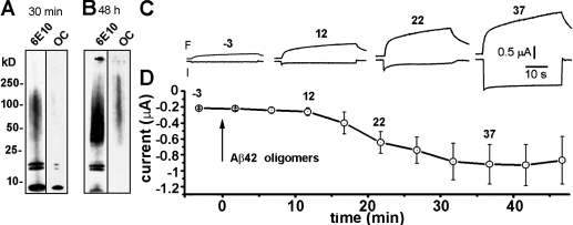
Figure 1.
Western blot analysis of Au03b21u221242 oligomer preparations and assay of potency to evoke Ca 2+ influx in Xenopus oocytes. (A and B) Western blots showing aggregation profiles of Au03b21u201342 at 3...
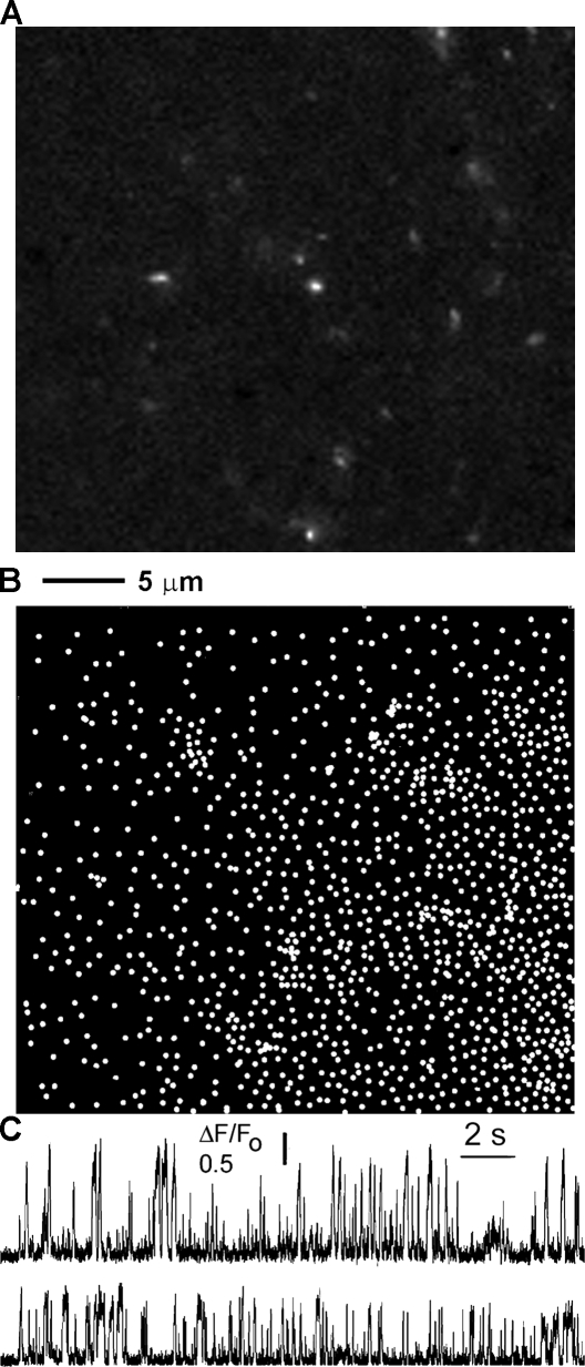
Figure 2.
Au03b21u201342 oligomers form Ca 2+ -permeable ion pores in the plasma membrane of Xenopus oocytes. (A) Representative image showing localized Ca 2+ transients (SCCaFTs) imaged by TIRF microscopy duri...
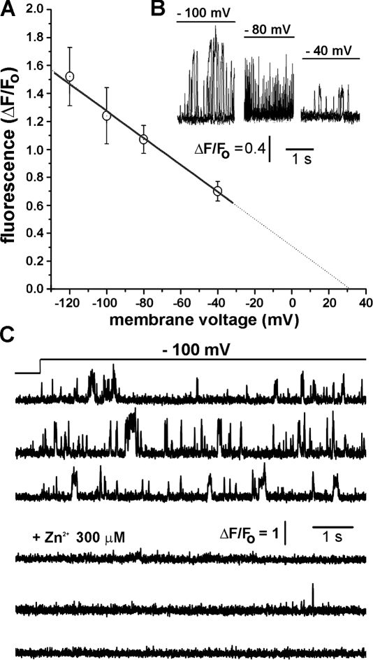
Figure 3.
Ca 2+ influx through Au03b21u221242 pores is voltage dependent and blocked by Zn 2+ . (A) Amplitudes of SCCaFTs from Au03b21u221242 pores plotted as function of membrane potential. Points show mean am...
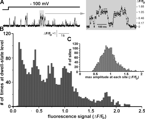
Figure 4.
SCCaFTs generated by Au03b21u221242 display multiple Ca 2+ flux levels. (A) The trace at the left shows a representative record of SCCaFTs induced by Au03b21u221242 oligomers, and the trace at the rig...
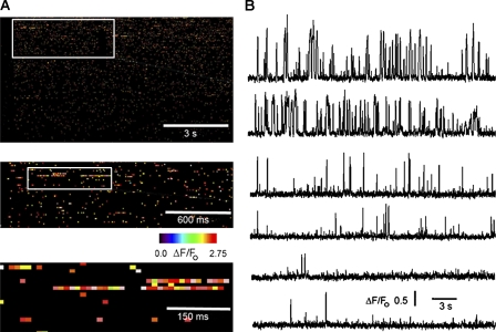
Figure 5.
Au03b21u221242 pores display wide variability in kinetics and P o . (A) Channel-chip representation of the activity of Au03b21u221242 pores. Pore openings (SCCaFTs) are represented as pseudocolored st...
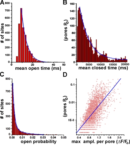
Figure 6.
Gating kinetics of Au03b21u221242 pores. (A) Distribution of mean open durations (SCCaFT durations) among the 2,820 pores. (B) Corresponding distribution of mean closed times (intervals between SCCaFT...
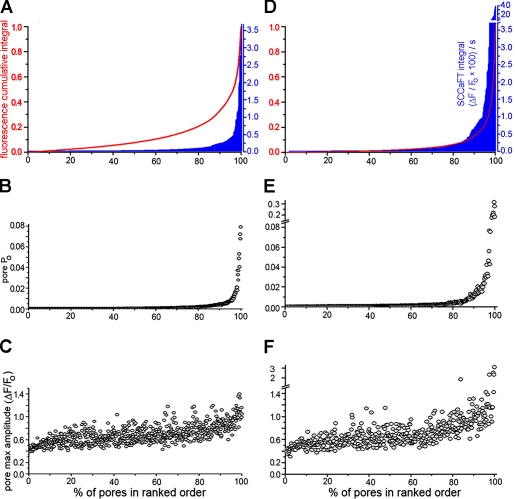
Figure 7.
Au03b21u221242 pores with high open probabilities contribute disproportionately to the total cellular Ca 2+ load. The Ca 2+ load contributed by a given pore was determined by integrating the area unde...
Figure images are served from the NIH/NLM PubMed Central Open Access Subset or Europe PMC; copyright remains with the publishers and authors.
💬 Discussion
0 commentsNo comments yet. Be the first to start a discussion!
Leave a Comment