Abstract
The structural maintenance of chromosomes (SMC) proteins constitute the core of critical complexes involved in structural organization of chromosomes. In yeast, the Smc5/6 complex is known to mediate repair of DNA breaks and replication of repetitive genomic regions, including ribosomal DNA loci and telomeres. In mammalian cells, which have diverse genome structure and scale from yeast, the Smc5/6 complex has also been implicated in DNA damage response, but its further function in unchallenged conditions remains elusive. In this study, we addressed the behavior and function of Smc5/6 during the cell cycle. Chromatin fractionation, immunofluorescence, and live-cell imaging analyses indicated that Smc5/6 associates with chromatin during interphase but largely dissociates from chromosomes when they condense in mitosis. Depletion of Smc5 and Smc6 resulted in aberrant mitotic chromosome phenotypes that were accompanied by the abnormal distribution of topoisomerase IIα (topo IIα) and condensins and by chromosome segregation errors. Importantly, interphase chromatin structure indicated by the premature chromosome condensation assay suggested that Smc5/6 is required for the on-time progression of DNA replication and subsequent binding of topo IIα on replicated chromatids. These results indicate an essential role of the Smc5/6 complex in processing DNA replication, which becomes indispensable for proper sister chromatid assembly in mitosis.
🔬 Techniques
🔭 Microscopes
🧬 Organisms
✨ Fluorophores
🧪 Sample Preparation
🔬 Cell Lines
🏭 Microscope Brands
🧪 Reagent Suppliers
📷 Detectors
🔎 Objectives
💻 Software Details
💾 Data Repositories
🏛️ Research Organizations (ROR)
Affiliated research institutions:
📋 Methods
Antibodies
Antibody to Smc2 was generously provided by Jan-Michael Peters (Research Institute of Molecular Pathology, Vienna, Austria). A mouse monoclonal antibody was raised against the peptide sequence MATPSKKTSTPSPQPSKRALPRDPSSEVPC of Smc5 (Monoclonal Antibody Research Institute, Sapporo, Japan) and then purified using a protein G affinity column (MabTrap kit; Amersham Biosciences, Piscataway, NJ). Antibodies against other proteins were as follows: SMC6L1 (M01, Cl7one 2E6; Abnova, Taipei, Taiwan), Smc6 (sc-365742, clone A-3; Santa Cruz Biotechnology, Santa Cruz, CA), SMC5 (18038; Abcam, Cambridge, UK), BLM (sc-7790, Santa Cruz Biotechnology), PICH (Clone 142-26-3; Millipore, Billerica, MA), DNA topoisomerase IIα (D081-1, clone 8D2; MBL, Aichi, Japan), DNA topoisomerase IIα (74715; Abcam), cyclin B (610219; BD PharMingen, Lexington, KY), α-tubulin (T6074, clone B-5-1-2; Sigma-Aldrich, St. Louis, MO), GFP (290; Abcam), and phospho–histone H3 (Ser-10) (9701; Cell Signaling Technology, Danvers, MA), cyclin B1 (K0128-3, clone V152; MBL), phospho–histone H3 (Ser-10) (9706, clone 6G3; Cell Signaling Technology), histone H2B (ab1790; Abcam), and γ-H2AX (A300-081A; Bethyl, Montgomery, TX). Chromatin fractionation For isolation of chromatin, cells were resuspended (4 × 10 7 cells/ml) in buffer A (10 mM HEPES, pH 7.9, 10 mM KCl, 1.5 mM MgCl 2 , 0.34 M sucrose, 10% glycerol, 1 mM dithiothreitol [DTT], 5 μg/ml aprotinin, 5 μg/ml leupeptin, 0.5 μg/ml pepstatin A, 0.1 mM phenylmethylsulfonyl fluoride). Triton X-100 (0.1%) was added, and the cells were incubated for 5 min on ice. Nuclei were pelleted by low-speed centrifugation (5 min, 1590 × g , 4°C). The supernatant containing the cytoplasmic fraction was further clarified by high-speed centrifugation (15 min, 3977 × g , 4°C) to remove cell debris and insoluble aggregates. Nuclei were washed once and then resuspended in buffer A containing Triton X-100 (0.1%) and analyzed by immunoblotting. Cell synchronization, inhibitor treatment, and cell lines HeLa and RPE-1 cells were cultured in DMEM (Wako, Tokyo, Japan) supplemented with 10% fetal calf serum (FBS; Equitech-Bio, Tokyo, Japan), 0.2 mM l -glutamine, 100 U/ml penicillin, and 100 μg/ml streptomycin (Meiji Seika, Tokyo, Japan) at 37°C in a 5% CO 2 environment. HeLa cells were synchronized by double thymidine arrest (24 h in the presence of 2 mM thymidine [T1895–1G; Sigma-Aldrich], 8 h release, 12 h in the presence of 2 mM thymidine) and were collected every 3 h after the second release for cell cycle progression analysis. RPE-1 cells were synchronized at G 0 /G 1 by serum starvation for 96 h and then released into the cell cycle with fresh medium containing 10% fetal calf serum for further analysis. For G 2 /M-phase arrest, asynchronous RPE-1 cells were treated with 9.0 μM RO-3306 (Enzo Life Sciences, Plymouth, PA) for 12 or 24 h. For enrichment of cells in prometaphase, 50 ng/ml nocodazole was added 7 h after release from the second thymidine block for 1 or 2 h; RPE-1 cells were treated with 15 mM STLC (TCI, Tokyo, Japan) for 2 h after release from RO-3306 arrest or 31 h after release from serum starvation for 2 or 3 h, and then recovered by mitotic shake-off. The HeLa cell line stably expressing EGFP-tagged Smc5 protein was generated by fusing EGFP in frame with the C terminus of Smc5 in a bacterial artificial chromosome vector (clone RP11; BACPAC Resources, Oakland, CA). The plasmid DNA was purified and transfected into HeLa cells using the FuGENE 6 reagent (Promega, Madison, WI). Stably expressing cell clones were selected in a complete medium containing G418, and expression of the tagged transgene was verified by fluorescence microscopy and immunoblotting. For visualization of regions with newly synthesized DNA, cells were cultured in medium supplemented with 10 μM EdU kit (Invitrogen, Carlsbad, CA) for 0.5 h before fixation.
Show full methods section
Antibodies
Antibody to Smc2 was generously provided by Jan-Michael Peters (Research Institute of Molecular Pathology, Vienna, Austria). A mouse monoclonal antibody was raised against the peptide sequence MATPSKKTSTPSPQPSKRALPRDPSSEVPC of Smc5 (Monoclonal Antibody Research Institute, Sapporo, Japan) and then purified using a protein G affinity column (MabTrap kit; Amersham Biosciences, Piscataway, NJ). Antibodies against other proteins were as follows: SMC6L1 (M01, Cl7one 2E6; Abnova, Taipei, Taiwan), Smc6 (sc-365742, clone A-3; Santa Cruz Biotechnology, Santa Cruz, CA), SMC5 (18038; Abcam, Cambridge, UK), BLM (sc-7790, Santa Cruz Biotechnology), PICH (Clone 142-26-3; Millipore, Billerica, MA), DNA topoisomerase IIα (D081-1, clone 8D2; MBL, Aichi, Japan), DNA topoisomerase IIα (74715; Abcam), cyclin B (610219; BD PharMingen, Lexington, KY), α-tubulin (T6074, clone B-5-1-2; Sigma-Aldrich, St. Louis, MO), GFP (290; Abcam), and phospho–histone H3 (Ser-10) (9701; Cell Signaling Technology, Danvers, MA), cyclin B1 (K0128-3, clone V152; MBL), phospho–histone H3 (Ser-10) (9706, clone 6G3; Cell Signaling Technology), histone H2B (ab1790; Abcam), and γ-H2AX (A300-081A; Bethyl, Montgomery, TX). Chromatin fractionation For isolation of chromatin, cells were resuspended (4 × 10 7 cells/ml) in buffer A (10 mM HEPES, pH 7.9, 10 mM KCl, 1.5 mM MgCl 2 , 0.34 M sucrose, 10% glycerol, 1 mM dithiothreitol [DTT], 5 μg/ml aprotinin, 5 μg/ml leupeptin, 0.5 μg/ml pepstatin A, 0.1 mM phenylmethylsulfonyl fluoride). Triton X-100 (0.1%) was added, and the cells were incubated for 5 min on ice. Nuclei were pelleted by low-speed centrifugation (5 min, 1590 × g , 4°C). The supernatant containing the cytoplasmic fraction was further clarified by high-speed centrifugation (15 min, 3977 × g , 4°C) to remove cell debris and insoluble aggregates. Nuclei were washed once and then resuspended in buffer A containing Triton X-100 (0.1%) and analyzed by immunoblotting. Cell synchronization, inhibitor treatment, and cell lines HeLa and RPE-1 cells were cultured in DMEM (Wako, Tokyo, Japan) supplemented with 10% fetal calf serum (FBS; Equitech-Bio, Tokyo, Japan), 0.2 mM l -glutamine, 100 U/ml penicillin, and 100 μg/ml streptomycin (Meiji Seika, Tokyo, Japan) at 37°C in a 5% CO 2 environment. HeLa cells were synchronized by double thymidine arrest (24 h in the presence of 2 mM thymidine [T1895–1G; Sigma-Aldrich], 8 h release, 12 h in the presence of 2 mM thymidine) and were collected every 3 h after the second release for cell cycle progression analysis. RPE-1 cells were synchronized at G 0 /G 1 by serum starvation for 96 h and then released into the cell cycle with fresh medium containing 10% fetal calf serum for further analysis. For G 2 /M-phase arrest, asynchronous RPE-1 cells were treated with 9.0 μM RO-3306 (Enzo Life Sciences, Plymouth, PA) for 12 or 24 h. For enrichment of cells in prometaphase, 50 ng/ml nocodazole was added 7 h after release from the second thymidine block for 1 or 2 h; RPE-1 cells were treated with 15 mM STLC (TCI, Tokyo, Japan) for 2 h after release from RO-3306 arrest or 31 h after release from serum starvation for 2 or 3 h, and then recovered by mitotic shake-off. The HeLa cell line stably expressing EGFP-tagged Smc5 protein was generated by fusing EGFP in frame with the C terminus of Smc5 in a bacterial artificial chromosome vector (clone RP11; BACPAC Resources, Oakland, CA). The plasmid DNA was purified and transfected into HeLa cells using the FuGENE 6 reagent (Promega, Madison, WI). Stably expressing cell clones were selected in a complete medium containing G418, and expression of the tagged transgene was verified by fluorescence microscopy and immunoblotting. For visualization of regions with newly synthesized DNA, cells were cultured in medium supplemented with 10 μM EdU kit (Invitrogen, Carlsbad, CA) for 0.5 h before fixation.
PCC assay
PCC was induced by adding calyculin A (Wako) into culture media at a final concentration of 160 nM. After 1 h, cells were hypotonically swollen in a 40:60 mix (vol/vol) of phosphate-buffered saline (PBS) and distilled water for 5 min at room temperature. Cells were fixed with freshly made Carnoy's solution (70% methanol, 30% acetic acid), dropped on glass slides, and dried. Slides were stained with 5% Giemsa stain, washed with water, air-dried, and mounted with Entellan embedding agent (Merck, Darmstadt, Germany). For detection of incorporated EdU, the Click-iT EdU imaging kit (Invitrogen) was used. Briefly, the glass slides were incubated with a reaction solution containing fluorochrome-azide (Alexa Fluor 488) for 1 h at room temperature, after which immunofluorescence staining was conducted. RNA interference siRNA sequences were as follows: Smc5 siRNA-1, 5′-GGAACUUCAGCAGGGCUUUAAUAGUA-3′; Smc5 siRNA-2, 5′-GGCAUUAUGUGAAGGCGAAAUAAUU-3′; Smc6 siRNA-1, 5′-CAAAUUCUUCAUGAAAGCAACGCAA-3′; Smc6 siRNA-2, 5′-GACCUAUCUUGAUCUGGAUAGUAAA-3′. Smc6 siRNA-1 was used for ChIP-Seq analysis of topo IIα. Cells were transfected by incubating 50 nM duplex siRNA with Lipofectamine RNAiMAX (Invitrogen) in antibiotic-free growth medium. RNA interference (RNAi) was performed concomitantly with the synchronization with thymidine or serum starvation, and 48 h of transfection with each siRNA was required for maximal knockdown of Smc5 and Smc6. For control transfections, the same annealing reaction was set up without the presence of siRNA oligonucleotides. Chromosome spreads RPE-1 cells treated with STLC (15 mM) for 2–3 h were collected by mitotic shake-off and hypotonically swollen in a 40:60 mix (vol/vol) of PBS and distilled water for 5 min at room temperature. Cells were fixed with freshly made Carnoy's solution, dropped on glass slides, and dried. Slides were stained with 5% Giemsa stain, washed with water, air-dried, and mounted with Entellan embedding agent.
Immunofluorescence microscopy
Cells grown on coverslips with or without preextraction with 0.2% PBS/Tween 100 were fixed with ice-chilled 100% methanol for 20 min at −20°C or with 4% paraformaldehyde in 0.137 M sodium phosphate buffer (pH 7.4). Fixed cells/chromosomes were permeabilized with 0.5% Triton X-100 in PBS and incubated with 3% bovine serum albumin in PBS for at least 1 h. Cells were incubated with the primary antibodies overnight at room temperature; this was followed by incubation with secondary antibodies for 35 min together with 0.1 μg/ml 4,6-diamidino-2-phenylindole (DAPI). The secondary antibodies used in this study were goat anti-rabbit immunoglobulin G (IgG) and goat anti-mouse IgG coupled to Alexa Fluor 488 or 568 and goat anti-human IgG coupled to Alexa Fluor 568 (Molecular Probes, Carlsbad, CA). Cells were washed twice and mounted in ProLong Gold anti-fade mounting reagent (Invitrogen). Images were acquired on a Zeiss Imager Z.1 microscope equipped with epifluorescence and a CoolSNAP HQ CCD camera (Photometrics, Tucson, AZ). Three-dimensional projections of the immunofluorescence images were carried out using the 3D Volume viewer tool in ImageJ software (National Institutes of Health, Bethesda, MD).
Live-cell imaging analysis
Cells were placed in CO 2 -independent medium without phenol red (Life Technologies-BRL, Grand Island, NY) on chambered coverslips (Lab-Tek; Nunc, Rochester, NY), and the chamber lids were sealed with silicone grease. Images were captured every 3 min, with 100-ms exposure times, through a 100×/1.40 NA Plan-Apochromat oil-objective lens mounted on an inverted microscope (IX-71; Olympus, Tokyo, Japan) equipped with a CoolSNAP HQ CCD camera. A series of projected images of three Z-sections with 5.0-μm intervals were analyzed. For data analysis, images were processed using ImageJ software.
Immunoprecipitation Asynchronized HeLa cells expressing EGFP-tagged
Smc5 were lysed in immunoprecipitation buffer (20 mM Tris-HCl, pH 7.5, 150 mM NaCl, 20 mM β-glycerophosphate, 5 mM MgCl 2 , 0.1% NP-40, protease inhibitors [Complete Mini EDTA-free; Roche, Indianapolis, IN], 1 mM DTT), supplemented with 100 nM okadaic acid, 2 mM Na 3 VO 4 , 10 mM NaF, and 0.25 U/l benzonase nuclease (Novagen, EMD Millipore, Billerica, MA), for 20 min on ice. Cell extracts, after the insoluble fraction was removed by centrifugation at 15,000 rpm for 30 min at 4°C, were used for immunoprecipitation. Ten microliters of protein A (Bio-Rad, Hercules, CA) beads coupled to antibodies was incubated with cell extracts for ∼2 h at 4°C and then washed three times with immunoprecipitation buffer and three times with 0.05% TBS-Tween20.
Fluorescence-activated cell sorting analysis
Cells were harvested by trypsinization, fixed in 70% ethanol, stained with propidium iodide solution at a final concentration of 50 μg/ml, and subjected to fluorescence-activated cell sorting (FACS) analysis on FaCScalibur using Cell Quest (Becton Dickinson, Mansfield, MA) software. ChIP Cells were cross-linked with 1% formaldehyde for 10 min, quenched with 125 mM glycine, and prepared for ChIP as previously described ( Wendt et al ., 2008 ). ChIP was performed as previously described using SMC6L1 (M01, Cl7one 2E6; Abnova) and DNA topoisomerase IIα (D081-1, clone 8D2; MBL; Wendt et al ., 2008 ). In brief, cross-linked cell lysates solubilized by sonication were incubated with Affi-prep protein A Support (Bio-Rad) and cross-linked with the antibodies for 14 h at 4°C. After this, beads were washed several times and eluted with elution buffer (50 mM Tris, 10 mM EDTA, and 1% SDS) for 20 min at 65°C. The eluates were incubated at 65°C overnight to reverse cross-links and were then treated with RNaseA and then with proteinase K. The samples were further purified by phenol–chloroform extraction and an extra purification step using a PCR purification kit (Qiagen, Valencia, CA).
ChIP-Seq analyses
DNA from whole-cell extracts
(WCE) and ChIP fractions was further sheared by sonication (Branson sonifier 250D), end-repaired, ligated to sequencing adaptors, and amplified according to the SOLiD Library Preparation kit Protocol (Applied Biosystems, Foster City, CA). DNA purified from gel and amplified between 100 and 150 base pairs was sequenced on the Applied Biosystems SOLiD platforms (SOLiD 3 and 5500) to generate single-end 50–base pair reads. Sequenced data for both ChIP fractions and WCE were aligned to the human genome (UCSC hg19) using Bowtie ( Langmead et al ., 2009 ), allowing three mismatches in the first 28 bases per read (-n3 option).
Software
DROMPA was used for visualization and statistical analysis of ChIP-Seq data sets ( Nakato et al ., 2013 ). All duplicate reads and those without unique alignment were removed from further analysis. ChIP-Seq and DNA sequencing data from this study have been deposited in the Sequence Read Archive database ( www.ncbi.nlm.nih.gov/sra ) under accession number SRX381015. We further analyzed only uniquely aligned reads. Each aligned read was extended to a predicted fragment length of 150 base pairs. Reads were summed in 100-kb windows along the chromosome, and the fold enrichment (ChIP/WCE) for each window was calculated.
Raw data availability
Original unprocessed versions of all figures have been uploaded to the server of IMCB, the University of Tokyo, and are available on request.
📊 Figures
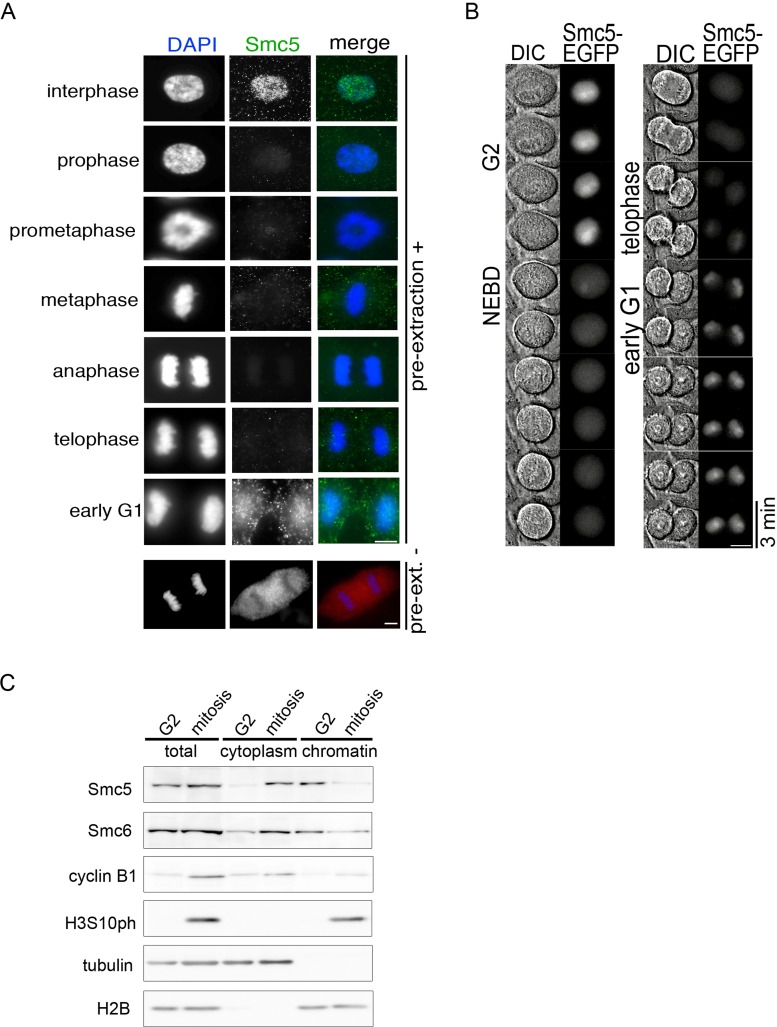
FIGURE 1:
Chromatin association of the Smc5/6 complex during the cell cycle. (A) RPE-1 cells with or without preextraction with 0.2% PBS/Triton X-100 were fixed in paraformaldehyde and stained with Smc5 antibod...
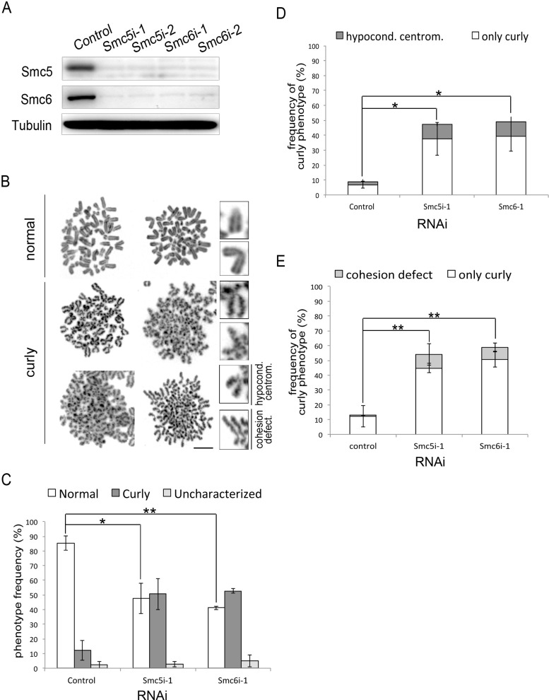
FIGURE 2:
RNAi-mediated depletion of Smc5 and Smc6 in RPE-1 cells. (A) Logarithmically proliferating RPE-1 cells were transfected with the indicated siRNA, and WCE were analyzed by immunoblotting 72 h after the...
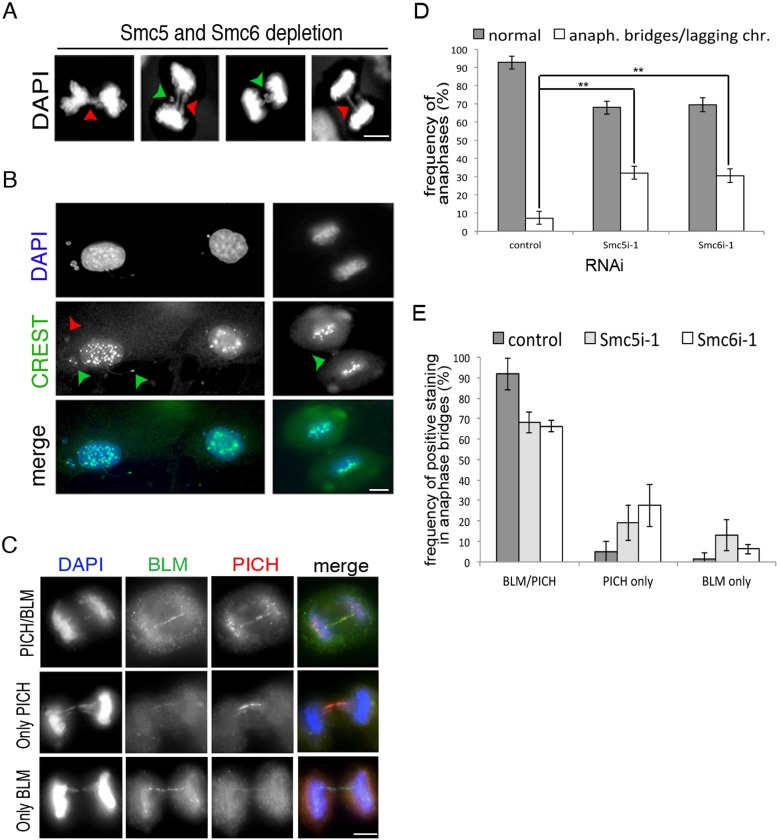
FIGURE 3:
Depletion of Smc5 or Smc6 disrupts chromosome segregation. (A) Example images of the anaphase bridges (red arrowheads) and lagging chromosomes (green arrowheads) frequently seen in Smc5/6-depleted cel...
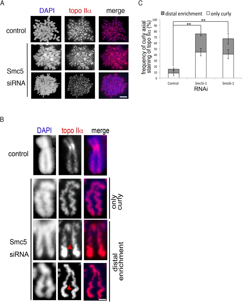
FIGURE 4:
Chromosome axis deformation in Smc5/6-depleted cells. (A) RPE-1 metaphase cells transfected with siRNAs against Smc5 or Smc6 or a control mock were subjected to hypotonic treatment, fixation, and stai...
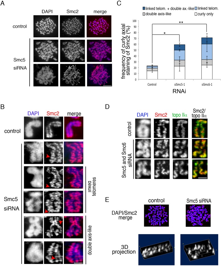
FIGURE 5:
(A) Metaphase chromosome spreads were prepared from control and Smc5-depleted cells and stained with DAPI (blue) and anti-Smc2 (red). Scale bar: 5 u03bcm. (B) Images of individual chromosomes showing ...
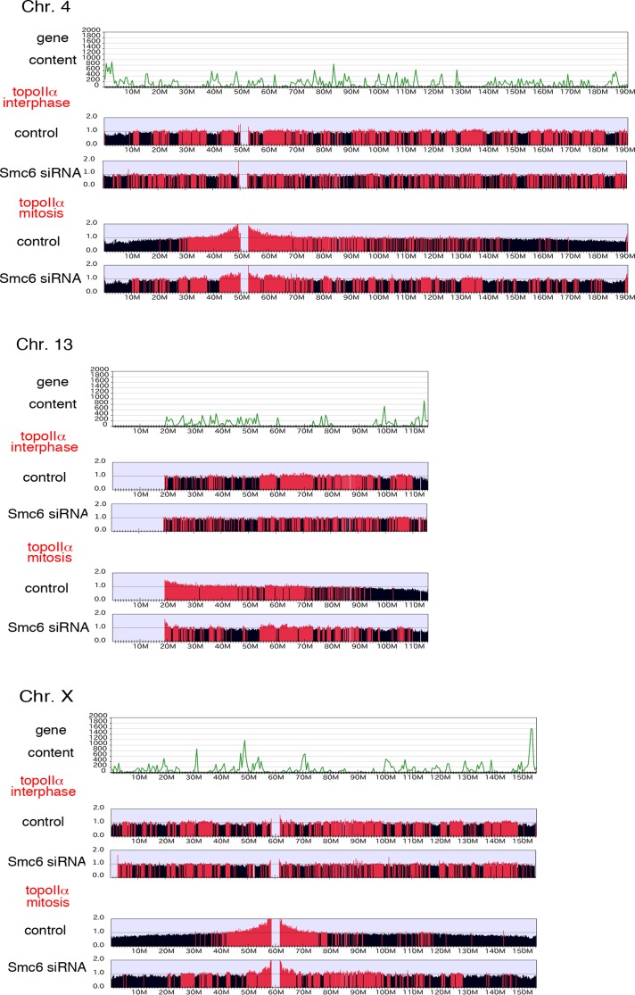
FIGURE 6:
Chromatin binding profile of topo IIu03b1 on human chromosomes in interphase and mitosis in the presence and absence of Smc6, as revealed by ChIP-Seq. Uniquely aligned reads are summed in 100-kb windo...
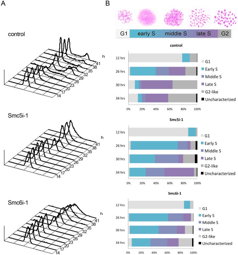
FIGURE 7:
Probing different stages of DNA replication using FACS and PCC assay. (A) RPE-1 cells transfected with an siRNA against Smc5 or Smc6 or a control mock were collected at indicated time points after rel...
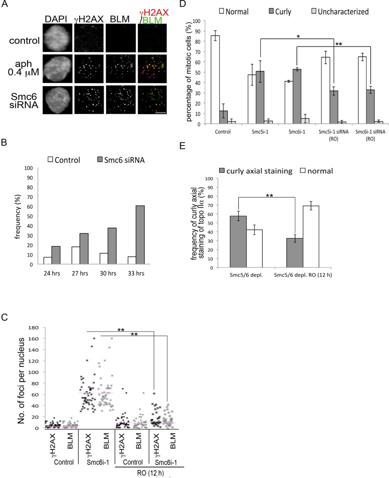
FIGURE 8:
Replication-related DNA damage. RPE-1 cells transfected with an siRNA against Smc5 or Smc6 or a control mock were analyzed at the specified time points after release from serum starvation. (A) Represe...
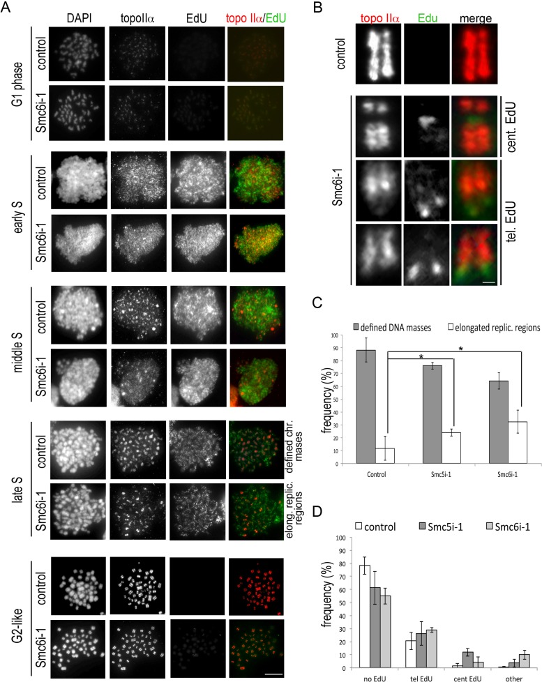
FIGURE 9:
Progression of DNA replication and loading of topo IIu03b1 onto sister chromatids in S phase. (A) PCC analysis with EdU pulse-labeling was carried out as described in Figure 8, E and F . Immunofluores...
Figure images are served from the NIH/NLM PubMed Central Open Access Subset or Europe PMC; copyright remains with the publishers and authors.
💬 Discussion
0 commentsNo comments yet. Be the first to start a discussion!
Leave a Comment