Abstract
BACKGROUND: The Na+,K+-ATPase plays an important role for ion homeostasis in virtually all mammalian cells, including neurons. Despite this, there is as yet little known about the isoform specific distribution in neurons. RESULTS: With help of superresolving stimulated emission depletion microscopy the spatial distribution of Na+,K+-ATPase in dendritic spines of cultured striatum neurons have been dissected. The found compartmentalized distribution provides a strong evidence for the confinement of neuronal Na+,K+-ATPase (α3 isoform) in the postsynaptic region of the spine. CONCLUSIONS: A compartmentalized distribution may have implications for the generation of local sodium gradients within the spine and for the structural and functional interaction between the sodium pump and other synaptic proteins. Superresolution microscopy has thus opened up a new perspective to elucidate the nature of the physiological function, regulation and signaling role of Na+,K+-ATPase from its topological distribution in dendritic spines.
🔬 Techniques
🧬 Organisms
✨ Fluorophores
🧪 Sample Preparation
🔬 Cell Lines
🏭 Microscope Brands
🧪 Reagent Suppliers
🏛️ Research Organizations (ROR)
Affiliated research institutions:
📋 Methods
Cell culture
Cultures of striatal neurons were prepared from embryonic day 18.5 old Sprauge dawley rat embryos. Striatum was dissected, incubated for 10 minutes at 37°C in Hank's balanced salt solution (Gibco Invitrogen) containing 20 mM HEPES (Sigma) and 0.25% Trypsin (Gibco Invitrogen) and dissociated in MEM (Gibco Invitrogen) by mechanical triturating using a fire polished Pasteur pipette. The cells were plated at a density of 2 × 10 5 cells on round 28 mm, No. 1.0, poly-ornithine (Gibco Invitrogen, 80 μg/mL over night at 37°C) coated coverslips and incubated for three hours in MEM media containing 10% Horse serum, 2 mM L-Glutamine and 1 mM NaPyruvate. The cells where then cultured in Neurobasal media (Gibco Invitrogen) containing 1× B27 (Gibco Invitrogen) and 2 mM L-Glutamine. Cells were maintained in culture for two weeks before experiments and culture media was changed once a week. Immunostaining Cells were fixed for 15 min in ice cold methanol, permeabilized with 0.1% triton and blocked with 7% normal goat serum and then incubated overnight at 4°C with primary antibody diluted 1:1000 in PBS containing 3% normal goat serum and 0.1% Triton X-100. The primary antibody was mouse monoclonal anti-α3 Na + ,K + -ATPase (Affinity bioreagents). Following incubation the cells were washed in PBS, 0.1% triton and incubated at room temperature for two hours with fluorescent secondary antibody, goat-anti-mouse Alexa-594 (Molecular Probes Inc.) diluted 1:500 in PBS with 3% serum and 0.1% Triton X-100. Following secondary incubation cells were washed again, PBS, 0.1% Triton X-100 and mounted using Immu-mount (Thermo scientific).
Show full methods section
Cell culture
Cultures of striatal neurons were prepared from embryonic day 18.5 old Sprauge dawley rat embryos. Striatum was dissected, incubated for 10 minutes at 37°C in Hank's balanced salt solution (Gibco Invitrogen) containing 20 mM HEPES (Sigma) and 0.25% Trypsin (Gibco Invitrogen) and dissociated in MEM (Gibco Invitrogen) by mechanical triturating using a fire polished Pasteur pipette. The cells were plated at a density of 2 × 10 5 cells on round 28 mm, No. 1.0, poly-ornithine (Gibco Invitrogen, 80 μg/mL over night at 37°C) coated coverslips and incubated for three hours in MEM media containing 10% Horse serum, 2 mM L-Glutamine and 1 mM NaPyruvate. The cells where then cultured in Neurobasal media (Gibco Invitrogen) containing 1× B27 (Gibco Invitrogen) and 2 mM L-Glutamine. Cells were maintained in culture for two weeks before experiments and culture media was changed once a week. Immunostaining Cells were fixed for 15 min in ice cold methanol, permeabilized with 0.1% triton and blocked with 7% normal goat serum and then incubated overnight at 4°C with primary antibody diluted 1:1000 in PBS containing 3% normal goat serum and 0.1% Triton X-100. The primary antibody was mouse monoclonal anti-α3 Na + ,K + -ATPase (Affinity bioreagents). Following incubation the cells were washed in PBS, 0.1% triton and incubated at room temperature for two hours with fluorescent secondary antibody, goat-anti-mouse Alexa-594 (Molecular Probes Inc.) diluted 1:500 in PBS with 3% serum and 0.1% Triton X-100. Following secondary incubation cells were washed again, PBS, 0.1% Triton X-100 and mounted using Immu-mount (Thermo scientific).
DNA constructs
The full length intracellular N-tail of α3 NKA was obtained from rat brain total RNA as RT-PCR fragment with additional CACC leading sequence and a stop codon introduced after the end of the particular domain. cDNA were then cloned into pENTR-D vector with the following exchange into pDEST15 expression vector by Gateway TOPO cloning system (Invitrogen, USA). The structure of GST-fusion plasmids was confirmed by sequencing with BigDye v.3.1 (Applied Biosystems, USA).
Antibodies
In both imaging and biochemical assays the well characterized mouse monoclonal anti-α3 Na + ,K + -ATPase (Affinity bioreagents, MA3-915) was used [ 30 ]. For Western blotting and immunoprecipitation, the following antibodies were used: mouse monoclonal anti-PSD-95 (Abcam, Cambridge, UK) and as control mouse IgG (Sigma Aldrich, St.Louis, MO, USA). The used antibodies show high specificity with single bands in Western blot experiments and very low unspecific labelling in imaging experiments.
Immunoprecipitation
Striatum from 20 days old rats was homogenized in the RIPA buffer containing 50 mM Tris HCl (pH 7,4), 150 mM NaCl, 2 mM EDTA, 1 mM phenylmethylsulfonyl fluoride, 0.25% sodium deoxycholate, 1% Triton X-100 and protease inhibitors (Pierce, Rockford, IL, USA) and centrifuged 30 min at 9000 g. Supernatant protein concentration was measured. 1 ml of the striatal lysate (300 μg of total protein) was pre-cleared with 60 μl of protein G Sepharose beads (GE Healthcare Bio-Sciences AB, Uppsala, Sweden) 1 hour at 4°C. Lysate was then incubated with 3 μg of α3 Na + ,K + -ATPase, PSD-95, or mouse IgG antibodies for 2 hours at 4°C. 30 μl of protein G Sepharose beads was added and incubation continued over night at 4°C. The samples were washed 5 times with RIPA buffer and the proteins were eluted with 50 μl of 2× Laemmli buffer for 15 min at 65°C. Samples were then subjected to SDS-PAGE for Western blotting. GST affinity pull-down Expression and purification of the GST-fused proteins were performed as described previously [ 31 ]. Rat striatal lysate (300 μg of total protein) was prepared in the RIPA buffer and added to the beads in a volume of 1 ml and incubated for 4 hours at 4°C. The beads were then washed four times with RIPA buffer. Proteins were eluted with 50 μl of 2× Laemmli buffer for 15 min at 65°C. Samples were then subjected to SDS-PAGE for Western blotting, using antibodies for: PSD-95 (1:1000) and α3 NKA (1:1000). The experimental procedure was repeated at a minimum of three times.
STED microscopy
The stimulated emission depletion microscope has been described in detail before [ 13 ]. Our setup has the following minor differences: The excitation and STED beams are coupled together using a dichroic mirror (z690SPRDC, Chroma Technology Corp., Bellow Falls, US) before being sent into the microscope objective; The fluorescence from the labeled sample is collected back through the objective and separated from the excitation and the STED beams by a customized dichroic mirror (Laseroptik, Garbsen, Germany); The sample is placed on a 3 D scanning piezo stage coupled to a closed loop controller unit (MAX311/M and BPC203, Thorlabs Sweden AB, Göteborg, Sweden) offering a positional resolution of 5 nm. Typically images between 7 μm × 7 μm to 15 μm × 15 μm were acquired with a pixel size of 10 - 30 nm and a pixel dwell time of 0.5-1 ms. The average excitation powers applied were 0.5-1.1 μW and the applied STED powers were 2.6-3.0 mW.
📊 Figures
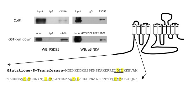
Figure 1
Co-immunoprecipitation and GST pull down assay of PSD-95 and u03b13 NKA . Co-immunoprecipitation (CoIP) shows interaction between PSD-95 and u03b13 NKA. GST pull down assays shows interaction of PSD-9...
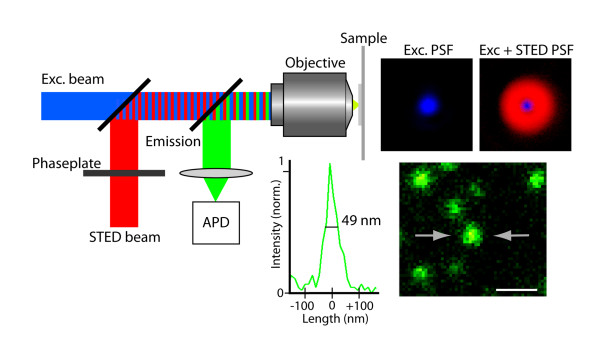
Figure 2
Principle of STED microscopy . The red depletion beam is phase-modulated to form a focal doughnut shown in the top right panel. Superimposition of this STED focus onto the diffraction limited excitati...
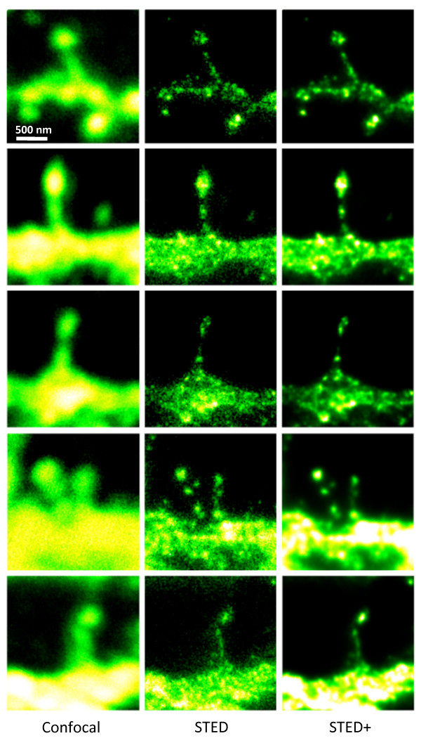
Figure 3
STED microscopy dissecting the localization of Na + ,K + -ATPase in cultured striatal neurons . The 5 u00d7 3 mosaic shows a comparision of the confocal (left) and STED images (middle) of the Alexa-59...
Figure images are served from the NIH/NLM PubMed Central Open Access Subset or Europe PMC; copyright remains with the publishers and authors.
💬 Discussion
0 commentsNo comments yet. Be the first to start a discussion!
Leave a Comment