Abstract
Mitosis is triggered by the activation of Cdk1-cyclin B1 and its translocation from the cytoplasm to the nucleus. Positive feedback loops regulate the activation of Cdk1-cyclin B1 and help make the process irreversible and all-or-none in character. Here we examine whether an analogous process, spatial positive feedback, regulates Cdk1-cyclin B1 redistribution. We used chemical biology approaches and live-cell microscopy to show that nuclear Cdk1-cyclin B1 promotes the translocation of Cdk1-cyclin B1 to the nucleus. Mechanistic studies suggest that cyclin B1 phosphorylation promotes nuclear translocation and, conversely, nuclear translocation promotes cyclin B1 phosphorylation, accounting for the feedback. Interfering with the abruptness of Cdk1-cyclin B1 translocation affects the timing and synchronicity of subsequent mitotic events, underscoring the functional importance of this feedback. We propose that spatial positive feedback ensures a rapid, complete, robust, and irreversible transition from interphase to mitosis and suggest that bistable spatiotemporal switches may be widespread in biological regulation.
🔬 Techniques
🔭 Microscopes
🧬 Organisms
💻 Software
✨ Fluorophores
🧪 Sample Preparation
🔬 Cell Lines
🏭 Microscope Brands
🧪 Reagent Suppliers
🔎 Objectives
💻 Software Details
💾 Data Repositories
🏛️ Research Organizations (ROR)
Affiliated research institutions:
📋 Methods
Cell Culture, Synchronization, and Transfection HeLa cells (ATCC) and Tet-On Advanced HeLa cells (Invitrogen) were cultured at 37°C, 5% CO2 in DMEM (Invitrogen) supplemented with 10% FCS, penicillin (100 U/ml), streptomycin (100 μg/ml) and G418 (100 μg/ml, for Tet-On Advanced HeLa cells). Cells were synchronized at the G1/S phase of the cell cycle by double thymidine block. Transfection of cDNAs was typically performed using either Fugene 6 (Roche) (for DNA transfection) or Gene Silencer (Genlantis) (for small interfering RNAs, siRNA, transfection or double DNA/siRNA transfection) according to the manufacturers’ instructions. Transfection of 50 μM dextran-70-kDa-Texas Red was done by suspended drop electroporation (SDE) as described in ( Guignet and Meyer, 2008 ).
Microscopy and Data Analysis
Time-lapse live cell imaging was performed on either an ImageXpress Micro inverted epifluorescence microscope (Molecular Devices) or on a Nikon Eclipse TI with perfect focus, both equipped with temperature, humidity and CO 2 control. ImageXpress images were acquired with 20x or 40x plan fluorescence objectives. Excitation (Ex) and emission (Em) filters sets (Chroma Technology Corporation) were as follows: CFP, 427-10nm (Ex), 483-32 nm (Em); YFP, 504-12 nm (Ex), 542-27 nm (Em); mCherry, 589-15 nm (Ex), 632-22 nm (Em). Nikon Eclipse TI images were acquired using either 40x NA 1.3 Plan Flour or 60x NA 1.3 Plan Apo objectives. Excitation (Ex) and emission (Em) filters sets were as follows: CFP, 430-24 nm (Ex), 470-24 nm (Em); YFP, 500-20 nm (Ex), 535-30 nm (Em); mCherry, 572-35 nm (Ex), 632-60 nm (Em). Micromanager 1.3 was used for acquisition of time-lapse images. A typical experiment would monitor cells from 15–20 h with images taken every 3–5 min. Confocal microscopy was performed on a Leica TCS SP2 ABOS equipped with a 63x 1.3 NA oil immersion objective. DAPI was excited using a 405 nm diode laser; Alexa 488 or YFP were excited with a 488 nm or 514 nm Ar laser and Alexa 647 was excited with a 633 nm HeNe laser line. All data analysis was done with scripts written in Matlab (Mathworks) or using Cell Profiler (Broad Institute) and ImageJ (National Institutes of Health). Cdk1-Cyclin B1 Autophosphorylation For cyclin B1 auto-phosphorylation experiments where we varied the concentration of Cdk1-cyclin B1, purified human Cdk1-cyclin B1 protein complex was incubated at the indicated concentrations at 25°C for 1, 2 or 5 min with kinase buffer (20 mM HEPES/NaOH pH 7.4, 10 mM MgCl 2, 1 mM DTT), 15 μM ATP and phosphatase inhibitor cocktail (Sigma) in a total volume of 20 μl. For Cyclin B1 auto-phosphorylation experiments where we varied kinase reaction volumes, purified human Cdk1-cyclin B1 protein complex was incubated at 25°C for 1, 2 or 5min with kinase buffer (20 mM HEPES/NaOH pH 7.4, 10 mM MgCl 2, 1 mM DTT), 15 μM ATP and phosphatase inhibitor cocktail (Sigma) in a total volume of 10, 20, 40, 80 or 160 μl. Reactions were stopped by addition of Laemmli sample buffer and analyzed by gel electrophoresis and blotting followed by phospho-imaging. Nuclear/Cytoplasmic Cell Fractionation HeLa cell nuclear and cytoplasmic fractions were isolated with NE-PER Nuclear and Cytoplasmic Extraction Kit (Thermo Scientific) according to the manufacturer’ instructions. Total protein amount in the nuclear and the cytosolic lysates was quantified by BCA assay for protein quantitation. Typically, 15–20 μg or 100–200 μg of total protein for each fraction was used for western blot analysis or immunoprecipitation, respectively. For Cdk1-cyclin B1 nuclear/cytosolic kinase assays, 100 μg of total protein was used to immunoprecipitate cyclin B1 using anti-cyclin B1 antibody (Santa Cruz). Kinase assays were performed using purified histone H1 (Upstate) as a general Cdk1-cyclin B1 substrate and the resulting 32 P-labeled histone was detected and quantified by SDS gel electrophoresis and blotting followed by phosphorimaging.
Show full methods section
Cell Culture, Synchronization, and Transfection HeLa cells (ATCC) and Tet-On Advanced HeLa cells (Invitrogen) were cultured at 37°C, 5% CO2 in DMEM (Invitrogen) supplemented with 10% FCS, penicillin (100 U/ml), streptomycin (100 μg/ml) and G418 (100 μg/ml, for Tet-On Advanced HeLa cells). Cells were synchronized at the G1/S phase of the cell cycle by double thymidine block. Transfection of cDNAs was typically performed using either Fugene 6 (Roche) (for DNA transfection) or Gene Silencer (Genlantis) (for small interfering RNAs, siRNA, transfection or double DNA/siRNA transfection) according to the manufacturers’ instructions. Transfection of 50 μM dextran-70-kDa-Texas Red was done by suspended drop electroporation (SDE) as described in ( Guignet and Meyer, 2008 ).
Microscopy and Data Analysis
Time-lapse live cell imaging was performed on either an ImageXpress Micro inverted epifluorescence microscope (Molecular Devices) or on a Nikon Eclipse TI with perfect focus, both equipped with temperature, humidity and CO 2 control. ImageXpress images were acquired with 20x or 40x plan fluorescence objectives. Excitation (Ex) and emission (Em) filters sets (Chroma Technology Corporation) were as follows: CFP, 427-10nm (Ex), 483-32 nm (Em); YFP, 504-12 nm (Ex), 542-27 nm (Em); mCherry, 589-15 nm (Ex), 632-22 nm (Em). Nikon Eclipse TI images were acquired using either 40x NA 1.3 Plan Flour or 60x NA 1.3 Plan Apo objectives. Excitation (Ex) and emission (Em) filters sets were as follows: CFP, 430-24 nm (Ex), 470-24 nm (Em); YFP, 500-20 nm (Ex), 535-30 nm (Em); mCherry, 572-35 nm (Ex), 632-60 nm (Em). Micromanager 1.3 was used for acquisition of time-lapse images. A typical experiment would monitor cells from 15–20 h with images taken every 3–5 min. Confocal microscopy was performed on a Leica TCS SP2 ABOS equipped with a 63x 1.3 NA oil immersion objective. DAPI was excited using a 405 nm diode laser; Alexa 488 or YFP were excited with a 488 nm or 514 nm Ar laser and Alexa 647 was excited with a 633 nm HeNe laser line. All data analysis was done with scripts written in Matlab (Mathworks) or using Cell Profiler (Broad Institute) and ImageJ (National Institutes of Health). Cdk1-Cyclin B1 Autophosphorylation For cyclin B1 auto-phosphorylation experiments where we varied the concentration of Cdk1-cyclin B1, purified human Cdk1-cyclin B1 protein complex was incubated at the indicated concentrations at 25°C for 1, 2 or 5 min with kinase buffer (20 mM HEPES/NaOH pH 7.4, 10 mM MgCl 2, 1 mM DTT), 15 μM ATP and phosphatase inhibitor cocktail (Sigma) in a total volume of 20 μl. For Cyclin B1 auto-phosphorylation experiments where we varied kinase reaction volumes, purified human Cdk1-cyclin B1 protein complex was incubated at 25°C for 1, 2 or 5min with kinase buffer (20 mM HEPES/NaOH pH 7.4, 10 mM MgCl 2, 1 mM DTT), 15 μM ATP and phosphatase inhibitor cocktail (Sigma) in a total volume of 10, 20, 40, 80 or 160 μl. Reactions were stopped by addition of Laemmli sample buffer and analyzed by gel electrophoresis and blotting followed by phospho-imaging. Nuclear/Cytoplasmic Cell Fractionation HeLa cell nuclear and cytoplasmic fractions were isolated with NE-PER Nuclear and Cytoplasmic Extraction Kit (Thermo Scientific) according to the manufacturer’ instructions. Total protein amount in the nuclear and the cytosolic lysates was quantified by BCA assay for protein quantitation. Typically, 15–20 μg or 100–200 μg of total protein for each fraction was used for western blot analysis or immunoprecipitation, respectively. For Cdk1-cyclin B1 nuclear/cytosolic kinase assays, 100 μg of total protein was used to immunoprecipitate cyclin B1 using anti-cyclin B1 antibody (Santa Cruz). Kinase assays were performed using purified histone H1 (Upstate) as a general Cdk1-cyclin B1 substrate and the resulting 32 P-labeled histone was detected and quantified by SDS gel electrophoresis and blotting followed by phosphorimaging.
Supplementary Material 01 02 03 04 05 06 07 08
📊 Figures
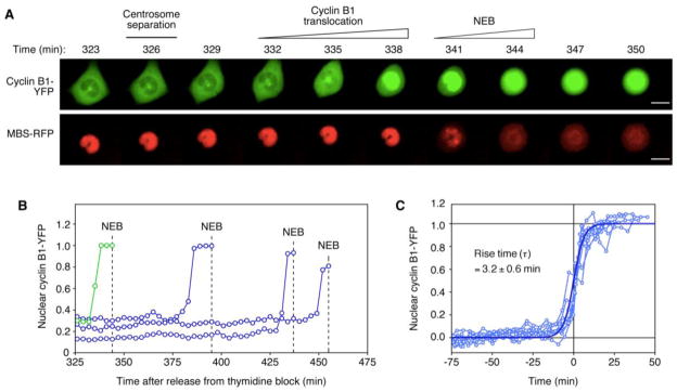
Figure 1
Abrupt Cyclin B1 Nuclear Translocation Prior to Nuclear Envelope Breakdown
(A) A representative HeLa cell expressing cyclin B1-YFP (top) and the mitotic biosensor (MBS-RFP) (bottom) after release from G1/S block. Centrosome separation (top), cyclin B1 translocation (top), an...
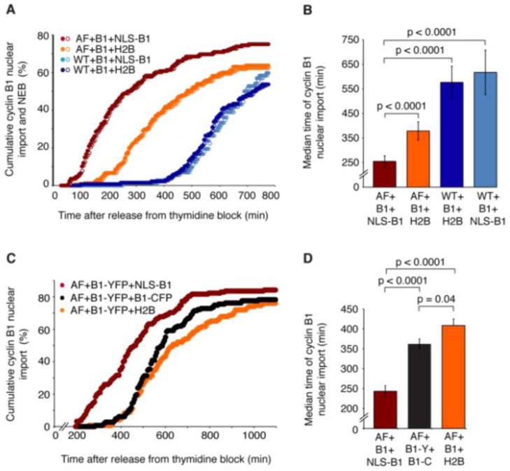
Figure 2
In Cdk1AF-Expressing Cells, NLS-Cyclin B1-CFP Expression Promotes Cyclin B1-YFP Translocation
(A) Cumulative percentage of cells that showed cyclin B1 nuclear translocation (closed circles) and NEB (open circles) after release from G1/S block. Cells were expressing Cdk1AF (AF) or Cdk1-WT (WT),...
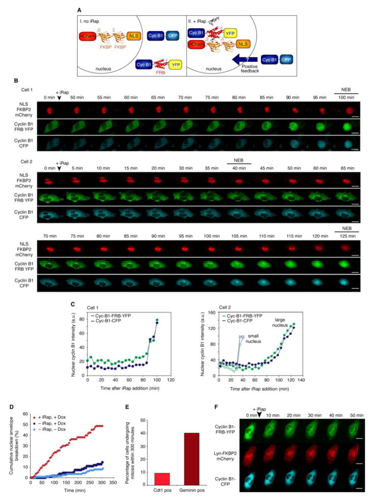
Figure 3
Induced Translocation of Active Cdk1-Cyclin B1 Complexes to the Nucleus Triggers Mitosis and Induces Spatial Positive Feedback
(A) Schematic of the experimental approach by which the rapamycin-analog iRap causes cyclin B1-FRB to accumulate in the nucleus (see also Figure S3 ). (B) Two representative cells expressing mCherry-N...
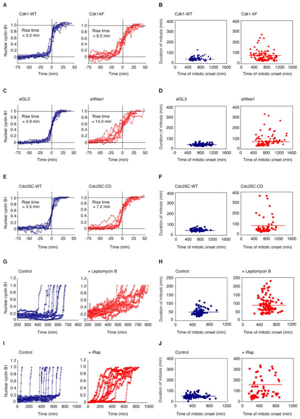
Figure 4
Timely Completion of Mitosis Requires Abrupt Activation and Redistribution of Cdk1-Cyclin B1
(A, B) Cdk1AF expression makes cyclin B1 translocation more graded and makes the duration of mitosis longer and more variable. As a proxy for the duration mitosis we measured the time of completion of...
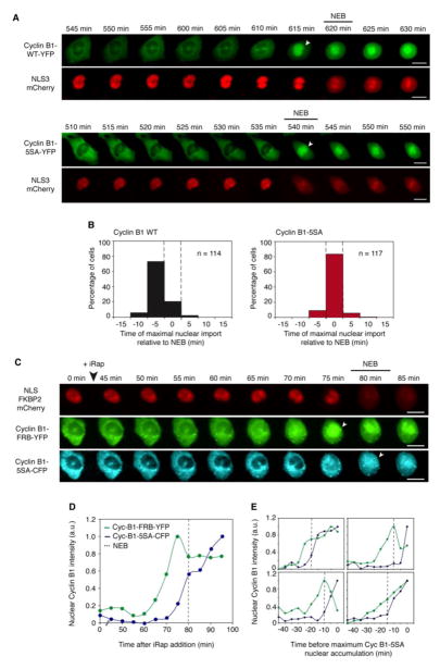
Figure 5
Cyclin B1 Phosphorylation is Required for the Pre-NEB Translocation of Cyclin B1 to the Nucleus and for Spatial Positive Feedback
(A, B) Cyclin B1-WT-YFP generally translocates to the nucleus ~5 min prior to NEB. In contrast, cyclin B1-5SA translocates to the nucleus concomitantly with NEB. Panel A shows representative cells. Th...
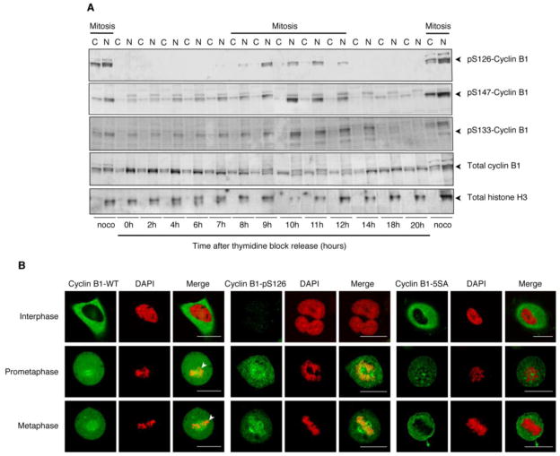
Figure 6
Cyclin B1 Phosphorylation Is Required for the Stable Association of Cyclin B1 with Mitotic Chromosomes
(A) Cell fractionation. Cells were released from a double thymidine block and lysed at various times. Mitotic cells were obtained by releasing double thymidine blocked cells into nocodazole for 12 hou...
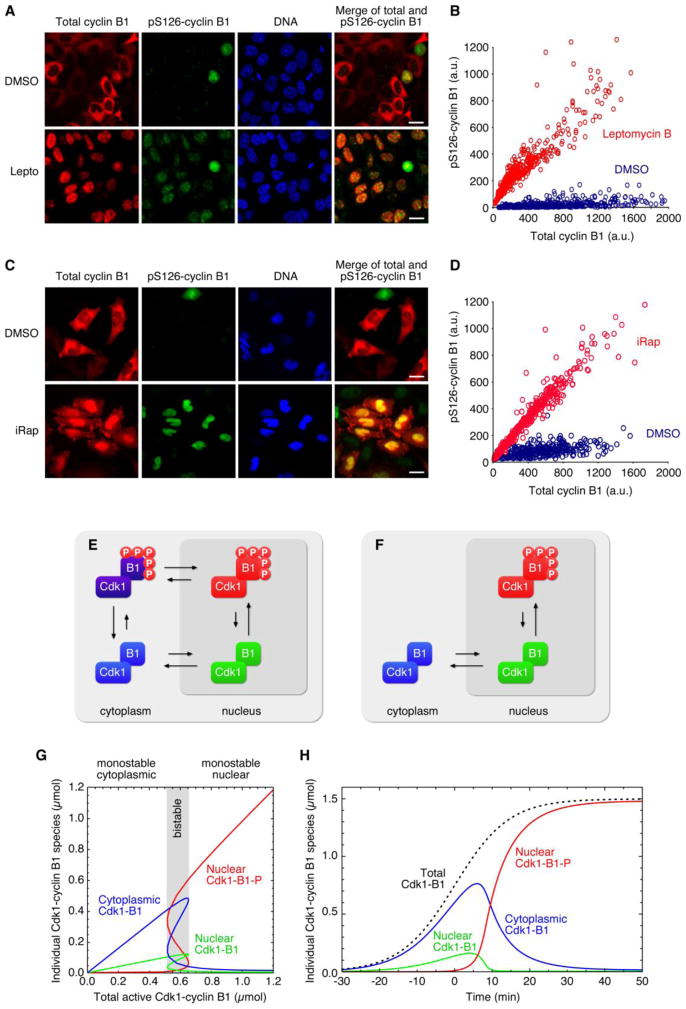
Figure 7
Translocation Promotes Cyclin B1 Phosphorylation, Completing the Positive Feedback Loop
(A) Control (DMSO-treated) HeLa cells (top) and leptomycin B-treated HeLa cells (bottom) 8 h post release from a double thymidine block. Cells were stained for total cyclin B1 (red), pS126-cyclin B1 (...
Figure images are served from the NIH/NLM PubMed Central Open Access Subset or Europe PMC; copyright remains with the publishers and authors.
💬 Discussion
0 commentsNo comments yet. Be the first to start a discussion!
Leave a Comment