Abstract
Frizzleds (Fzd) are the primary receptors for Wnt morphogens, which are essential regulators of stem cell biology, yet the structural basis of Wnt signaling through Fzd remains poorly understood. Here we report the structure of an unliganded human Fzd5 determined by single-particle cryo-EM at 3.7 Å resolution, with the aid of an antibody chaperone acting as a fiducial marker. We also analyzed the topology of low-resolution XWnt8/Fzd5 complex particles, which revealed extreme flexibility between the Wnt/Fzd-CRD and the Fzd-TM regions. Analysis of Wnt/β-catenin signaling in response to Wnt3a versus a 'surrogate agonist' that cross-links Fzd to LRP6, revealed identical structure-activity relationships. Thus, canonical Wnt/β-catenin signaling appears to be principally reliant on ligand-induced Fzd/LRP6 heterodimerization, versus the allosteric mechanisms seen in structurally analogous class A G protein-coupled receptors, and Smoothened. These findings deepen our mechanistic understanding of Wnt signal transduction, and have implications for harnessing Wnt agonism in regenerative medicine.
🔬 Techniques
🧬 Organisms
✨ Fluorophores
🧪 Sample Preparation
🔬 Cell Lines
🏭 Microscope Brands
🧪 Reagent Suppliers
📷 Detectors
💻 Software Details
💾 Data Repositories
🏷️ Research Resource Identifiers (RRIDs)
Verified research resources used in this paper:
🏛️ Research Organizations (ROR)
Affiliated research institutions:
📋 Methods
Key resources table
Reagent type (species) or resource Designation Source or reference Identifiers Additional information Cell line ( Spodoptera frugiperda ) Sf9 ATCC CTL-1711 (RRID: CVCL_0549 ) Insect cells used for baculovirus production Cell line ( Homo sapiens ) HEK293S GnTI - Gift from Prof. H Gobind Khorana (PMID: 12370423 ) Mammalian cells used for baculovirus expression of hFzd5FL and hFzd5 ICL3 BRIL Cell line ( Homo sapiens ) HEK293T with Fzd1/2/4/5/7/8 KO Gift from Prof. Michael Boutros (PMID: 28733458 ) For signaling assay Cell line ( Mus musculus ) Mouse monoclonal M1 hybridoma Gift from Prof. Brian K Kobilka (PMID: 17962520 ) To purify anti-FLAG M1 (mouse IgG2a) antibody to prepare FLAG affinity column (15 mg/ml resin). This was used for purification of hFzd5FL and hFzd5 ICL3 BRIL. Strain, strain background ( Escherichia coli ) BL21(DE3) New England Biolabs C2527I E. coli strain for expression of the nanobody Strain, strain background ( Escherichia coli ) BL21 (gold) Agilent Technologies 230130 E. coli strain for expression of the Fabs Transfected construct ( Homo sapiens ) pRK5 hFzd5 wild-type Yu et al., 2012 (PMID: 23095888 ) For signaling assay Transfected construct ( Homo sapiens ) pRK5 hFzd5 mutants this study For signaling assay Recombinant DNA reagent 7xTCF-Ffluc Addgene 24308 For signaling assay Recombinant DNA reagent BestBac Linearized Baculovirus DNA 2.0 Expression Systems 91–002 For baculovirus production Peptide, recombinant protein Wnt3a R and D Systems 5036-WN For signaling assay Peptide, recombinant protein Surrogate Wnt Miao et al. Cell Stem Cell 2020 For signaling assay Peptide, recombinant protein DRPB_Fz8 Dang et al., 2019 (PMID: 31086346 ) For signaling assay Commercial assay or kit Luciferase assay kit Promega E152A For signaling assay Chemical compound, drug n-Dodecyl-β-D-Maltoside (DDM) Anatrace D310S Membrane protein purification Chemical compound, drug Lauryl Maltose Neopentyl Glycol (LMNG) Anatrace NG310 Membrane protein purification Chemical compound, drug Glyco-Diosgenin (GDN) Anatrace GDN101 Membrane protein purification Chemical compound, drug Cholesterol Hemisuccinate tris Salt (CHS) Anatrace CH210 Membrane protein purification Chemical compound, drug Digitonin EMD Millipore 300410 Cryo-EM specimen freezing additive Software, algorithm DigitalMicrograph Gatan Microscope alignment and cryo-EM data collection Software, algorithm serialEM https://bio3d.colorado.edu/SerialEM/ (PMID: 16182563 ) version 3.6 Cryo-EM data collection Software, algorithm MotionCor2 https://emcore.ucsf.edu/ucsf-motioncor2/ (PMID: 28250466 ) Motion correction of cryo-EM movies Software, algorithm cryoSPARC https://cryosparc.com/ (PMID: 28165473 ) version 2.12.14 Cryo-EM data processing Software, algorithm Coot https://www2.mrc-lmb.cam.ac.uk/personal/pemsley/coot/ (PMID: 20383002 ) Structure model building Software, algorithm Isolde https://isolde.cimr.cam.ac.uk/ (PMID: 29872003 ) Structure model building/refinement Software, algorithm Phenix suite https://www.phenix-online.org/ (PMID: 20124702 ) Structure refinement Software, algorithm UCSF Chimera https://www.cgl.ucsf.edu/chimera/ (PMID: 15264254 ) Initial homology model docking Software, algorithm UCSF ChimeraX https://www.rbvi.ucsf.edu/chimerax/ (PMID: 28710774 ) Structural visualization/figure preparation/Isolde execution Software, algorithm PyMol Schrödinger Structural visualization/figure preparation Software, algorithm GraphPad Prism 7 GraphPad analysis of signaling data Other CNBr-Activated Sepharose 4 Fast Flow GE Healthcare 17098101 For preparation of anti-FLAG sepharose Other Lipofectamine 2000 Invitrogen 11668030 Transfection reagent for signaling assay Cell lines Sf9 cells were obtained from ATCC (CTL-1711, RRID: CVCL_0549 ), HEK293S GnTI - cells were provided by Prof. H Gobind Khorana, Fzd1/2/4/5/7/8 knockout HEK293T cells were provided by Prof. Michael Boutros and M1 hybridoma cells were provided by Prof. Brian K Kobilka. Cell line authentication was guaranteed by the sources where the cells were obtained. For Sf9, ATCC does not indicate the authentication method. For HEK293S GnTI - cells, clonal selection was performed by the provider based on their GnTI activity, and the authentication was regularly monitored by purified protein's sensitivity to Endo H enzyme. For Fzd1/2/4/5/7/8 knockout HEK293T cells, the original cell authentication was confirmed with NGS sequencing by the provider, and the authentication was regularly monitored by their response to Wnt3a as shown in this study (negative and positive controls). For M1 hybridoma, clonal selection was performed by the provider based on the antibody expression levels, and the authentication was monitored in the same way. Routine mycoplasma tests were not performed for each cell line. All cell lines were kept at low passages in order to maintain their health and identity. Protein expression and purification for cryo-EM study The expression construct for human Frizzled five was designed with insertion of cytochrome b562 RIL (BRIL) in the intracellular loop 3 (termed hFzd5 ICL3 BRIL) with no additional thermostabilizing mutation. The chimeric protein was made using the inactive BRIL-fusion Smoothened with V329F 3.40 mutation in TM3 (PDB: ID 5L7D) as a reference, which yielded well-diffracting crystals in the report ( Byrne et al., 2016 ). Wild-type hFzd5 (residue 27–428 and 441–546, UniProt ID: Q13467 ) was connected to BRIL using two short linkers derived from A 2A adenosine receptor (ARRQL between residue 428 and N-terminus of BRIL, and ARSTL between C-terminus of BRIL and residue 441) and cloned with an N-terminal HA signal peptide and a FLAG epitope into the mammalian baculovirus expression (BacMam) vector pVLAD6 ( Dukkipati et al., 2008 ), that has a C-terminal 3C protease site followed by a protein C epitope and an octahistidine tag. While we did not have to test multiple constructs for cryo-EM screening, we noticed that the extended helical linker was suitable for the Fab marker, which would have otherwise led to a ‘collision’ between the Fab/Nb unit and the detergent micelle. We also note that hFzd5 ICL3 BRIL had an overall 5-fold higher yield after expression and purification compared to hFzd5FL, indirectly suggesting its superior stability. P0 virus was prepared in Sf9 by co-transfecting the expression vector and BestBac Linearized Baculovirus DNA (Expression Systems, CA, USA) using a standard method, and the virus was amplified from P0 to P2. hFzd5 ICL3 BRIL was expressed in HEK293S GnTI - cells with BacMam baculovirus transduction. 4% (v/v) P2 virus was added to cells at a density of 2 × 10 6 cells per ml and culture flasks were shaken at 37 °C for 24 hr with 5% CO 2 . After centrifugation, cells were washed with phosphate-buffered saline (PBS) supplemented with 5 mM EDTA and 1:1000 protease inhibitor cocktail (PIC, Sigma Aldrich, MO, USA), weigh and stored at −20°C. Approx. 30 grams of cell pellet was thawed and lysed with a Dounce homogenizer in a lysis buffer composed of 20 mM Tris-HCl pH 8.0, 5 mM EDTA, 2 mg/ml iodoacetamide, and 1:1000 PIC. The lysate was centrifuged at 48,000 x g for 1 hr and the membrane pellet was resuspended and nutated for 2 hr in 400 ml of a solubilization buffer consisting of 10 mM HEPES pH 7.2, 150 mM NaCl (HBS), 1% (w/v) n-dodecyl-β-D-maltopyranoside (DDM, Sol-Grade, Anatrace), 0.2% (w/v) cholesterol hemisuccinate (CHS, Anatrace), 10% (v/v) glycerol, 2 mg/ml iodoacetamide, and cOmplete PIC (Roche, Basel, Switzerland). After centrifugation at 48,000 x g for 1 hr, 10 ml Ni-NTA resin (Qiagen, Hilden, Germany) was added to the supernatant, and the mixture was stirred at 4°C overnight. The resin was collected in a column, washed with HBS with 0.1% (w/v) DDM, 0.02% (w/v) CHS, 10% (v/v) glycerol, and 20 mM imidazole (wash buffer) and eluted in wash buffer supplemented with 230 mM imidazole. 2 mM CaCl 2 was then added to the eluate, which was further purified over the in-house anti-FLAG M1 affinity sepharose column. The resin was washed with HBS + 2 mM CaCl 2 supplemented with detergents, which was gradually exchanged from 0.1% DDM to 0.01% GDN. The receptor was eluted in HBS with 0.002% (w/v) GDN, 0.0002% (w/v) CHS, 0.2 mg/ml FLAG peptide and 5 mM EDTA, and further purified by a size-exclusion chromatography (SEC) column Superdex 200 10/300 GL equilibrated with SEC buffer comprised of HBS, 0.002% (w/v) GDN and 0.0002% (w/v) CHS. The anti-BRIL Fab was expressed in E. coli and purified as described ( Mukherjee et al., 2020 ). The anti-Fab Nb ( Ereño-Orbea et al., 2018 ; Hermans et al., 2017 ) was cloned in pET26b+ vector with an N-terminal hexahistidine tag followed by a TEV protease site. The protein was expressed in E. coli BL21(DE3) cells and purified by Ni-NTA chromatography, followed by removal of the His-tag by TEV digestion. The final step in the purification was a SEC on a Superdex 75 prep grade column whereby monodisperse Nb was obtained. The purified receptor was incubated with 1.4-fold and 2-fold molar excess of anti-BRIL Fab and anti-Fab Nb, respectively, at 4°C for overnight. The mixture was concentrated to 500 µL and injected into a SEC column Superdex 200 10/300 GL equilibrated with the SEC buffer. The peak fractions were concentrated to ~2 mg/ml and 0.01% digitonin was added at this point. The solution was further concentrated to ~8 mg/ml for cryo-EM analysis. To prepare the Wnt/Fzd complex, we expressed XWnt8 and hFzd5FL separately from Drosophila S2 cells or HEK293S GnTI - cells, respectively. For solubilized XWnt8, we employed the XWnt8/mFzd8CRD co-expression system developed previously ( Janda et al., 2012 ). Untagged XWnt8 and C-terminal Fc fusion mFzd8CRD were stably co-expressed in cell culture media, and secreted XWnt8/mFzd8CRD-Fc was captured on Protein A sepharose resin (Sigma). After washing the resin with HBS on column, only XWnt8 was eluted from the column with Wnt elution buffer containing 10 mM Hepes-Na pH 7.2, 500 mM NaCl and 0.1% DDM, in which DDM ‘wash out’ lipidated Wnt from CRD with competition. The eluted XWnt8 was then captured by ConA glycan affinity resin (Vector Laboratories) for detergent exchange. The detergent composition was gradually changed as follow; 0.1% DDM, 0.09% DDM/0.01% GDN, 0.05% DDM/0.05% GDN, 0.01% DDM/0.09% GDN, 0.1% GDN and 0.01% GDN. XWnt8 in GDN is eluted with HBS containing 0.01% GDN and 250 mM each Methyl α-D-glucopyranoside and Methyl α-D-mannopyranoside (Sigma). hFzd5FL was prepared by essentially the same scheme as hFzd5 ICL3 BRIL. Briefly, wild-type hFzd5FL with N-terminal FLAG and C-terminal octahistidine tags was expressed on cell membrane using BacMam system, extracted in 1% DDM/0.2% CHS and first purified with Ni-NTA resin. Pool fractions of hFzd5FL from Ni-NTA resin in 0.1% DDM/0.02% CHS was further purified with anti-FLAG M1 affinity sepharose with detergent exchange to 0.01% GDN/0.002% CHS, and a Superdex 200 10/300 GL column equilibrated with the SEC buffer. 1.2 molar excess XWnt8 was added to the SEC purified hFzd5FL, and incubated at 4°C for overnight. The mixture was concentrated to 500 µL and injected into a Superdex 200 10/300 GL column equilibrated with the SEC buffer. The peak fractions were concentrated to 5–15 mg/ml with final
Show full methods section
Key resources table
Reagent type (species) or resource Designation Source or reference Identifiers Additional information Cell line ( Spodoptera frugiperda ) Sf9 ATCC CTL-1711 (RRID: CVCL_0549 ) Insect cells used for baculovirus production Cell line ( Homo sapiens ) HEK293S GnTI - Gift from Prof. H Gobind Khorana (PMID: 12370423 ) Mammalian cells used for baculovirus expression of hFzd5FL and hFzd5 ICL3 BRIL Cell line ( Homo sapiens ) HEK293T with Fzd1/2/4/5/7/8 KO Gift from Prof. Michael Boutros (PMID: 28733458 ) For signaling assay Cell line ( Mus musculus ) Mouse monoclonal M1 hybridoma Gift from Prof. Brian K Kobilka (PMID: 17962520 ) To purify anti-FLAG M1 (mouse IgG2a) antibody to prepare FLAG affinity column (15 mg/ml resin). This was used for purification of hFzd5FL and hFzd5 ICL3 BRIL. Strain, strain background ( Escherichia coli ) BL21(DE3) New England Biolabs C2527I E. coli strain for expression of the nanobody Strain, strain background ( Escherichia coli ) BL21 (gold) Agilent Technologies 230130 E. coli strain for expression of the Fabs Transfected construct ( Homo sapiens ) pRK5 hFzd5 wild-type Yu et al., 2012 (PMID: 23095888 ) For signaling assay Transfected construct ( Homo sapiens ) pRK5 hFzd5 mutants this study For signaling assay Recombinant DNA reagent 7xTCF-Ffluc Addgene 24308 For signaling assay Recombinant DNA reagent BestBac Linearized Baculovirus DNA 2.0 Expression Systems 91–002 For baculovirus production Peptide, recombinant protein Wnt3a R and D Systems 5036-WN For signaling assay Peptide, recombinant protein Surrogate Wnt Miao et al. Cell Stem Cell 2020 For signaling assay Peptide, recombinant protein DRPB_Fz8 Dang et al., 2019 (PMID: 31086346 ) For signaling assay Commercial assay or kit Luciferase assay kit Promega E152A For signaling assay Chemical compound, drug n-Dodecyl-β-D-Maltoside (DDM) Anatrace D310S Membrane protein purification Chemical compound, drug Lauryl Maltose Neopentyl Glycol (LMNG) Anatrace NG310 Membrane protein purification Chemical compound, drug Glyco-Diosgenin (GDN) Anatrace GDN101 Membrane protein purification Chemical compound, drug Cholesterol Hemisuccinate tris Salt (CHS) Anatrace CH210 Membrane protein purification Chemical compound, drug Digitonin EMD Millipore 300410 Cryo-EM specimen freezing additive Software, algorithm DigitalMicrograph Gatan Microscope alignment and cryo-EM data collection Software, algorithm serialEM https://bio3d.colorado.edu/SerialEM/ (PMID: 16182563 ) version 3.6 Cryo-EM data collection Software, algorithm MotionCor2 https://emcore.ucsf.edu/ucsf-motioncor2/ (PMID: 28250466 ) Motion correction of cryo-EM movies Software, algorithm cryoSPARC https://cryosparc.com/ (PMID: 28165473 ) version 2.12.14 Cryo-EM data processing Software, algorithm Coot https://www2.mrc-lmb.cam.ac.uk/personal/pemsley/coot/ (PMID: 20383002 ) Structure model building Software, algorithm Isolde https://isolde.cimr.cam.ac.uk/ (PMID: 29872003 ) Structure model building/refinement Software, algorithm Phenix suite https://www.phenix-online.org/ (PMID: 20124702 ) Structure refinement Software, algorithm UCSF Chimera https://www.cgl.ucsf.edu/chimera/ (PMID: 15264254 ) Initial homology model docking Software, algorithm UCSF ChimeraX https://www.rbvi.ucsf.edu/chimerax/ (PMID: 28710774 ) Structural visualization/figure preparation/Isolde execution Software, algorithm PyMol Schrödinger Structural visualization/figure preparation Software, algorithm GraphPad Prism 7 GraphPad analysis of signaling data Other CNBr-Activated Sepharose 4 Fast Flow GE Healthcare 17098101 For preparation of anti-FLAG sepharose Other Lipofectamine 2000 Invitrogen 11668030 Transfection reagent for signaling assay Cell lines Sf9 cells were obtained from ATCC (CTL-1711, RRID: CVCL_0549 ), HEK293S GnTI - cells were provided by Prof. H Gobind Khorana, Fzd1/2/4/5/7/8 knockout HEK293T cells were provided by Prof. Michael Boutros and M1 hybridoma cells were provided by Prof. Brian K Kobilka. Cell line authentication was guaranteed by the sources where the cells were obtained. For Sf9, ATCC does not indicate the authentication method. For HEK293S GnTI - cells, clonal selection was performed by the provider based on their GnTI activity, and the authentication was regularly monitored by purified protein's sensitivity to Endo H enzyme. For Fzd1/2/4/5/7/8 knockout HEK293T cells, the original cell authentication was confirmed with NGS sequencing by the provider, and the authentication was regularly monitored by their response to Wnt3a as shown in this study (negative and positive controls). For M1 hybridoma, clonal selection was performed by the provider based on the antibody expression levels, and the authentication was monitored in the same way. Routine mycoplasma tests were not performed for each cell line. All cell lines were kept at low passages in order to maintain their health and identity. Protein expression and purification for cryo-EM study The expression construct for human Frizzled five was designed with insertion of cytochrome b562 RIL (BRIL) in the intracellular loop 3 (termed hFzd5 ICL3 BRIL) with no additional thermostabilizing mutation. The chimeric protein was made using the inactive BRIL-fusion Smoothened with V329F 3.40 mutation in TM3 (PDB: ID 5L7D) as a reference, which yielded well-diffracting crystals in the report ( Byrne et al., 2016 ). Wild-type hFzd5 (residue 27–428 and 441–546, UniProt ID: Q13467 ) was connected to BRIL using two short linkers derived from A 2A adenosine receptor (ARRQL between residue 428 and N-terminus of BRIL, and ARSTL between C-terminus of BRIL and residue 441) and cloned with an N-terminal HA signal peptide and a FLAG epitope into the mammalian baculovirus expression (BacMam) vector pVLAD6 ( Dukkipati et al., 2008 ), that has a C-terminal 3C protease site followed by a protein C epitope and an octahistidine tag. While we did not have to test multiple constructs for cryo-EM screening, we noticed that the extended helical linker was suitable for the Fab marker, which would have otherwise led to a ‘collision’ between the Fab/Nb unit and the detergent micelle. We also note that hFzd5 ICL3 BRIL had an overall 5-fold higher yield after expression and purification compared to hFzd5FL, indirectly suggesting its superior stability. P0 virus was prepared in Sf9 by co-transfecting the expression vector and BestBac Linearized Baculovirus DNA (Expression Systems, CA, USA) using a standard method, and the virus was amplified from P0 to P2. hFzd5 ICL3 BRIL was expressed in HEK293S GnTI - cells with BacMam baculovirus transduction. 4% (v/v) P2 virus was added to cells at a density of 2 × 10 6 cells per ml and culture flasks were shaken at 37 °C for 24 hr with 5% CO 2 . After centrifugation, cells were washed with phosphate-buffered saline (PBS) supplemented with 5 mM EDTA and 1:1000 protease inhibitor cocktail (PIC, Sigma Aldrich, MO, USA), weigh and stored at −20°C. Approx. 30 grams of cell pellet was thawed and lysed with a Dounce homogenizer in a lysis buffer composed of 20 mM Tris-HCl pH 8.0, 5 mM EDTA, 2 mg/ml iodoacetamide, and 1:1000 PIC. The lysate was centrifuged at 48,000 x g for 1 hr and the membrane pellet was resuspended and nutated for 2 hr in 400 ml of a solubilization buffer consisting of 10 mM HEPES pH 7.2, 150 mM NaCl (HBS), 1% (w/v) n-dodecyl-β-D-maltopyranoside (DDM, Sol-Grade, Anatrace), 0.2% (w/v) cholesterol hemisuccinate (CHS, Anatrace), 10% (v/v) glycerol, 2 mg/ml iodoacetamide, and cOmplete PIC (Roche, Basel, Switzerland). After centrifugation at 48,000 x g for 1 hr, 10 ml Ni-NTA resin (Qiagen, Hilden, Germany) was added to the supernatant, and the mixture was stirred at 4°C overnight. The resin was collected in a column, washed with HBS with 0.1% (w/v) DDM, 0.02% (w/v) CHS, 10% (v/v) glycerol, and 20 mM imidazole (wash buffer) and eluted in wash buffer supplemented with 230 mM imidazole. 2 mM CaCl 2 was then added to the eluate, which was further purified over the in-house anti-FLAG M1 affinity sepharose column. The resin was washed with HBS + 2 mM CaCl 2 supplemented with detergents, which was gradually exchanged from 0.1% DDM to 0.01% GDN. The receptor was eluted in HBS with 0.002% (w/v) GDN, 0.0002% (w/v) CHS, 0.2 mg/ml FLAG peptide and 5 mM EDTA, and further purified by a size-exclusion chromatography (SEC) column Superdex 200 10/300 GL equilibrated with SEC buffer comprised of HBS, 0.002% (w/v) GDN and 0.0002% (w/v) CHS. The anti-BRIL Fab was expressed in E. coli and purified as described ( Mukherjee et al., 2020 ). The anti-Fab Nb ( Ereño-Orbea et al., 2018 ; Hermans et al., 2017 ) was cloned in pET26b+ vector with an N-terminal hexahistidine tag followed by a TEV protease site. The protein was expressed in E. coli BL21(DE3) cells and purified by Ni-NTA chromatography, followed by removal of the His-tag by TEV digestion. The final step in the purification was a SEC on a Superdex 75 prep grade column whereby monodisperse Nb was obtained. The purified receptor was incubated with 1.4-fold and 2-fold molar excess of anti-BRIL Fab and anti-Fab Nb, respectively, at 4°C for overnight. The mixture was concentrated to 500 µL and injected into a SEC column Superdex 200 10/300 GL equilibrated with the SEC buffer. The peak fractions were concentrated to ~2 mg/ml and 0.01% digitonin was added at this point. The solution was further concentrated to ~8 mg/ml for cryo-EM analysis. To prepare the Wnt/Fzd complex, we expressed XWnt8 and hFzd5FL separately from Drosophila S2 cells or HEK293S GnTI - cells, respectively. For solubilized XWnt8, we employed the XWnt8/mFzd8CRD co-expression system developed previously ( Janda et al., 2012 ). Untagged XWnt8 and C-terminal Fc fusion mFzd8CRD were stably co-expressed in cell culture media, and secreted XWnt8/mFzd8CRD-Fc was captured on Protein A sepharose resin (Sigma). After washing the resin with HBS on column, only XWnt8 was eluted from the column with Wnt elution buffer containing 10 mM Hepes-Na pH 7.2, 500 mM NaCl and 0.1% DDM, in which DDM ‘wash out’ lipidated Wnt from CRD with competition. The eluted XWnt8 was then captured by ConA glycan affinity resin (Vector Laboratories) for detergent exchange. The detergent composition was gradually changed as follow; 0.1% DDM, 0.09% DDM/0.01% GDN, 0.05% DDM/0.05% GDN, 0.01% DDM/0.09% GDN, 0.1% GDN and 0.01% GDN. XWnt8 in GDN is eluted with HBS containing 0.01% GDN and 250 mM each Methyl α-D-glucopyranoside and Methyl α-D-mannopyranoside (Sigma). hFzd5FL was prepared by essentially the same scheme as hFzd5 ICL3 BRIL. Briefly, wild-type hFzd5FL with N-terminal FLAG and C-terminal octahistidine tags was expressed on cell membrane using BacMam system, extracted in 1% DDM/0.2% CHS and first purified with Ni-NTA resin. Pool fractions of hFzd5FL from Ni-NTA resin in 0.1% DDM/0.02% CHS was further purified with anti-FLAG M1 affinity sepharose with detergent exchange to 0.01% GDN/0.002% CHS, and a Superdex 200 10/300 GL column equilibrated with the SEC buffer. 1.2 molar excess XWnt8 was added to the SEC purified hFzd5FL, and incubated at 4°C for overnight. The mixture was concentrated to 500 µL and injected into a Superdex 200 10/300 GL column equilibrated with the SEC buffer. The peak fractions were concentrated to 5–15 mg/ml with final
📊 Figures
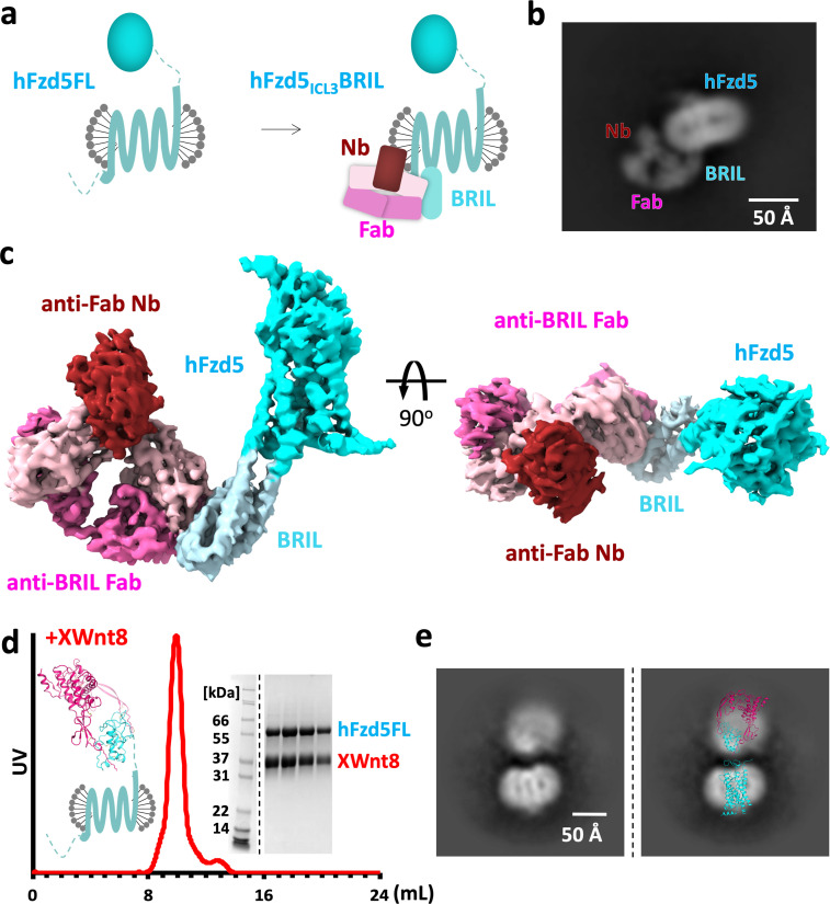
Figure 1.
Design scheme and cryo-EM analysis of hFzd5.
( a ) Cartoon representation of the strategy for hFzd5 particle decoration by anti-BRIL Fab and anti-Fab Nb. These chaperones double hFzd5 molecular weight and render it asymmetric in detergent micell...
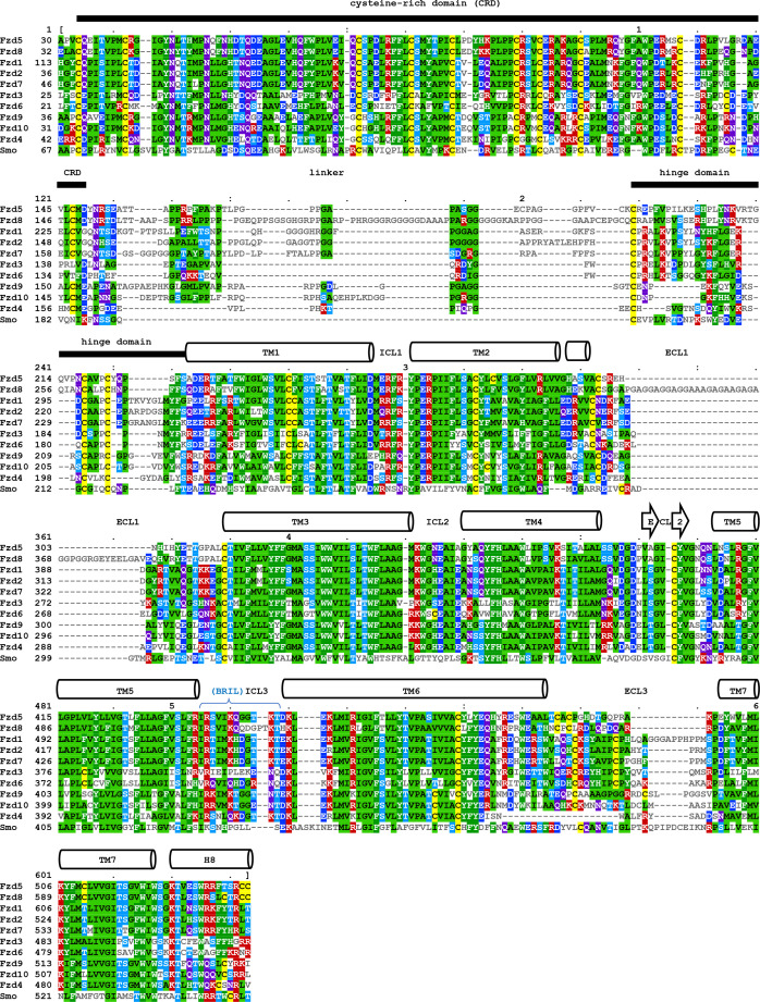
Figure 1u2014figure supplement 1.
Multiple sequence alignment of human class F GPCRs.
Sequence alignment of CRD and transmembrane regions of human class F GPCRs. The CRD region and individual transmembrane helix is labelled above the amino acid sequence. The alignments for CRD-hinge do...
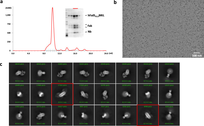
Figure 1u2014figure supplement 2.
Purification, data collection and 2D classification of hFzd5 ICL3 BRIL/Fab/Nb.
( a ) Representative size-exclusion chromatography profile of the hFzd5 ICL3 BRIL/Fab/Nb complex with the Coomassie stained SDS-PAGE around the peak fractions. SDS samples were prepared in reducing co...
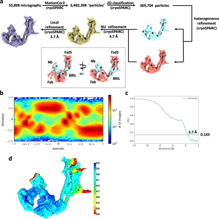
Figure 1u2014figure supplement 3.
Cryo-EM data analysis of hFzd5 ICL3 BRIL/Fab/Nb.
( a ) Cryo-EM data processing scheme. The resulting maps from the final heterogeneous refinement are superimposed for comparison (cyan and brick). ( b ) Orientation distribution of particles for the f...
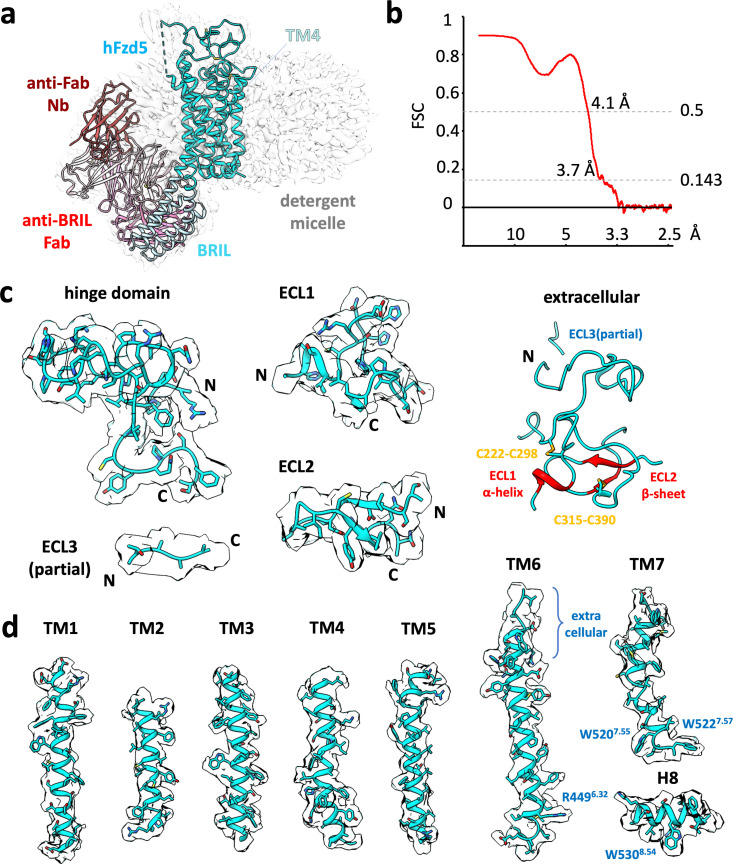
Figure 1u2014figure supplement 4.
Cryo-EM map of representative built in model.
( a ) Cryo-EM map (gray) and structural model of hFzd5 ICL3 BRIL in detergent complexed with anti-BRIL Fab and anti-Fab Nb. The model is colored as follow: the sequences from Fzd5 (cyan), BRIL and the...
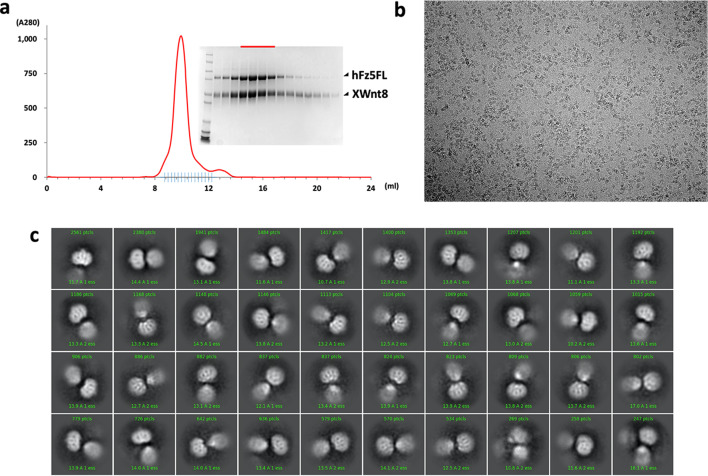
Figure 1u2014figure supplement 5.
Purification, data collection and 2D class averages of the XWnt8/hFzd5FL complex.
( a ) A size-exclusion chromatography profile of the XWnt8/hFzd5FL complex with the Coomassie stained SDS-PAGE. SDS samples were prepared in reducing condition. The peak fractions for the EM analysis ...
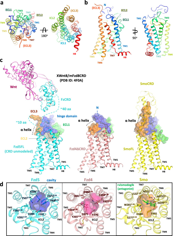
Figure 2.
Overall structure of hFzd5 and structural comparison with hFzd4 and hSmo.
( a ) Top (left) and bottom (right) views, and ( b ) side views of hFzd5 colored by rainbow with blue on the N-terminus and red on the C-terminus of the structural model. ( c ) Comparison of the extra...
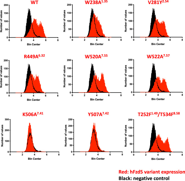
Figure 2u2014figure supplement 1.
Confirmation of hFzd5 receptor expression in HEK293T transfected cells.
Cell-surface hFzd5 variants were fluorescently labeled, and frequency distribution of FACS data are presented for each variant (red) with a negative control (black). Figure 2u2014figure supplement 1u2...
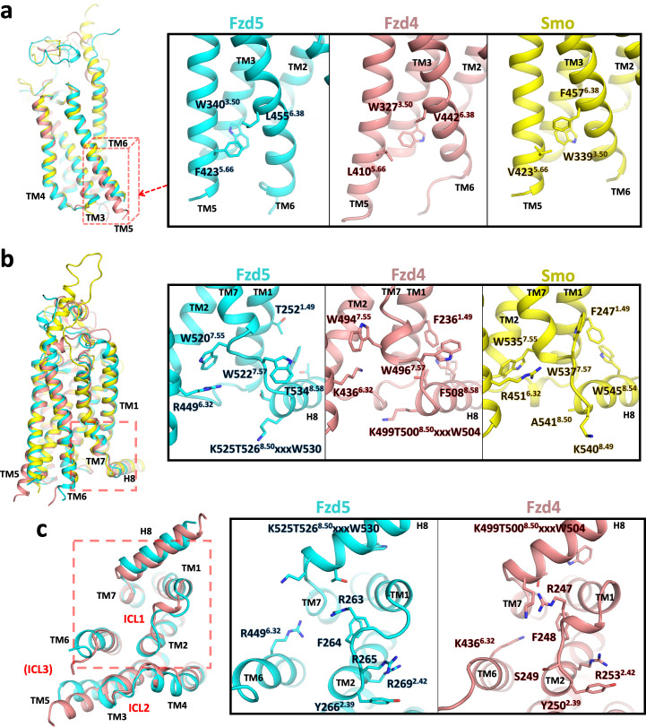
Figure 3.
Conformational differences between hFzd5, hFzd4 and hSmo.
( a, b ) Structural comparison of hFzd5, hFzd4 and hSmou00a0( a ) at the cytoplasmic end of TM5, andu00a0(b) the K/R 6.32 -W 7.55 ionic lock with the aromatic network around H8. ( c ) Bottom views of ...
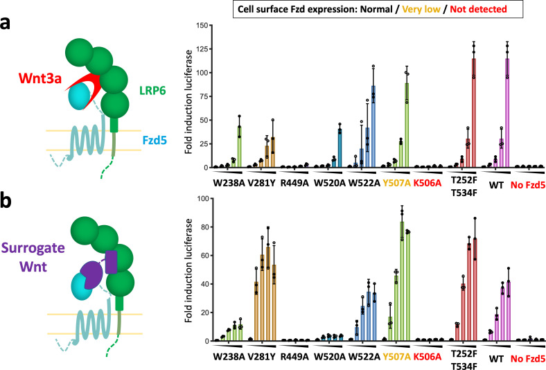
Figure 4.
Structure-guided hFzd5 mutant signaling assays.
Schematic presentation of the ternary complex formation, and hFzd5-mediated signaling upon stimulation by ( a ) Wnt3a and ( b ) surrogate Wnt agonist. Non-lipidated, water-soluble surrogate Wnt would ...
Figure images are served from the NIH/NLM PubMed Central Open Access Subset or Europe PMC; copyright remains with the publishers and authors.
💬 Discussion
0 commentsNo comments yet. Be the first to start a discussion!
Leave a Comment