Abstract
Preribosomal particles evolve in the nucleus through transient interaction with biogenesis factors before export to the cytoplasm. Here, we report the architecture of the late pre-60S particle, purified from Saccharomyces cerevisiae, through Arx1, a nuclear export factor with structural homology to methionine aminopeptidases, or its binding partner Alb1. Cryo-EM reconstruction of the Arx1 particle at 11.9-Å resolution reveals regions of extra density on the pre-60S particle attributed to associated biogenesis factors, confirming the immature state of the nascent subunit. One of these densities could be unambiguously assigned to Arx1. Immunoelectron microscopy and UV cross-linking localize Arx1 close to the ribosomal exit tunnel, in direct contact with ES27, a highly dynamic eukaryotic rRNA expansion segment. The binding of Arx1 at the exit tunnel may position this export factor to prevent premature recruitment of ribosome-associated factors active during translation.
🔬 Techniques
🔭 Microscopes
🧬 Organisms
🏭 Microscope Brands
🧪 Reagent Suppliers
📷 Detectors
💻 Software Details
💾 Data Repositories
🏛️ Research Organizations (ROR)
Affiliated research institutions:
📋 Methods
Yeast strains
Yeast strains used in this study are listed in Supplementary Table 1 . The genotype of the DS1-2b strain is MATalpha his3-Δ00 leu2-Δ1 trp1-Δ63 ura3-Δ52 derived from FY23xFY86 26 . TAP-tag or 3HA-tag were genomically integrated at the C-terminus as described 49 , 50 . For genomic integration of the Flag-tag the sequence of the Flag-epitope was integrated into the F3 primer 50 . For genomic integration of C-terminal His 6 -TEV-ProtA-tag the integration cassette plasmid pFA6a-HTpA-HIS3MX4 (kindly provided by D. Kressler) was used. CRAC analysis of Arx1 The CRAC method and bioinformatics analysis were performed as described 35 using the Arx1-HTP (His 6 -TEV-ProtA) strain for CRAC analysis of Arx1 and the parental strain DS1-2b as a negative control. Two independent experiments were performed and the results merged. Biochemical purification of pre-60S particles Particles were essentially purified from 2-4 liters YPD liquid cultures grown to log phase at 30°C via TAP (tandem affinity purification) in standard buffer (50 mM Tris-HCl [pH 7.5], 100 mM NaCl, 1.5 mM or 5 mM MgCl 2 [where indicated], 0.075 % (v/v) NP-40) as described 26 with the following modifications. NP-40 was present only during cell lysis. Final eluates were in standard buffer without NP-40, with 0.5 mM DTT and 3.5 mM EGTA. For split-tag purifications the TEV-protease eluate from the first affinity purification step of a TAP purification exploiting the ProtA-tag present on the bait protein was applied to anti-Flag M2-agarose from mouse (Sigma) for 1 hour at 4°C, followed by elution for 45 minutes at 4°C with 3xFlag peptide (Sigma) in standard buffer without NP-40 with 0.5 mM DTT. Immuno-labeling, negative stain EM, single particle analysis and random conical tilt Anti-HA antibody (monoclonal HA.11, Covance) was present in 1:50 (used for negative stain EM) or 1:200 dilution during the incubation on Calmodulin beads. Binding time was extended to 1.5 hours at 4°C. The tandem affinity-purified, split-tag purified, or TAP-purified and immuno-labeled particles were negatively stained with 2% uranyl acetate using the sandwich technique, imaged under low dose at 200 kV at 27,500 fold magnification (5.2 Å/pixel) with a Philips CM200 FEG transmission electron microscope supplied with a 2k x 2k CCD camera (TVIPS-GmbH) or at 100 kV at 33,000 or 53,000 fold magnification (3.6 Å/pixel or 2.2 Å/pixel) with a Philips CM120 BioTWIN LaB6 cathode transmission electron microscope supplied with a 4k x 4k CCD camera (FEI slow scan; used for negative stain of Fig. 4 ), all essentially as described 14 . Boxing of particles using “Boxer” 51 and image processing using IMAGIC-5 52 for single particle analysis were also essentially done as described 14 . For random conical tilt 53 , micrographs were taken at 0° and −55° tilt using the CM200 FEG microscope. A map was calculated from the tilted particles belonging to one class of untilted particles using IMAGIC-5 53 and SPIDER 54 software and displayed using the UCSF Chimera 55 software. Image and particle numbers analyzed were 176/4201 for Arx1-TAP, 250/8449 for Alb1-TAP Arx1-HA without anti-HA, 201/5236 for Alb1-TAP Rpl3-HA, 213/3310 for Alb1-TAP Rpl5-HA, 259/5933 for Alb1-TAP Rpl8-HA, 224/4331 for Alb1-TAP Rpl26-HA, 200/3835 for Alb1-TAP Arx1-HA, 162/4983 for Alb1-TAP Nsa2-HA, 116/3216 for Alb1-TAP Tif6-HA, 200/1476 for Rix1-TAP Arx1-HA, 170/2141 for Rix1-TAP, all with anti-HA, and 55/7219 for Alb1-TAP, all at 1.5 mM MgCl 2 , and 60/11,922 for Alb1-TAP at 5 mM MgCl 2 .
Show full methods section
Yeast strains
Yeast strains used in this study are listed in Supplementary Table 1 . The genotype of the DS1-2b strain is MATalpha his3-Δ00 leu2-Δ1 trp1-Δ63 ura3-Δ52 derived from FY23xFY86 26 . TAP-tag or 3HA-tag were genomically integrated at the C-terminus as described 49 , 50 . For genomic integration of the Flag-tag the sequence of the Flag-epitope was integrated into the F3 primer 50 . For genomic integration of C-terminal His 6 -TEV-ProtA-tag the integration cassette plasmid pFA6a-HTpA-HIS3MX4 (kindly provided by D. Kressler) was used. CRAC analysis of Arx1 The CRAC method and bioinformatics analysis were performed as described 35 using the Arx1-HTP (His 6 -TEV-ProtA) strain for CRAC analysis of Arx1 and the parental strain DS1-2b as a negative control. Two independent experiments were performed and the results merged. Biochemical purification of pre-60S particles Particles were essentially purified from 2-4 liters YPD liquid cultures grown to log phase at 30°C via TAP (tandem affinity purification) in standard buffer (50 mM Tris-HCl [pH 7.5], 100 mM NaCl, 1.5 mM or 5 mM MgCl 2 [where indicated], 0.075 % (v/v) NP-40) as described 26 with the following modifications. NP-40 was present only during cell lysis. Final eluates were in standard buffer without NP-40, with 0.5 mM DTT and 3.5 mM EGTA. For split-tag purifications the TEV-protease eluate from the first affinity purification step of a TAP purification exploiting the ProtA-tag present on the bait protein was applied to anti-Flag M2-agarose from mouse (Sigma) for 1 hour at 4°C, followed by elution for 45 minutes at 4°C with 3xFlag peptide (Sigma) in standard buffer without NP-40 with 0.5 mM DTT. Immuno-labeling, negative stain EM, single particle analysis and random conical tilt Anti-HA antibody (monoclonal HA.11, Covance) was present in 1:50 (used for negative stain EM) or 1:200 dilution during the incubation on Calmodulin beads. Binding time was extended to 1.5 hours at 4°C. The tandem affinity-purified, split-tag purified, or TAP-purified and immuno-labeled particles were negatively stained with 2% uranyl acetate using the sandwich technique, imaged under low dose at 200 kV at 27,500 fold magnification (5.2 Å/pixel) with a Philips CM200 FEG transmission electron microscope supplied with a 2k x 2k CCD camera (TVIPS-GmbH) or at 100 kV at 33,000 or 53,000 fold magnification (3.6 Å/pixel or 2.2 Å/pixel) with a Philips CM120 BioTWIN LaB6 cathode transmission electron microscope supplied with a 4k x 4k CCD camera (FEI slow scan; used for negative stain of Fig. 4 ), all essentially as described 14 . Boxing of particles using “Boxer” 51 and image processing using IMAGIC-5 52 for single particle analysis were also essentially done as described 14 . For random conical tilt 53 , micrographs were taken at 0° and −55° tilt using the CM200 FEG microscope. A map was calculated from the tilted particles belonging to one class of untilted particles using IMAGIC-5 53 and SPIDER 54 software and displayed using the UCSF Chimera 55 software. Image and particle numbers analyzed were 176/4201 for Arx1-TAP, 250/8449 for Alb1-TAP Arx1-HA without anti-HA, 201/5236 for Alb1-TAP Rpl3-HA, 213/3310 for Alb1-TAP Rpl5-HA, 259/5933 for Alb1-TAP Rpl8-HA, 224/4331 for Alb1-TAP Rpl26-HA, 200/3835 for Alb1-TAP Arx1-HA, 162/4983 for Alb1-TAP Nsa2-HA, 116/3216 for Alb1-TAP Tif6-HA, 200/1476 for Rix1-TAP Arx1-HA, 170/2141 for Rix1-TAP, all with anti-HA, and 55/7219 for Alb1-TAP, all at 1.5 mM MgCl 2 , and 60/11,922 for Alb1-TAP at 5 mM MgCl 2 .
Cryo-EM and 3D-reconstruction
As previously described 56 , samples were applied to carbon-coated holey grids, and micrographs were recorded under low-dose conditions (20 e − /Å 2 ) on a Titan Krios TEM (FEI Company) microscope at 200 kV in a defocus range of 1.0–3.5 μm with a TemCam F416 camera (4,096 × 4,096 pixel, TVIPS GmbH) resulting in a pixel size of 1.049 Å on the object scale. The data were processed with the SPIDER software package 54 . Images were manually inspected for good areas and power spectra. Particles were automatically picked from 6,359 micrographs using projections of the crystal structure of the S. cerevisiae 60S ribosomal subunit (PDB: 3O58 7 ) as a template resulting in 222,229 particles. After initial alignment to the 60S ribosomal subunit, remaining non-ribosomal particles and 80S contaminations were removed by semi-supervised classification using iterative multi-reference 3D projection alignment. Templates were provided only for the initial classification step. Subsequently, the respective output maps of the previous classification rounds were used as new templates. Non-ribosomal particles were identified by their higher cross-correlation to an essentially featureless high-contrast density than to ribosomal references. An 80S-containing population of particles accumulated when the prominent orange-colored ligand was removed from the density and the resulting map was supplied as alternative reference during classification. 54,196 pre-ribosomal particles were then classified using maps of consecutive refinement rounds as templates. Using these quasi identical templates, the dataset was classified according to the most relevant intrinsic heterogeneity (e.g. density in the intersubunit space) minimizing the risk of introducing bias by artificial templates. Subsequently, the data were analyzed by focused sorting according to the density below the exit tunnel using a cylindrical binary mask (removing 6,795 particles) 47 , 57 . 8,322 particles were used for an initial reconstruction of the Arx1 pre-60S ribosomal particle at 14.3 Å resolution. In an effort to improve the reconstruction the entire data set was then re-processed. Considering the substantial additional mass identified on the pre-60S subunit in the previous reconstruction, this reconstruction was now used as template for the initial alignment. This increased the number of identified pre-ribosomal particles to 112,632. These particles were again classified using maps of consecutive refinement rounds as templates, resulting in a stable set of 63,943 particles that was used for reconstruction of the Arx1-particle. The final contrast transfer function corrected reconstruction has a resolution of 11.9 Å, based on the Fourier Shell Correlation with a cutoff value of 0.5. The density for the conserved 60S core and additional densities were isolated manually using the UCSF Chimera 55 software. Miscellaneous Antibodies used for Western analysis in the following dilutions were anti-HA 1:3,000 (HA.11 mouse monoclonal antibody, clone 16B12, Cat.-No. MMS-101R, Covance, Berkeley, California, USA), anti-Arx1 32 1:2,000, anti-Nmd3 58 1:5,000, polyclonal rabbit anti-Mex67 1:5,000 (gift from Catherine Dargemont), anti-Mtr2 59 1:500, anti-Rpl3 60 1:4,000, anti-Rei1 25 1:5,000, anti-CBP 1:2,000 (Cat.-No. CAB1001, Thermo Scientific Open Biosystems, Rockford, Illinois, USA), goat-anti-mouse 1:6,000 (Cat.-No. 170-6516) and mouse-anti-rabbit horse radish peroxidase conjugated antibodies 1:6,000 (Cat.-No. 170-6515, both BIORAD, Munich, Germany). Page Ruler Unstained Protein Ladder (Thermo Scientific, Rockford, Illinois, USA) was used as a protein marker, Brillant Blue G-Colloidal Concentrate Electrophoresis Reagent (Sigma-Aldrich, Munich, Germany) was used for Coomassie stain, and 4-12% NuPAGE Bis-Tris Gels (Novex, Darmstadt, Germany) together with NuPAGE MOPS SDS Running Buffer (Invitrogen, Darmstadt, Germany) were used for SDS-PAGE.
Supplementary Material 1
📊 Figures
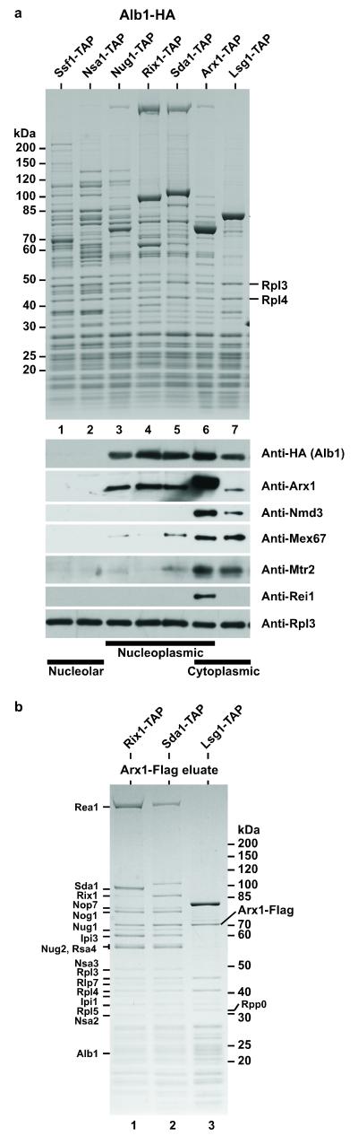
Figure 1
Arx1 and Alb1 are present on the same pre-60S particles
( a ) Tandem affinity purification of different pre-60S particles (nucleolar, nucleoplasmic, cytoplasmic) using the indicated TAP-tagged bait proteins from the respective yeast strains harboring chrom...
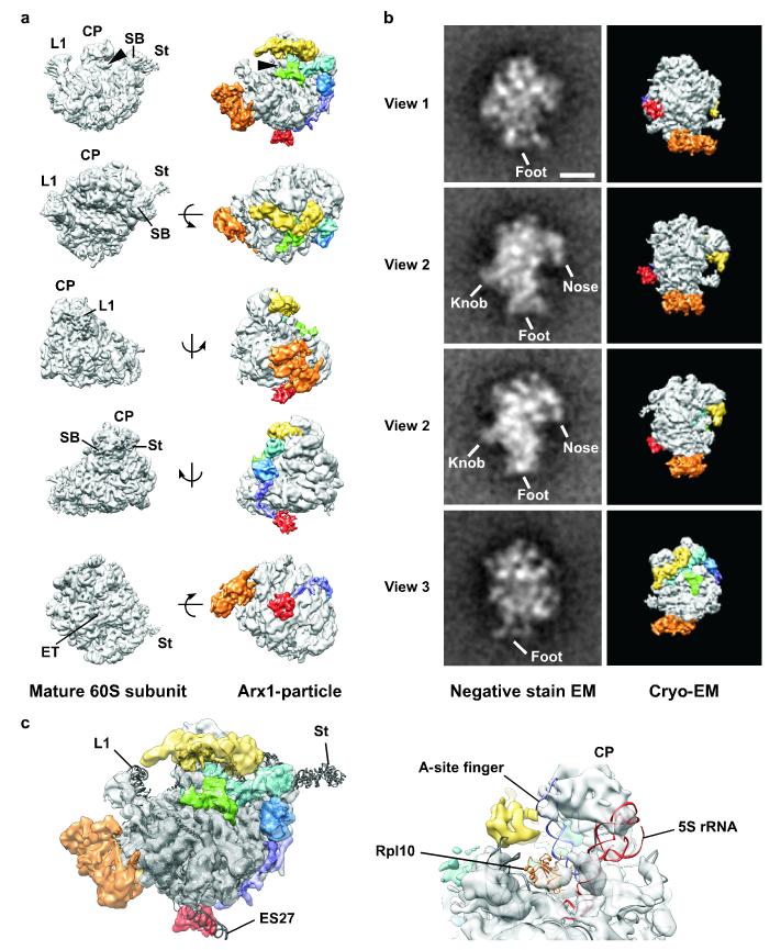
Figure 2
Cryo-EM reconstruction of the Arx1-particle
( a ) Cryo-EM reconstruction of the Alb1-TAP affinity-purified pre-60S at 11.9 u00c5 resolution (right) in comparison to mature 60S subunit, isolated from S. cerevisiae RNC-Ssh1 47 (left). Labeled are...
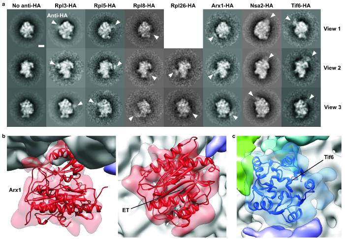
Figure 3
Immuno-EM reveals the relative position of biogenesis factors and r-proteins on the Arx1-particle
( a ) Class averages of negative-stained Arx1-particles affinity-purified via Alb1-TAP with anti-HA antibody bound to the indicated C-terminally integrated HA-tagged proteins of interest. The three ty...
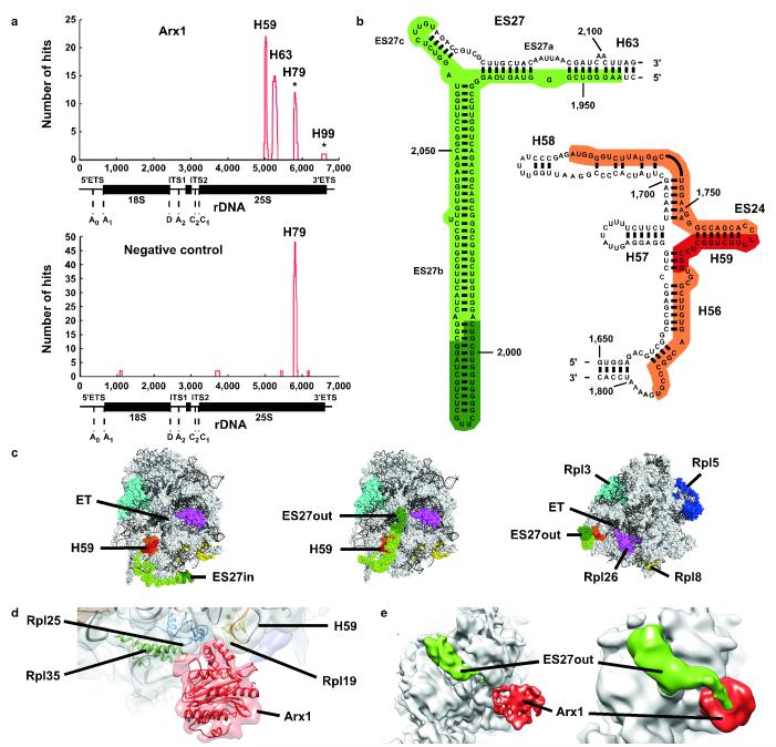
Figure 4
CRAC analysis confirms the location of Arx1 near the exit tunnel
( a ) CRAC analysis on Arx1-HTP and untagged strain. Total number of hits plotted against the relative location along the rDNA sequence. Arx1 binds helix 59 (H59; 22 hits) and helix 63 (15 hits; upper...
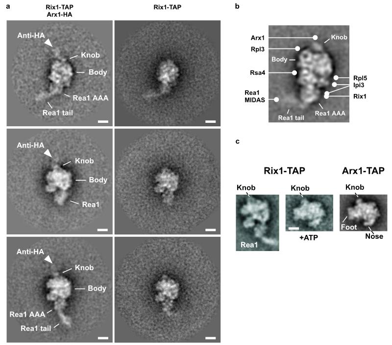
Figure 5
Arx1 localizes to the knob structure of the Rix1-particle, a precursor of the Arx1-particle
( a ) Immuno-EM analysis reveals the position of Arx1 on the Rix1-particle. Rix1-TAP was affinity-purified in the presence of an anti-HA antibody from a strain with genomically integrated Arx1-HA (lef...
Figure images are served from the NIH/NLM PubMed Central Open Access Subset or Europe PMC; copyright remains with the publishers and authors.
💬 Discussion
0 commentsNo comments yet. Be the first to start a discussion!
Leave a Comment