Abstract
AbstractPalladium nanoparticles have been immobilized into an amino‐functionalized metal–organic framework (MOF), MIL‐101Cr‐NH2, to form Pd@MIL‐101Cr‐NH2. Four materials with different loadings of palladium have been prepared (denoted as 4‐, 8‐, 12‐, and 16 wt %Pd@MIL‐101Cr‐NH2). The effects of catalyst loading and the size and distribution of the Pd nanoparticles on the catalytic performance have been studied. The catalysts were characterized by using scanning electron microscopy (SEM), transmission electron microscopy (TEM), Fourier‐transform infrared (FTIR) spectroscopy, powder X‐ray diffraction (PXRD), N2‐sorption isotherms, elemental analysis, and thermogravimetric analysis (TGA). To better characterize the palladium nanoparticles and their distribution in MIL‐101Cr‐NH2, electron tomography was employed to reconstruct the 3D volume of 8 wt %Pd@MIL‐101Cr‐NH2 particles. The pair distribution functions (PDFs) of the samples were extracted from total scattering experiments using high‐energy X‐rays (60 keV). The catalytic activity of the four MOF materials with different loadings of palladium nanoparticles was studied in the Suzuki–Miyaura cross‐coupling reaction. The best catalytic performance was obtained with the MOF that contained 8 wt % palladium nanoparticles. The metallic palladium nanoparticles were homogeneously distributed, with an average size of 2.6 nm. Excellent yields were obtained for a wide scope of substrates under remarkably mild conditions (water, aerobic conditions, room temperature, catalyst loading as low as 0.15 mol %). The material can be recycled at least 10 times without alteration of its catalytic properties.
🔬 Techniques
🔭 Microscopes
💻 Software
✨ Fluorophores
🧪 Sample Preparation
🏭 Microscope Brands
🧪 Reagent Suppliers
💻 Software Details
🏛️ Research Organizations (ROR)
Affiliated research institutions:
📋 Methods
General : All chemicals and solvents were used as received without further purification. Powder X-ray diffraction (PXRD) was performed in the Bragg–Brentano geometry on a PANalytical X’Pert PRO diffractometer that was equipped with a Pixel detector and Cu Kα1 radiation ( λ =1.5406 Å). The samples were dispersed over zero-background Si plates.
SEM and energy-dispersive spectroscopy
(EDS) were performed on a JEOL-7000F field-emission scanning electron microscope operating at 12 kV. The samples were precoated with a thin carbon film. The presence of possible byproducts was investigated by using EDS. TEM observations were performed on a JEOL TEM (JEM-2100F) operating at 200 kV. The MIL-101Cr samples were dispersed in absolute EtOH and then treated by ultrasonication for 2 min. A droplet of the suspension was transferred onto a copper grid and dried in air. A tilt series of TEM images for electron tomography was recorded with a tilt step of 0.5° and an angular range of 41° (83 images in total). TomoJ[ 37 , 38 ] was employed for the tomographic reconstruction. The images were aligned by using the Pd particles themselves as markers. Ten iterations of the ART[ 39 ] algorithm were performed in TomoJ with a relaxation coefficient of 0.1. ImageJ[ 40 ] was used to preprocess the tilt series of TEM images.
Thermogravimetric analysis
(TGA) was performed under a flow of air between 20 and 500 °C with a heating rate of 2 °C min −1 on a thermogravimetric analyzer (PERKIN ELMER TGA 7) that was equipped with a platinum pan. N 2 -adsorption isotherms were recorded at 77 K on a Micromeritics ASAP2020 analyzer. The samples for N 2 adsorption were first exchanged with absolute EtOH before activation. The samples were degassed by heating at 120 °C overnight prior to the sorption measurements. Specific surface areas were calculated from the data in the adsorption branch at p / p 0 =0.05–0.30. The total pore volume was calculated from the uptake at p / p 0 =0.99. FTIR spectroscopy was performed on a Varian 670-IR spectrometer. Elemental analysis was performed on a Carlo Erba Flash 1112 elemental analyzer and the Cr and Pd contents were determined by inductively coupled plasma-optical emission spectrometry (ICP-OES) on a Varian Vista MPX ICP-OES at Medac Ltd, Chobham, UK. High-energy X-ray diffraction experiments were carried out at a fixed energy of approximately 60 keV at a wavelength of 0.2073 Å (determined by using a CeO 2 standard) at the beamline P02.1 of Petra III, Desy, Hamburg. The 2D diffraction images were integrated into a linear scattering signal by using the Fit2D software.[ 41 ] The pair distribution function (PDF), G ( r ), was obtained by sine Fourier transform of the normalized scattering intensity to a maximum Q value, Q max4 =19 Å −1 , by using the PDFgetX2 software.[ 42 ] The PDF of the Pd nanoparticles was calculated with the PDFgui software[ 43 ] (for a detailed procedure, see the Supporting Information). 1 H NMR spectra were recorded at 400 MHz; 13 C NMR spectra were recorded at 100 MHz on a Bruker Advance spectrometer. 1 H and 13 C NMR chemical shifts ( δ ) are reported in ppm relative to tetramethylsilane, with the solvent resonance as an internal standard (CDCl 3 : δ H =7.26 ppm, δ C =77.16 ppm; [D 6 ]DMSO: δ H =2.50 ppm, δ C =39.5 ppm). Coupling constants ( J ) are given in Hz. High-resolution mass spectra (HRMS) were recorded on a Bruker microTOF ESI-TOF mass spectrometer. Synthesis of Pd@MIL-101Cr-NH 2 : Samples of MIL-101Cr (Cr 3 F(H 2 O) 2 O[(CO 2 )-C 6 H 4 -(CO 2 )] 3 ⋅ n H 2 O),[ 9 , 44 ] MIL-101Cr-NO 2 (Cr 3 (H 2 O) 2 O[(CO 2 )-C 6 H 3 NO 2 -(CO 2 )] 3 ⋅ n H 2 O),[ 24 ] and MIL-101Cr-NH 2 (Cr 3 (H 2 O) 2 O[(CO 2 )-C 6 H 3 NH 2 -(CO 2 )] 3 ⋅ n H 2 O)[ 24 ] were prepared according to literature procedures. Samples of Pd@MIL-101Cr-NH 2 with four different loadings of Pd nanoparticles that were intended to be 4, 8, 12, and 16 wt % Pd were prepared by using analogous procedures, as given below. Synthesis of MIL-101Cr-NH 2 -PdCl 2 : For MIL-101Cr-NH 2 -PdCl 2 affording 8 wt %Pd@MIL-101Cr-NH 2 , the following procedure was used: A mixture of MIL-101Cr-NH 2 (0.240 g, 3.15×10 −4 mol), [PdCl 2 (MeCN) 2 ] (0.055 g, 2.11×10 −4 mmol), and anhydrous CH 2 Cl 2 (30 mL) was stirred overnight at RT (18 h). The solid was obtained by centrifugation, washed with anhydrous CH 2 Cl 2 (2×10 mL), and dried at 40 °C for 2 h under vacuum. To obtain samples with 4, 12, and 16 wt % Pd loading, the amount of [PdCl 2 (MeCN) 2 ] that was used to afford 8 wt % Pd loading was multiplied by 0.5, 2, and 3, respectively; the amount of solvent was kept the same. Synthesis of 8 wt %Pd@MIL-101Cr-NH 2 : NaBH 4 (0.076 g, 2 mmol) was added portion-wise to a mixture of MIL-101Cr-NH 2 -PdCl 2 (0.140 g) and EtOH (30 mL) at 0 °C. The green mixture became darker after about 2 min. The reaction was stirred for an additional 2 h at 0 °C to complete the reduction. The solid was obtained by centrifugation, washed twice with water (45 mL) and once with EtOH (45 mL), and the dark-green solid was dried at 85 °C overnight (18 h) in air. Digestion of MIL-101Cr : In a typical experiment, the material (10 mg) was suspended in an aqueous solution of NaOH (2.0 m , 1 mL) and ultrasonicated for 30 min. The water was evaporated under vacuum and the residue was analyzed by 1 H NMR spectroscopy in deuterated DMSO (Supporting Information, Figure S1 ). General procedure for the Suzuki–Miyaura cross-coupling reactions : Unless otherwise stated, pinacol phenylboronate (24.5 mg, 0.12 mmol), the aryl bromide (0.1 mmol), K 2 CO 3 (27.6 mg, 0.2 mmol), and 8 wt %Pd@MIL-101Cr-NH 2 (4 mg, 0.003 mmol) were suspended in deionized water (2 mL) and the mixture was stirred vigorously at RT for 6 h. Then, the mixture was extracted into CH 2 Cl 2 (4 mL), the organic phase was separated, the volatile compounds were evaporated, and the residue was analyzed by 1 H NMR spectroscopy. To isolate the final product, the reaction was repeated on a larger scale (×4) and the crude reaction mixture was purified by column chromatography on silica gel (pentane or pentane/EtOAc mixtures).
Show full methods section
General : All chemicals and solvents were used as received without further purification. Powder X-ray diffraction (PXRD) was performed in the Bragg–Brentano geometry on a PANalytical X’Pert PRO diffractometer that was equipped with a Pixel detector and Cu Kα1 radiation ( λ =1.5406 Å). The samples were dispersed over zero-background Si plates.
SEM and energy-dispersive spectroscopy
(EDS) were performed on a JEOL-7000F field-emission scanning electron microscope operating at 12 kV. The samples were precoated with a thin carbon film. The presence of possible byproducts was investigated by using EDS. TEM observations were performed on a JEOL TEM (JEM-2100F) operating at 200 kV. The MIL-101Cr samples were dispersed in absolute EtOH and then treated by ultrasonication for 2 min. A droplet of the suspension was transferred onto a copper grid and dried in air. A tilt series of TEM images for electron tomography was recorded with a tilt step of 0.5° and an angular range of 41° (83 images in total). TomoJ[ 37 , 38 ] was employed for the tomographic reconstruction. The images were aligned by using the Pd particles themselves as markers. Ten iterations of the ART[ 39 ] algorithm were performed in TomoJ with a relaxation coefficient of 0.1. ImageJ[ 40 ] was used to preprocess the tilt series of TEM images.
Thermogravimetric analysis
(TGA) was performed under a flow of air between 20 and 500 °C with a heating rate of 2 °C min −1 on a thermogravimetric analyzer (PERKIN ELMER TGA 7) that was equipped with a platinum pan. N 2 -adsorption isotherms were recorded at 77 K on a Micromeritics ASAP2020 analyzer. The samples for N 2 adsorption were first exchanged with absolute EtOH before activation. The samples were degassed by heating at 120 °C overnight prior to the sorption measurements. Specific surface areas were calculated from the data in the adsorption branch at p / p 0 =0.05–0.30. The total pore volume was calculated from the uptake at p / p 0 =0.99. FTIR spectroscopy was performed on a Varian 670-IR spectrometer. Elemental analysis was performed on a Carlo Erba Flash 1112 elemental analyzer and the Cr and Pd contents were determined by inductively coupled plasma-optical emission spectrometry (ICP-OES) on a Varian Vista MPX ICP-OES at Medac Ltd, Chobham, UK. High-energy X-ray diffraction experiments were carried out at a fixed energy of approximately 60 keV at a wavelength of 0.2073 Å (determined by using a CeO 2 standard) at the beamline P02.1 of Petra III, Desy, Hamburg. The 2D diffraction images were integrated into a linear scattering signal by using the Fit2D software.[ 41 ] The pair distribution function (PDF), G ( r ), was obtained by sine Fourier transform of the normalized scattering intensity to a maximum Q value, Q max4 =19 Å −1 , by using the PDFgetX2 software.[ 42 ] The PDF of the Pd nanoparticles was calculated with the PDFgui software[ 43 ] (for a detailed procedure, see the Supporting Information). 1 H NMR spectra were recorded at 400 MHz; 13 C NMR spectra were recorded at 100 MHz on a Bruker Advance spectrometer. 1 H and 13 C NMR chemical shifts ( δ ) are reported in ppm relative to tetramethylsilane, with the solvent resonance as an internal standard (CDCl 3 : δ H =7.26 ppm, δ C =77.16 ppm; [D 6 ]DMSO: δ H =2.50 ppm, δ C =39.5 ppm). Coupling constants ( J ) are given in Hz. High-resolution mass spectra (HRMS) were recorded on a Bruker microTOF ESI-TOF mass spectrometer. Synthesis of Pd@MIL-101Cr-NH 2 : Samples of MIL-101Cr (Cr 3 F(H 2 O) 2 O[(CO 2 )-C 6 H 4 -(CO 2 )] 3 ⋅ n H 2 O),[ 9 , 44 ] MIL-101Cr-NO 2 (Cr 3 (H 2 O) 2 O[(CO 2 )-C 6 H 3 NO 2 -(CO 2 )] 3 ⋅ n H 2 O),[ 24 ] and MIL-101Cr-NH 2 (Cr 3 (H 2 O) 2 O[(CO 2 )-C 6 H 3 NH 2 -(CO 2 )] 3 ⋅ n H 2 O)[ 24 ] were prepared according to literature procedures. Samples of Pd@MIL-101Cr-NH 2 with four different loadings of Pd nanoparticles that were intended to be 4, 8, 12, and 16 wt % Pd were prepared by using analogous procedures, as given below. Synthesis of MIL-101Cr-NH 2 -PdCl 2 : For MIL-101Cr-NH 2 -PdCl 2 affording 8 wt %Pd@MIL-101Cr-NH 2 , the following procedure was used: A mixture of MIL-101Cr-NH 2 (0.240 g, 3.15×10 −4 mol), [PdCl 2 (MeCN) 2 ] (0.055 g, 2.11×10 −4 mmol), and anhydrous CH 2 Cl 2 (30 mL) was stirred overnight at RT (18 h). The solid was obtained by centrifugation, washed with anhydrous CH 2 Cl 2 (2×10 mL), and dried at 40 °C for 2 h under vacuum. To obtain samples with 4, 12, and 16 wt % Pd loading, the amount of [PdCl 2 (MeCN) 2 ] that was used to afford 8 wt % Pd loading was multiplied by 0.5, 2, and 3, respectively; the amount of solvent was kept the same. Synthesis of 8 wt %Pd@MIL-101Cr-NH 2 : NaBH 4 (0.076 g, 2 mmol) was added portion-wise to a mixture of MIL-101Cr-NH 2 -PdCl 2 (0.140 g) and EtOH (30 mL) at 0 °C. The green mixture became darker after about 2 min. The reaction was stirred for an additional 2 h at 0 °C to complete the reduction. The solid was obtained by centrifugation, washed twice with water (45 mL) and once with EtOH (45 mL), and the dark-green solid was dried at 85 °C overnight (18 h) in air. Digestion of MIL-101Cr : In a typical experiment, the material (10 mg) was suspended in an aqueous solution of NaOH (2.0 m , 1 mL) and ultrasonicated for 30 min. The water was evaporated under vacuum and the residue was analyzed by 1 H NMR spectroscopy in deuterated DMSO (Supporting Information, Figure S1 ). General procedure for the Suzuki–Miyaura cross-coupling reactions : Unless otherwise stated, pinacol phenylboronate (24.5 mg, 0.12 mmol), the aryl bromide (0.1 mmol), K 2 CO 3 (27.6 mg, 0.2 mmol), and 8 wt %Pd@MIL-101Cr-NH 2 (4 mg, 0.003 mmol) were suspended in deionized water (2 mL) and the mixture was stirred vigorously at RT for 6 h. Then, the mixture was extracted into CH 2 Cl 2 (4 mL), the organic phase was separated, the volatile compounds were evaporated, and the residue was analyzed by 1 H NMR spectroscopy. To isolate the final product, the reaction was repeated on a larger scale (×4) and the crude reaction mixture was purified by column chromatography on silica gel (pentane or pentane/EtOAc mixtures).
Supporting Information As a service to our authors and readers, this journal provides supporting information supplied by the authors. Such materials are peer reviewed and may be re-organized for online delivery, but are not copy-edited or typeset. Technical support issues arising from supporting information (other than missing files) should be addressed to the authors. 1 2 miscellaneous_information
📊 Figures
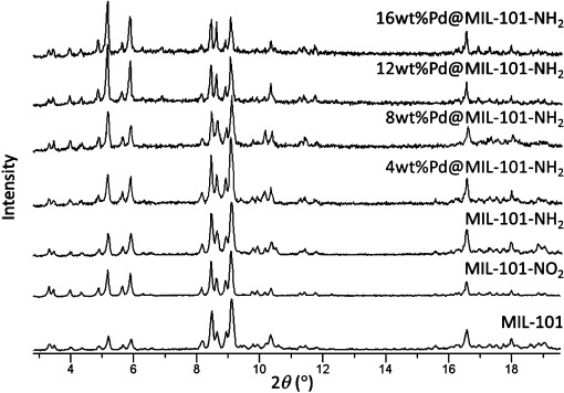
Figure 1
PXRD patterns of MIL-101Cr, MIL-101Cr-NO 2 , MIL-101Cr-NH 2 , and 4-, 8-, 12-, and 16 wt %Pd@MIL-101Cr-NH 2 . The palladium in all four Pd-loaded samples is Pd 0 .
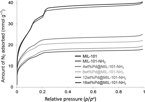
Figure 2
Nitrogen-adsorption isotherms of MIL-101Cr, MIL-101Cr-NH 2 , and 4-, 8-, 12-, and 16 wt %Pd@MIL-101Cr-NH 2 . The measurements were performed at 77 K.
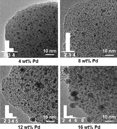
Figure 3
TEM images of samples of 4-, 8-, 12-, and 16 wt %Pd@MIL-101Cr-NH 2 with different Pd loadings (4.4, 8.1, 11.3, and 15.1 wt % Pd, respectively). Insets show the size distributions of the Pd particles a...
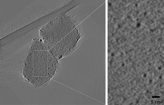
Figure 4
2D section of the 3D tomographic image of two Pd@MIL-101Cr-NH 2 crystals with 8 wt % Pd loading, as reconstructed by the electron tomography from a tilt series of 83 TEM images with a tilt range of 41...
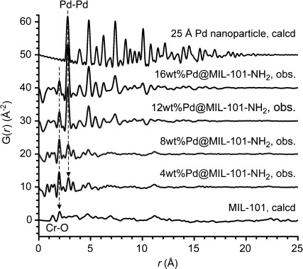
Figure 5
PDFs of samples of 4-, 8-, 12-, and 16 wt %Pd@MIL-101Cr-NH 2 , compared with the calculated PDFs of MIL-101Cr (bottom) and of a Pd nanoparticle (diameter: 25 u00c5, top). For the sake of clarity, the ...
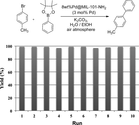
Figure 6
Recycling experiments on a 1 mmol scale; reaction time of each run: 0.5 h.
Figure images are served from the NIH/NLM PubMed Central Open Access Subset or Europe PMC; copyright remains with the publishers and authors.
💬 Discussion
0 commentsNo comments yet. Be the first to start a discussion!
Leave a Comment