Abstract
The adult human brain consists of approximately a hundred billion neurons, which are connected via synapses. The pattern and strength of the synaptic connections are constantly changing (synaptic plasticity), and these changes are considered to underlie learning, memory, and personality. Many psychiatric disorders have been related to disturbances in synaptogenesis and subsequent plasticity. In this review, we summarize findings of synaptic disturbance and its involvement in the pathogenesis and/or pathophysiology of psychiatric disorders. We will focus on schizophrenia, because this condition has a high proven heritability, which offers more unambiguous insights into the biological origins of not only schizophrenia but also related psychiatric disorders. To demonstrate the involvement of synaptopathy in psychiatric disorders, we discuss what knowledge is missing at the circuits level, and what new technologies are needed to achieve a comprehensive understanding of synaptopathy in psychiatric disorders.
🔬 Techniques
🧬 Organisms
✨ Fluorophores
🧪 Sample Preparation
🔬 Cell Lines
🏛️ Research Organizations (ROR)
Affiliated research institutions:
📊 Figures
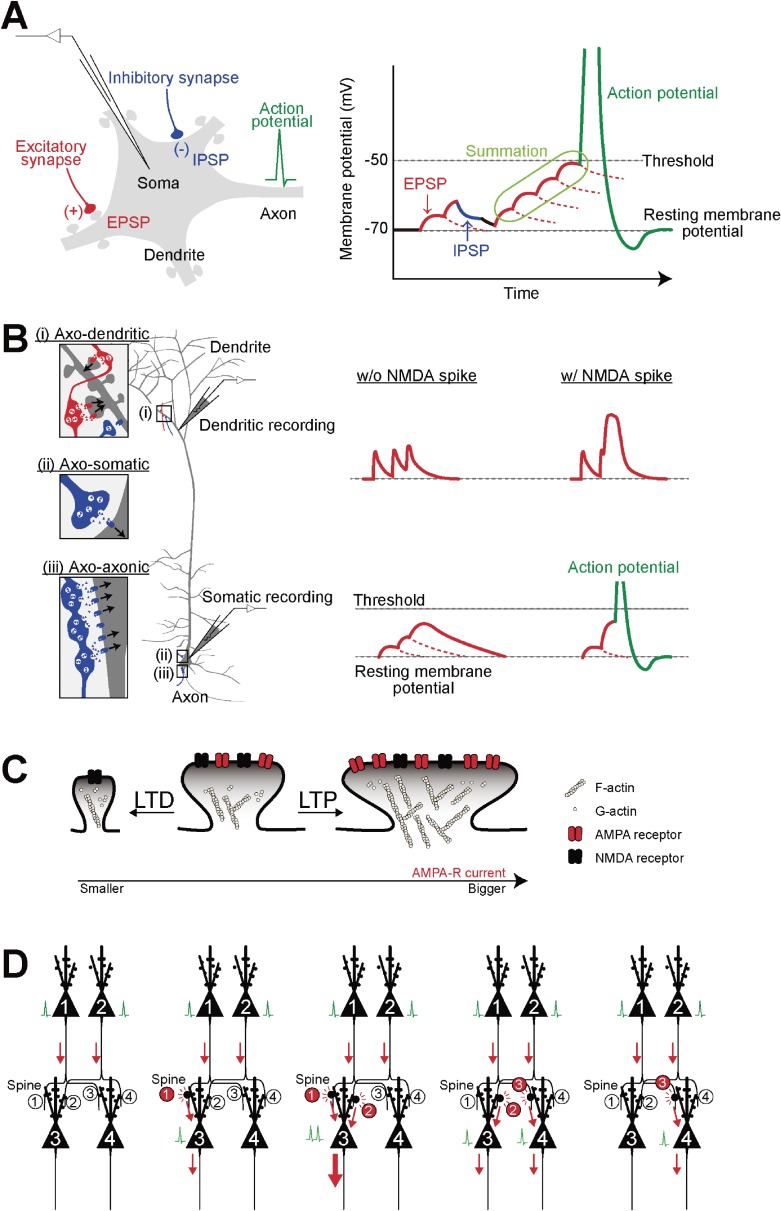
Figure 1.
Effect of EPSP and IPSP on the generation of an action potential. (A) The EPSP/IPSP signal is propagated down the dendrite and is summed with other synaptic inputs at the soma. A single EPSP cannot de...
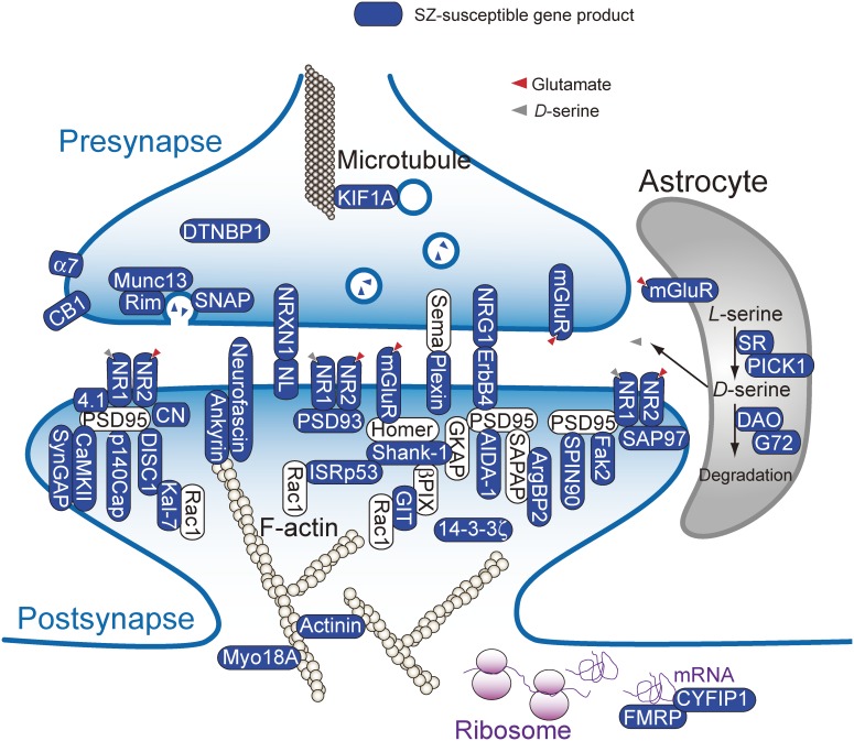
Figure 2.
A synaptic network is a significant schizophrenia-susceptible signal pathway. Schizophrenia-susceptibility genes, which have been repeatedly reported, are depicted as black icons. u03b17, alpha-7 nico...
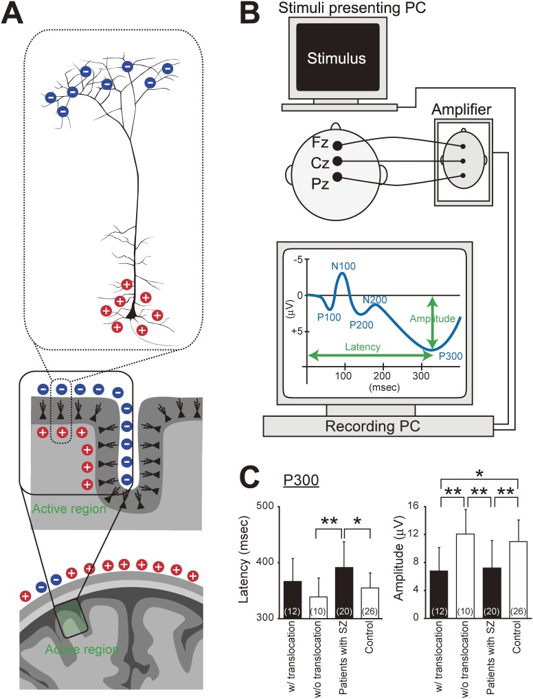
Figure 3.
ERP abnormalities during information processing in schizophrenia. (A) An excitatory transmitter released on apical dendrites causes the discharge of positively charged ions into dendrites, resulting i...
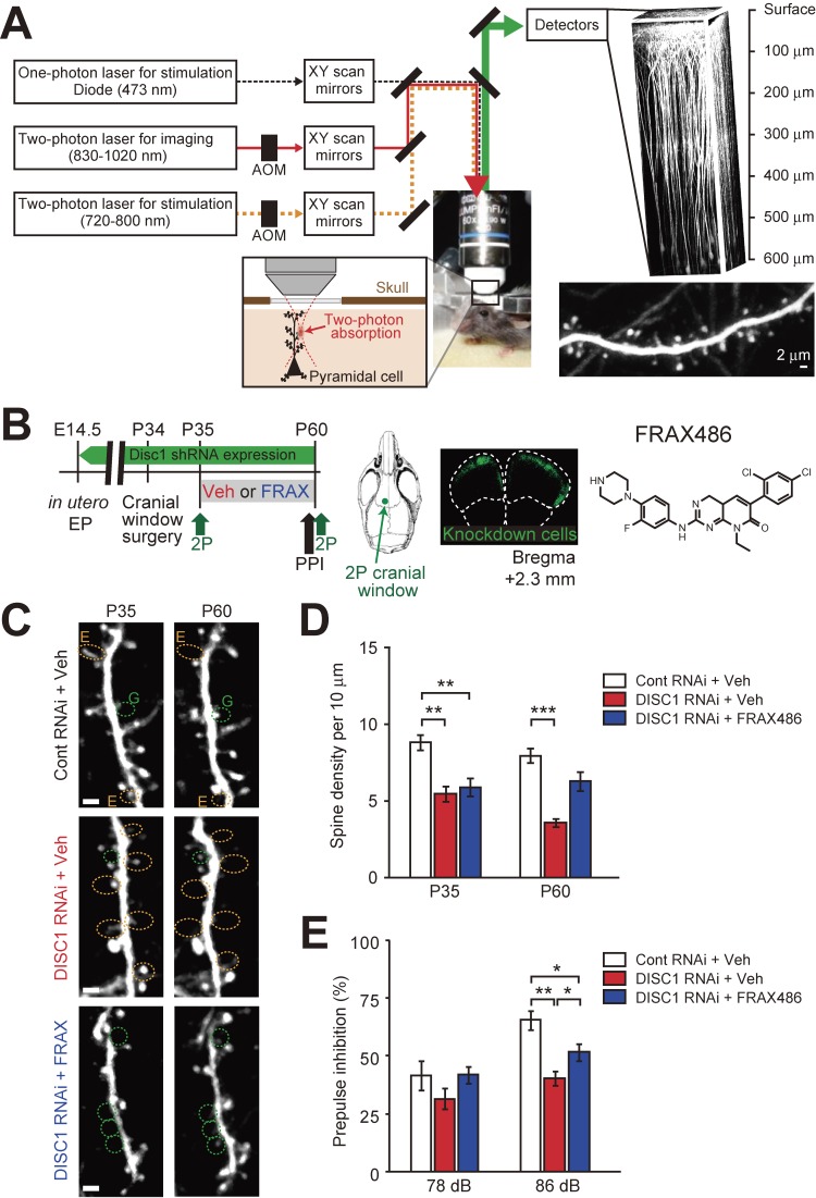
Figure 4.
Progressive synapse loss in a Disc1 knockdown mouse model. (A) Experimental setup for in vivo 2-photon imaging, which allows longitudinal imaging, as well as a specific pattern of photostimulation, in...
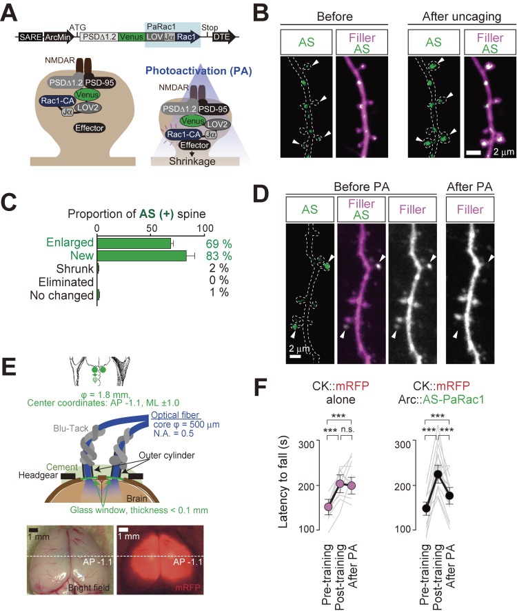
Figure 5.
Optical erasure of acquired skills by synaptic optogenetics. (A) Schematic of the AS-PaRac1 vector, which is transcribed in a synaptic activity-dependent manner. The LOV2 domain was attached to the co...
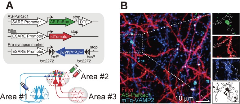
Figure 6.
Multi-scale functional imaging allows simultaneous visualization of both presynaptic and postsynaptic activation. (A) Construct design. For the projection-specific labeling of the presynaptic terminal...
Figure images are served from the NIH/NLM PubMed Central Open Access Subset or Europe PMC; copyright remains with the publishers and authors.
💬 Discussion
0 commentsNo comments yet. Be the first to start a discussion!
Leave a Comment