Abstract
AbstractMitochondrial dysfunction contributes to cardiac pathologies. Barriers to new therapies include an incomplete understanding of underlying molecular culprits and a lack of effective mitochondria-targeted medicines. Here, we test the hypothesis that the cardiolipin-binding peptide elamipretide, a clinical-stage compound under investigation for diseases of mitochondrial dysfunction, mitigates impairments in mitochondrial structure-function observed after rat cardiac ischemia-reperfusion. Respirometry with permeabilized ventricular fibers indicates that ischemia-reperfusion induced decrements in the activity of complexes I, II, and IV are alleviated with elamipretide. Serial block face scanning electron microscopy used to create 3D reconstructions of cristae ultrastructure reveals that disease-induced fragmentation of cristae networks are improved with elamipretide. Mass spectrometry shows elamipretide did not protect against the reduction of cardiolipin concentration after ischemia-reperfusion. Finally, elamipretide improves biophysical properties of biomimetic membranes by aggregating cardiolipin. The data suggest mitochondrial structure-function are interdependent and demonstrate elamipretide targets mitochondrial membranes to sustain cristae networks and improve bioenergetic function.
🔬 Techniques
💻 Software
✨ Fluorophores
🧪 Sample Preparation
🧪 Reagent Suppliers
💻 Software Details
🏛️ Research Organizations (ROR)
Affiliated research institutions:
📋 Methods
Animals Male Sprague–Dawley rats (aged 2–3 months) were used in the study. All procedures received prior approval from the Institutional Animal Care and Use Committees at East Carolina University, Latvian Animal Protection Ethical Committee of Food and Veterinary Service, and Virginia Tech. Animals were housed in a temperature and light-controlled environment and received food and water ad libitum. Prior to excision of the heart, animals received an intraperitoneal (ip) injection of a ketamine/xylazine solution (90 mg/kg/10 mg/kg, respectively), and hearts were excised following the diminution of animal reflexes via midline thoracotomy and placed in ice-cold saline. Materials All phospholipids were purchased from Avanti Polar Lipids Inc. Elamipretide and the TAMRA-elamipretide conjugate were synthesized by New England Peptide. All organic solvents were HPLC grade and all other reagents were purchased from either Fisher Scientific or Sigma. I/R and respirometry of permeabilized ventricular fibers Excised hearts were perfused on one of four modified Langendorff apparatus (AD Instruments) per our established protocols 99 , 100 . Hearts were exposed to 20/120 min of global ischemia/reperfusion, respectively. For the elamipretide treatment, hearts received 10μM elamipretide beginning at the onset of reperfusion, which is a well-established cardioprotective paradigm in our models 18 , 78 , 80 , 101 . Myocardial oxygen consumption was measured at the end of reperfusion in a subset of hearts per our established protocols 102 . At the end of reperfusion, hearts were split into the experimental groups described below. Two different protocols were employed to determine mitochondrial function after ischemic stress in ventricular muscle fibers. The first set determined mitochondrial function in fibers isolated after cardiac ischemia- reperfusion (“Post I/R Studies”). The second set of studies isolated cardiac fibers from a freshly isolated (normoxic) heart, and then induced anoxia-reoxygenation on the isolated fibers (“A/R Studies”). Detailed methods for the fiber studies are provided in the Methods Supplement. Below (Supplemental Fig. 5 ) is an overview of the permeabilized experimental flow. Isolated mitochondria and BN-PAGE Mitochondria were isolated from the left ventricle and succinate-derived reverse electron transport determined using our established protocols 103 . Detailed protocols for the separation of native mitochondrial respiratory chain complexes by BN-PAGE are described in the Methods Supplement. Supercomplex flux control coupling factor was measured as described 97 .
Show full methods section
Animals Male Sprague–Dawley rats (aged 2–3 months) were used in the study. All procedures received prior approval from the Institutional Animal Care and Use Committees at East Carolina University, Latvian Animal Protection Ethical Committee of Food and Veterinary Service, and Virginia Tech. Animals were housed in a temperature and light-controlled environment and received food and water ad libitum. Prior to excision of the heart, animals received an intraperitoneal (ip) injection of a ketamine/xylazine solution (90 mg/kg/10 mg/kg, respectively), and hearts were excised following the diminution of animal reflexes via midline thoracotomy and placed in ice-cold saline. Materials All phospholipids were purchased from Avanti Polar Lipids Inc. Elamipretide and the TAMRA-elamipretide conjugate were synthesized by New England Peptide. All organic solvents were HPLC grade and all other reagents were purchased from either Fisher Scientific or Sigma. I/R and respirometry of permeabilized ventricular fibers Excised hearts were perfused on one of four modified Langendorff apparatus (AD Instruments) per our established protocols 99 , 100 . Hearts were exposed to 20/120 min of global ischemia/reperfusion, respectively. For the elamipretide treatment, hearts received 10μM elamipretide beginning at the onset of reperfusion, which is a well-established cardioprotective paradigm in our models 18 , 78 , 80 , 101 . Myocardial oxygen consumption was measured at the end of reperfusion in a subset of hearts per our established protocols 102 . At the end of reperfusion, hearts were split into the experimental groups described below. Two different protocols were employed to determine mitochondrial function after ischemic stress in ventricular muscle fibers. The first set determined mitochondrial function in fibers isolated after cardiac ischemia- reperfusion (“Post I/R Studies”). The second set of studies isolated cardiac fibers from a freshly isolated (normoxic) heart, and then induced anoxia-reoxygenation on the isolated fibers (“A/R Studies”). Detailed methods for the fiber studies are provided in the Methods Supplement. Below (Supplemental Fig. 5 ) is an overview of the permeabilized experimental flow. Isolated mitochondria and BN-PAGE Mitochondria were isolated from the left ventricle and succinate-derived reverse electron transport determined using our established protocols 103 . Detailed protocols for the separation of native mitochondrial respiratory chain complexes by BN-PAGE are described in the Methods Supplement. Supercomplex flux control coupling factor was measured as described 97 .
Electron microscopy of mitochondria
A subset of hearts exposed to ischemia-reperfusion (as described above) were utilized for transmission electron microscopy imaging (Virginia Tech Morphology Service Core Laboratory, Virginia-Maryland College of Veterinary Medicine) using modifications to established protocols 104 , 105 . Serial block-face scanning electron microscopy was done in collaboration with Renovo Neural, Inc. (Cleveland, Ohio). Detailed methods are provided in the Methods Supplement.
Mass spectrometry
A subset of hearts was taken immediately at the end of reperfusion and the left ventricle was snap frozen, pulverized using a liquid nitrogen-cooled mortar/pestle, and analyzed for CL content and composition using shotgun lipidomics per our established methods 106 . Detailed protocols for the mass spectroscopy studies are provided in the Methods Supplement.
Construction of biomimetic membranes
A detailed description of the methods for biophysical CL aggregation studies are provided in the Methods Supplement. Pressure-area isotherm experiments were performed using differing biomimetic lipid compositions. For pressure-area isotherms displayed in Fig. 5 c, biomimetic mitochondrial lipid monolayers were generated by co-dissolving 40 mol% (18:0-22:6) PC, 30.0 mol% (16:0-20:4) PE, 20 mol% (18:2-18:2-18:2-18:2) CL, 5 mol% (18:1-18:1) PI, 3 mol% (18:1-18:1) PS, and 2 mol% cholesterol in chloroform (10 μg/μL) per our established methods 107 . Pressure-area isotherms displayed in Fig. 5d relied on biomimetic monolayers containing 40 mol% (18:0-22:6) PC, 33.0 mol% (16:0-20:4) PE, 20 mol% (18:2-18:2-18:2-18:2) CL, 5 mol% (18:1-18:1) PS, and 2 mol% cholesterol in chloroform (10 μg/μL). The area per molecule was determined at a physiological surface pressure of 30 mN/m as previously shown 108 . Giant unilamellar vesicles were constructed via electroformation as previously described 30 and contained 39.9 mol% (18:0-22:6) PC, 30.0 mol% (16:0-20:4) PE, 20 mol% (18:2-18:2-18:2-18:2) CL, 5 mol% (18:1-18:1) PI, 3 mol% (18:1-18:1) PS, 2 mol% cholesterol, and 0.1 mol% NAO. For select experiments, the total CL concentration was decreased 25-50% by mass to reflect changes seen from mass spectroscopy studies. In a subset of studies peptide was added immediately after spotting the lipid monolayer or prior to imaging.
Statistics and reproducibilty
All data were analyzed using and GraphPad Prism 8 and are presented as mean ± sem. Data were distributed normally, which allowed for parametric analyses. Statistical analyses were conducted using a one-way ANOVA followed by a Bonferroni post-hoc, with P -values ≤ 0.05 considered significant. One trace from RET studies was excluded because the hydrogen peroxide emission was greater than two standard deviations away from the mean. Reporting summary Further information on research design is available in the Nature Research Reporting Summary linked to this article.
Materials All phospholipids were purchased from Avanti Polar Lipids Inc. Elamipretide and the TAMRA-elamipretide conjugate were synthesized by New England Peptide. All organic solvents were HPLC grade and all other reagents were purchased from either Fisher Scientific or Sigma.
Supplementary information Supplementary Information Description of Additional Supplementary Files Supplementary Data 1 Supplementary Data 2 Supplementary Movie 1 Supplementary Movie 2 Supplementary Movie 3 Supplementary Movie 4 Supplementary Movie 5 Supplementary Movie 6 Reporting Summary
📊 Figures
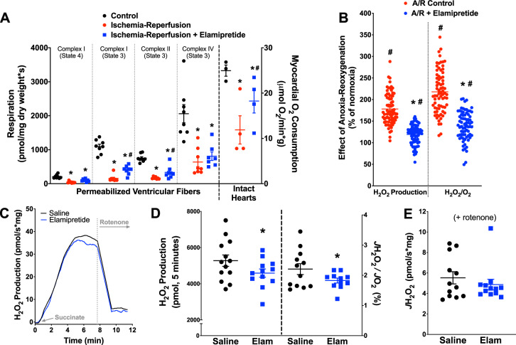
Fig. 1
Improvement of mitochondrial function with elamipretide.
a Decrements in mitochondrial respiration were seen across different mitochondrial complexes and substrate conditions in permeabilized fibers ( N =u20097u20138 for all groups) and intact hearts ( N =u...
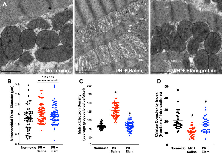
Fig. 2
TEM data from hearts in the study.
a Representative images from experimental groups. b Images were analyzed for mitochondrial Feret diameter ( N =u200959 for normoxic, N =u200970 for I/Ru2009+u2009saline, and N =u200962 for I/Ru2009+u2...
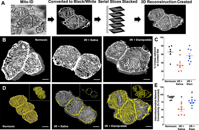
Fig. 3
Serial block-face scanning electron microscopy images of mitochondrial ultrastructure in the experimental groups.
Original SBFu2013SEM serial images were acquired and processed into 3D reconstructions using ImageJ ( a ). Mitochondria were then analyzed for contract site analysis ( b ) and intermitochondrial netwo...
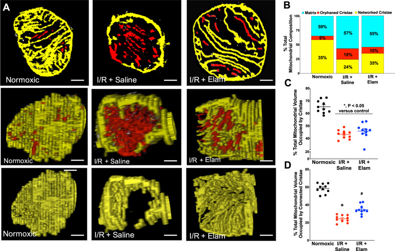
Fig. 4
Serial block-face scanning electron microscopy (SBFu2013SEM) images of single mitochondrial ultrastructure in the experimental groups.
Mitochondria were analyzed for cristae connectivity. a Top panel: individual slices from SBFu2013SEM imaging showing cristae networking. Middle panel: cross-section of composite stacks indicating netw...
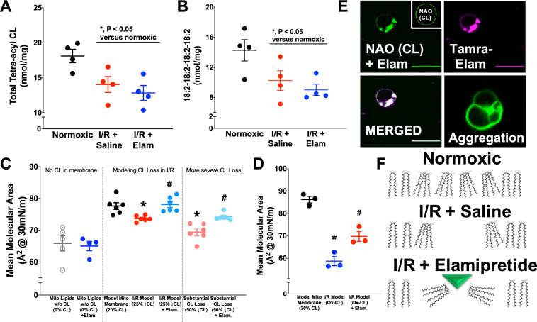
Fig. 5
Studies ofu00a0elamipretideu00a0interactions with CL.
Lipidomics indicated declines in total ( a ) and tetra-linoleoyl CL ( b ) after ischemia-reperfusion. Biomimetic models of the mitochondrial inner membrane lipids presented in c u2013 f . d GUV imagin...
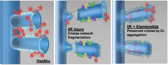
Fig. 6
Proposed model in which elamipretide (depicted in magentau2014right panel) aggregates CL (depicted in yellow) to preserve cristae ultrastructure in diseased mitochondria.
Preserved cristae integrity was associated with protection of complex V (red) band density. The other electron transport chain complexes are depicted in green, yellow, and purple (middle panel). Thera...
Figure images are served from the NIH/NLM PubMed Central Open Access Subset or Europe PMC; copyright remains with the publishers and authors.
💬 Discussion
0 commentsNo comments yet. Be the first to start a discussion!
Leave a Comment