Abstract
Kinetochores must remain associated with microtubule ends, as they undergo rapid transitions between growth and shrinkage. The molecular basis for this essential activity that ensures correct chromosome segregation is unclear. In this study, we have used reconstitution of dynamic microtubules and total internal reflection fluorescence microscopy to define the functional relationship between two important budding yeast kinetochore complexes. We find that the Dam1 complex is an autonomous plus end-tracking complex. The Ndc80 complex, despite being structurally related to the general tip tracker EB1, fails to recognize growing ends efficiently. Dam1 oligomers are necessary and sufficient to recruit Ndc80 to dynamic microtubule ends, where both complexes remain continuously associated. The interaction occurs specifically in the presence of microtubules and is subject to regulation by Ipl1 phosphorylation. These findings can explain how the force harvested by Dam1 is transmitted to the rest of the kinetochore via the Ndc80 complex.
🔬 Techniques
🧬 Organisms
💻 Software
✨ Fluorophores
🧪 Sample Preparation
🏭 Microscope Brands
🧪 Reagent Suppliers
📷 Detectors
🔎 Objectives
💻 Software Details
🏛️ Research Organizations (ROR)
Affiliated research institutions:
📋 Methods
Yeast genetics All Westermann laboratory strains are derived from strain S288C. Standard procedures were applied to genetically modify strains.
Live cell imaging
Cells expressing Dam1-3xGFP and mCherry-Tub1 (FLY26: MAT α , lys2-801 , ade2-1 , leu2 , cdc20::TRP1::GAL1/10-CDC20 , mCherry-tubulin::URA3 , and Dam1-3xGFP::HIS3 ; or FLY48: MAT a , lys2-801 , ade2-1 , leu2 , cdc20::TRP1::GAL1/10-CDC20 , mCherry-tubulin::URA3 , Dam1-3xGFP::HIS3 , and pGal-3xHA-Bim1::HIS3 ) were arrested in metaphase for 5 h in synthetic medium containing 2% glucose. Time-lapse images were collected at ambient temperature (21°C) using live cell microscopy (DeltaVision; Applied Precision), a UPlanSApo 100× NA 1.40 oil immersion objective lens (Olympus), and a charge-coupled device (CCD) camera (CoolSnap HQ; Photometrics). Z stacks (0.5 µm) were acquired in 20–30-s intervals, subsequently deconvoluted, projected to two-dimensional images (SoftWoRx software; Applied Precision), and further analyzed by MetaMorph (MDS Analytical Technologies) or ImageJ (National Institutes of Health) software.
Purification of recombinant complexes from bacteria
Expression, purification, and labeling of the Dam1 complex were performed as previously described ( Westermann et al., 2005 ). The two subcomplexes of the Ndc80 complex (Ndc80p–6xHis/Nuf2p–EGFP or ΔN1-116 –Ndc80p–6xHis/Nuf2p–EGFP, Spc24p–6xHis/Spc25p) were separately expressed from the pETDuett or pACYCDuet-1 vectors (EMD). Both plasmids were cotransfected into BL21 DE3 (EMD). Bacteria were grown to OD 600 = 0.6 at 37°C, induced with 0.2 mM IPTG, and grown for 12–15 h at 18°C. The two subcomplexes were eluted with 150 mM NaCl, 10 mM Hepes, pH 7.0, and 250 mM imidazol from the Ni-NTA beads then subjected to in vitro reconstitution of the full-length Ndc80 complex and further purified on the Superdex 200 HiLoad 16/60 (GE Healthcare). Gel filtration was conducted in 150 mM NaCl and 10 mM Hepes, pH 7.
Show full methods section
Yeast genetics All Westermann laboratory strains are derived from strain S288C. Standard procedures were applied to genetically modify strains.
Live cell imaging
Cells expressing Dam1-3xGFP and mCherry-Tub1 (FLY26: MAT α , lys2-801 , ade2-1 , leu2 , cdc20::TRP1::GAL1/10-CDC20 , mCherry-tubulin::URA3 , and Dam1-3xGFP::HIS3 ; or FLY48: MAT a , lys2-801 , ade2-1 , leu2 , cdc20::TRP1::GAL1/10-CDC20 , mCherry-tubulin::URA3 , Dam1-3xGFP::HIS3 , and pGal-3xHA-Bim1::HIS3 ) were arrested in metaphase for 5 h in synthetic medium containing 2% glucose. Time-lapse images were collected at ambient temperature (21°C) using live cell microscopy (DeltaVision; Applied Precision), a UPlanSApo 100× NA 1.40 oil immersion objective lens (Olympus), and a charge-coupled device (CCD) camera (CoolSnap HQ; Photometrics). Z stacks (0.5 µm) were acquired in 20–30-s intervals, subsequently deconvoluted, projected to two-dimensional images (SoftWoRx software; Applied Precision), and further analyzed by MetaMorph (MDS Analytical Technologies) or ImageJ (National Institutes of Health) software.
Purification of recombinant complexes from bacteria
Expression, purification, and labeling of the Dam1 complex were performed as previously described ( Westermann et al., 2005 ). The two subcomplexes of the Ndc80 complex (Ndc80p–6xHis/Nuf2p–EGFP or ΔN1-116 –Ndc80p–6xHis/Nuf2p–EGFP, Spc24p–6xHis/Spc25p) were separately expressed from the pETDuett or pACYCDuet-1 vectors (EMD). Both plasmids were cotransfected into BL21 DE3 (EMD). Bacteria were grown to OD 600 = 0.6 at 37°C, induced with 0.2 mM IPTG, and grown for 12–15 h at 18°C. The two subcomplexes were eluted with 150 mM NaCl, 10 mM Hepes, pH 7.0, and 250 mM imidazol from the Ni-NTA beads then subjected to in vitro reconstitution of the full-length Ndc80 complex and further purified on the Superdex 200 HiLoad 16/60 (GE Healthcare). Gel filtration was conducted in 150 mM NaCl and 10 mM Hepes, pH 7.
Microtubule-binding assays
The microtubule cosedimentation assay was performed as described previously ( Cheeseman et al., 2001a ). Purified Ndc80–EGFP and –Dam1 complexes were precleared and added to a final concentration of 0.4–0.5 µM to taxol-stabilized microtubules. The salt concentration was adjusted to 20, 25, 50, or 100 mM NaCl. Quantification was performed as previously described ( Zimniak et al., 2009 ). To determine apparent K D for Ndc80–EGFP complex binding to microtubules, data points were fitted into the equation Y = B max × X/(K D + X) using Prism (version 4.0; GraphPad Software, Inc.). Analytical gel filtration Ndc80 and the Dam1 complex were mixed in an equimolar ratio at 5.5 µM and incubated for 10 min at RT. 0.5 ml was loaded onto the Superdex 200 HiLoad 16/60. Proteins were eluted in buffer containing 100 mM NaCl and 10 mM Hepes, pH 7. In vitro reconstitution of a plus end–tracking system Flow cells were constructed using silanized glass slides, two double-sided tapes, and precleaned coverslips. Resulting chambers were blocked with 5 mg/ml biotinylated BSA (Vector Laboratories) for at least 3 h and subsequently washed with BRB80 buffer. Afterward, a solution of 0.3 mg/ml avidin DN (Vector Laboratories) in BRB80 was introduced for at least 20 min and exchanged for a blocking solution containing 0.1% pluronic F-127 (Sigma-Aldrich) in BRB80 ( Westermann et al., 2006 ; Zimniak et al., 2009 ). Rhodamine-labeled short GMPCPP (2'-deoxy-guanosince-5'-[(α,β)-methyleno]triphosphate) microtubule seeds were introduced and incubated for 30 s followed by a wash with wash buffer (BRB80 supplemented with 150 mM KCl, 1 mM GTP, 0.5% [vol/vol] β-mercaptoethanol, 4.5 µg/ml glucose, 200 µg/ml glucose oxidase, and 35 µg/ml catalase). Microtubule growth was induced by introducing a reaction mix composed of 14 µM tubulin (unlabeled or 1 µM labeled with rhodamine), 150 mM KCl, 0.6 mM GTP, 0.13% (wt/vol) methylcellulose, 0.33 mg/ml casein, 0.5% (vol/vol) β-mercaptoethanol, 4.5 µg/ml glucose, 200 µg/ml glucose oxidase, 35 µg/ml catalase, and 100 nM Dam1–Alexa Fluor 488 complex. Time-lapse videos were recorded at 30°C using 4–5-s intervals between frames. Image acquisition was performed using the TIRF3 microscopy system operated with AxioVision software (Carl Zeiss, Inc.), a 100× Plan Apochromat 1.46 NA objective, and an EM CCD camera (C9100-02; Hamamatsu Photonics). For streaming videos, concentration of free tubulin was increased to 20 µM and the time resolution to 400 ms/frame. For Ndc80–EGFP (300 nM) and Dam1–Alexa Fluor 594 (10 nM labeled + 90 nM unlabeled Dam1 complex) experiments, we used coverslips grafted with polyethylene glycol biotin, 100 mM KCl, and an EM CCD camera (Cascade II; Photometrics). In vitro phosphorylation of the Dam1 complex 0.5 µM recombinant Ipl1–Sli15 complex was incubated with 2.5 µM recombinant Dam1 wild-type complex in kinase buffer (20 mM Hepes, pH 7.5, 100 mM KCl, 10 mM MgCl 2 , 25 mM β-glycerophosphate, and 1 mM DTT) in the presence or absence of 1 mM ATP. The reaction was performed at 30°C for 40 min. EM Short GMPCPP microtubule seeds were incubated for 5 min with a solution containing 14 µM tubulin, 150 mM KCl, 0.6 mM GTP, 0.33 mg/ml casein, 0.5% (vol/vol) β-mercaptoethanol, 4.5 µg/ml glucose, 200 µg/ml glucose oxidase, 35 µg/ml catalase, and variable concentrations of the Dam1–Alexa Fluor 488 complex in BRB80 buffer. The reaction mix was placed on a carbon-coated grid, negatively stained with 2% uranyl acetate, and imaged immediately with an electron microscope operating at 80 kV (Morgagni; FEI) equipped with a CCD camera (Morada SIS; Olympus). Online supplemental material Fig. S1 addresses the oligomerization status of the Dam1 complex. Fig. S2 demonstrates that the N-terminal tail of Ndc80p is not essential for Dam1-dependent recruitment to microtubules. Fig. S3 shows that there is no interaction between the Ndc80 complex and the Dam1 complex in solution. Videos 1 and 2 visualize plus end tracking of the Dam1 complex in vivo. Video 3 shows that Dam1 tip localization is independent of the +TIP Bim1p. Videos 4–6 demonstrate autonomous microtubule plus end–tracking activity of the Dam1 complex in vitro. Video 7 displays plus end tracking of the Ndc80–EGFP complex in the presence of unlabeled Dam1 complex. Video 8 shows continuous tip tracking of the Ndc80–EGFP complex in the presence of unlabeled Dam1 complex. Video 9 demonstrates that the Dam1 complex and the Ndc80 complex colocalize on dynamic microtubule plus ends. Online supplemental material is available at http://www.jcb.org/cgi/content/full/jcb.200912021/DC1 .
Online supplemental material Fig. S1 addresses the oligomerization status of the Dam1 complex. Fig. S2 demonstrates that the N-terminal tail of Ndc80p is not essential for Dam1-dependent recruitment to microtubules. Fig. S3 shows that there is no interaction between the Ndc80 complex and the Dam1 complex in solution. Videos 1 and 2 visualize plus end tracking of the Dam1 complex in vivo. Video 3 shows that Dam1 tip localization is independent of the +TIP Bim1p. Videos 4–6 demonstrate autonomous microtubule plus end–tracking activity of the Dam1 complex in vitro. Video 7 displays plus end tracking of the Ndc80–EGFP complex in the presence of unlabeled Dam1 complex. Video 8 shows continuous tip tracking of the Ndc80–EGFP complex in the presence of unlabeled Dam1 complex. Video 9 demonstrates that the Dam1 complex and the Ndc80 complex colocalize on dynamic microtubule plus ends. Online supplemental material is available at http://www.jcb.org/cgi/content/full/jcb.200912021/DC1 .
📊 Figures
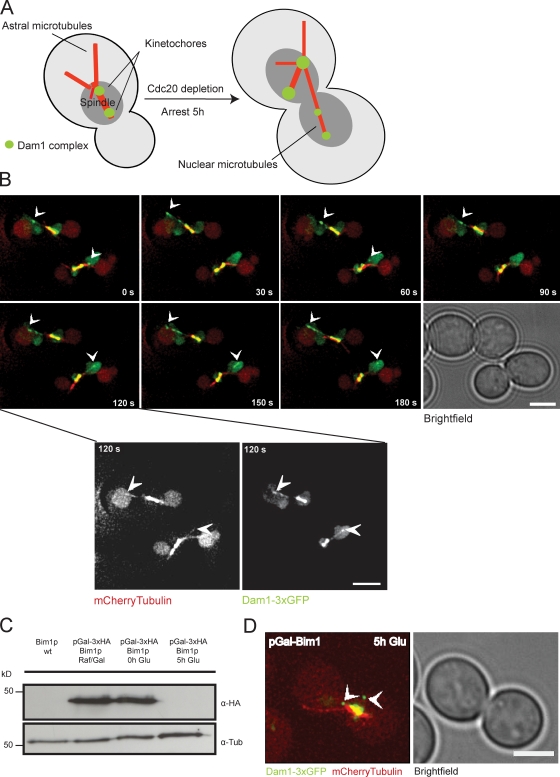
Figure 1.
The Dam1 complex tracks plus ends independently of Bim1p in vivo. (A) Schematic illustration of the live cell imaging strategy ( Tanaka et al., 2005 ). (B) Time-lapse live cell microscopy showing plus...
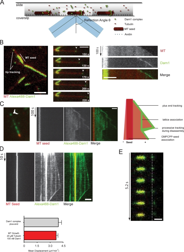
Figure 2.
Reconstitution of Dam1 plus end tracking in vitro using TIRF microscopy. (A) Schematic illustration of the in vitro imaging setup. (B, left) Still image of the Alexa Fluor 488u2013labeled Dam1 complex...
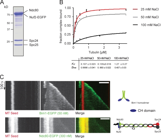
Figure 3.
Ndc80 is not an autonomous end-binding complex. (A) Coomassie-stained SDS-PAGE showing the reconstituted S. cerevisiae Ndc80 complex. The Nuf2 subunit is fused to EGFP. (B) The Ndc80 complex was teste...
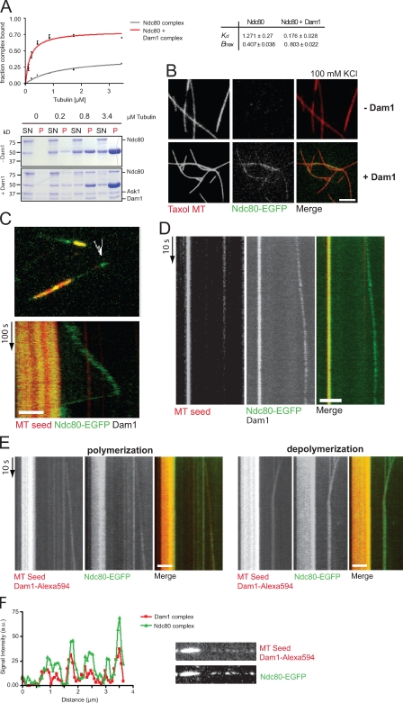
Figure 4.
The Dam1 complex recruits the Ndc80 complex to dynamic microtubule plus ends. (A) Microtubule-binding activity of Ndc80 was tested in the absence and presence of equimolar amounts of Dam1 complex and ...
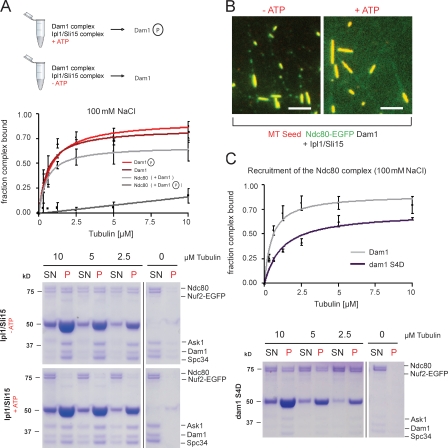
Figure 5.
Phosphorylation of Dam1 by Ipl1u2013Sli15 prevents Ndc80 recruitment to microtubules. (A) The Ndc80 complex was tested for microtubule binding in the presence of phosphorylated and unphosphorylated Da...
Figure images are served from the NIH/NLM PubMed Central Open Access Subset or Europe PMC; copyright remains with the publishers and authors.
💬 Discussion
0 commentsNo comments yet. Be the first to start a discussion!
Leave a Comment