Abstract
Nanodiamond (ND) has emerged as a promising carbon nanomaterial for therapeutic applications. In previous studies, ND has been reported to have outstanding biocompatibility and high uptake rate in various cell types. ND containing nitrogen-vacancy centers exhibit fluorescence property is called fluorescent nanodiamond (FND), and has been applied for bio-labeling agent. However, the influence and application of FND on the nervous system remain elusive. In order to study the compatibility of FND on the nervous system, neurons treated with FNDs in vitro and in vivo were examined. FND did not induce cytotoxicity in primary neurons from either central (CNS) or peripheral nervous system (PNS); neither did intracranial injection of FND affect animal behavior. The neuronal uptake of FNDs was confirmed using flow cytometry and confocal microscopy. However, FND caused a concentration-dependent decrease in neurite length in both CNS and PNS neurons. Time-lapse live cell imaging showed that the reduction of neurite length was due to the spatial hindrance of FND on advancing axonal growth cone. These findings demonstrate that FNDs exhibit low neuronal toxicity but interfere with neuronal morphogenesis, and should be taken into consideration when applications involve actively growing neurites (e.g. nerve regeneration).
🔬 Techniques
🔭 Microscopes
✨ Fluorophores
🧪 Sample Preparation
🔬 Cell Lines
🏭 Microscope Brands
🧪 Reagent Suppliers
📷 Detectors
🔎 Objectives
💻 Software Details
🏛️ Research Organizations (ROR)
Affiliated research institutions:
📋 Methods
FND preparation
NDs with an average size of 100 nm were purchased from Element Six (Micron+ MDA, Element Six, Ireland). ND powders were radiation-damaged by using either a 40-keV He + beam at a dose of ~1 × 10 14 ions/cm 2 or a 3-MeV H + beam at a dose of ~1 × 10 16 ions/cm 2 to create the optimum amount of vacancies in the diamond crystal lattice, as previously described 10 . ND particles were subsequently annealed in vacuum at 800°C for 2 hours to form FND. The nitrogen-vacancy-containing particles were extensively rinsed in distilled deionized water and stored at room temperature prior to use. The particle size and morphology of FNDs were examined by a scanning electron microscope (S6700, JEOL, Japan) ( Figure S2 ). The average size of FNDs was around 114.7 ± 8.4 nm when analyzed by dynamic light scattering (BI-200SM, Brookhaven Instruments, Holtsville, NY) ( Figure S3 ). We also observed that our FNDs carried negative charge around −20.65 ± 1.63 mV as determined by zeta potential analysis (ZetaPALS, Brookhaven Instruments, Holtsville, NY).
Primary neuron cultures
All experimental procedures were approved by the Institutional Animal Care and Use Committee (IACUC) of National Chiao Tung University (NCTU) and in accordance with the Guide for the Care and Use of Laboratory Animals. Hippocampal neurons from E18 mouse embryos (C57BL/6) were prepared as described 35 . Dissociated hippocampal neurons were seeded into poly-L-lysine-coated 96-well optical bottom plates at the density of 2 × 10 4 cells per well in 200 μL serum-containing neuronal plating medium (minimum essential medium supplemented with 5% fetal bovine serum, 0.6% D-glucose, and 2 mM L-glutamine; Life Technologies, Carlsbad, CA). The serum-containing medium was replace with 100 μL serum-free neuronal maintenance medium (neurobasal medium with B27 supplement and 0.5 mM L-glutamine; Life Technologies) 3 hours after seeding. 100 μL of 2× FND-containing serum-free neuronal maintenance medium was added 4 hours after seeding. Neurons were treated with 1, 5, 10, 25, 50, 100, or 250 μg/mL of FND. FND stock solution was vortexed for 30 seconds and sonicated for 30 mins before diluted in serum-free neuronal maintenance medium to minimize aggregation. DRG neurons from E14 mouse embryos (C57BL/6) were prepared as described 36 . Dissociated DRG neurons were seeded into poly-L-lysine- and laminin-coated 96-well optical bottom plates at the density of 3 × 10 3 cells per well in serum-free DRG growth medium (neurobasal medium with 25 ng/mL NGF, B27 supplement, 2 mM L-glutamine, Life Technologies). Neurons were treated with 1, 5, 10, 25, 50, 100, or 250 μg/mL of FND at the time of seeding. FND stock solution was vortexed for 30 seconds and sonicated for 30 mins before diluted in serum-free DRG growth medium to minimize aggregation.
Show full methods section
FND preparation
NDs with an average size of 100 nm were purchased from Element Six (Micron+ MDA, Element Six, Ireland). ND powders were radiation-damaged by using either a 40-keV He + beam at a dose of ~1 × 10 14 ions/cm 2 or a 3-MeV H + beam at a dose of ~1 × 10 16 ions/cm 2 to create the optimum amount of vacancies in the diamond crystal lattice, as previously described 10 . ND particles were subsequently annealed in vacuum at 800°C for 2 hours to form FND. The nitrogen-vacancy-containing particles were extensively rinsed in distilled deionized water and stored at room temperature prior to use. The particle size and morphology of FNDs were examined by a scanning electron microscope (S6700, JEOL, Japan) ( Figure S2 ). The average size of FNDs was around 114.7 ± 8.4 nm when analyzed by dynamic light scattering (BI-200SM, Brookhaven Instruments, Holtsville, NY) ( Figure S3 ). We also observed that our FNDs carried negative charge around −20.65 ± 1.63 mV as determined by zeta potential analysis (ZetaPALS, Brookhaven Instruments, Holtsville, NY).
Primary neuron cultures
All experimental procedures were approved by the Institutional Animal Care and Use Committee (IACUC) of National Chiao Tung University (NCTU) and in accordance with the Guide for the Care and Use of Laboratory Animals. Hippocampal neurons from E18 mouse embryos (C57BL/6) were prepared as described 35 . Dissociated hippocampal neurons were seeded into poly-L-lysine-coated 96-well optical bottom plates at the density of 2 × 10 4 cells per well in 200 μL serum-containing neuronal plating medium (minimum essential medium supplemented with 5% fetal bovine serum, 0.6% D-glucose, and 2 mM L-glutamine; Life Technologies, Carlsbad, CA). The serum-containing medium was replace with 100 μL serum-free neuronal maintenance medium (neurobasal medium with B27 supplement and 0.5 mM L-glutamine; Life Technologies) 3 hours after seeding. 100 μL of 2× FND-containing serum-free neuronal maintenance medium was added 4 hours after seeding. Neurons were treated with 1, 5, 10, 25, 50, 100, or 250 μg/mL of FND. FND stock solution was vortexed for 30 seconds and sonicated for 30 mins before diluted in serum-free neuronal maintenance medium to minimize aggregation. DRG neurons from E14 mouse embryos (C57BL/6) were prepared as described 36 . Dissociated DRG neurons were seeded into poly-L-lysine- and laminin-coated 96-well optical bottom plates at the density of 3 × 10 3 cells per well in serum-free DRG growth medium (neurobasal medium with 25 ng/mL NGF, B27 supplement, 2 mM L-glutamine, Life Technologies). Neurons were treated with 1, 5, 10, 25, 50, 100, or 250 μg/mL of FND at the time of seeding. FND stock solution was vortexed for 30 seconds and sonicated for 30 mins before diluted in serum-free DRG growth medium to minimize aggregation.
Immunofluorescence staining and microscopy
Hippocampal and DRG neurons were fixed with 3.7% formaldehyde in 1xPBS at 37°C for 15 mins and 30 mins, respectively. Fixed neurons were permeabilized with 0.25% triton X-100 for 5 mins at room temperature, and blocked for 30 mins at 37°C with 10% BSA. Neurons were incubated for 1 hour at 37°C with primary antibodies (TUJ1, 1:4000, Covence; cleaved caspase-3 antibody, 1:400, Cell Signaling) in 2% BSA. Alexa Fluor 488-labeled secondary antibodies (1:1000, Life Technologies) were incubated for 1 hour at 37°C in the dark. Fluorescence images were acquired with a Nikon Eclipse-Ti inverted microscope equipped with a Photometrics CoolSNAP HQ 2 CCD camera. A 10× 0.45 N.A. Plan Apochromat objective lens and the Nikon NIS-Element imaging software were used to automatically acquire fluorescence images for neuronal morphology quantification. A 20× 0.75 N.A. Plan Apochromat objective lens was used to automatically acquire fluorescence images for activated caspase-3 quantification. Live cell imaging was performed on the same Nikon microscope equipped with a Tokai Hit TIZHB live cell chamber. Time-lapse series were acquired using a 20× 0.45 N.A. Ph1 Plan Fluor objective. Time-lapse series were acquired at 5 mins intervals. Dissociated DRG neurons were cultured with or without 1 μg/mL FNDs in a poly-L-lysine- and laminin-coated 12-well plate for 18 hours. Live cell imaging was conducted for the subsequent 22 hours. For confocal microscopy of dissociated neurons, 5 × 10 5 mouse cortical neurons were seeded into a 35 mm optical bottomed dish (μ-dish, ibidi, Germany) in serum-containing neuronal plating medium. The serum-containing medium was replaced by freshly made serum-free neurobasal medium containing 20 μg/mL of FND 4 hours afterwards. Neurons were fixed 2 days after seeding with 3.7% formaldehyde in 1xPBS for 15 mins at 37°C. After washing with 1xPBS three times, neurons were incubated with Alexa Fluor 488-labeled concanavalin-A (50 μg/mL, Life Technologies) and DAPI (5 μg/mL, Life Technologies) for 1 hour at 37°C. FND particles were excited with wavelength centered at 580 nm and the emission was collected in the wavelength range of 610 ~ 750 nm using a Leica TCS-SP5-X microscopy system.
Cleaved caspase-3 quantification
Dissociated hippocampal neurons treated with FNDs were fixed and immunofluorescence stained as previously described. DAPI stained images were used to identify the location of the nuclei, and the signal of the activated (cleaved) caspase-3 inside the nuclei was quantified using ImageJ. To eliminate the spectral bleed through of FND into the caspase-3 channel, signal before immunofluorescence staining was subtracted from the final caspase-3 signal.
Intracranial injection
All experimental procedures were approved by the IACUC of NCTU and in accordance with the Guide for the Care and Use of Laboratory Animals. A total of 8 post-weaned juvenile rats (3 weeks old, 40–50 g, from BioLASCO, Taiwan) were used in this experiment. Each pair of control and experimental rats was selected from the same litter to minimize genetic variations. Both control and experimental rats were anesthetized with isofluorane (5% for induction and 2% for maintenance) and mounted on the standard stereotaxic instrument (Stoelting, Wood Dale, IL). A small holed was drilled according to landmarks in Paxinos and Watson's standard brain atlas 37 . 10 μL of FNDs (100 μg/mL) containing saline were injected into the experimental group hippocampi with a 31-gauge injection cannula that was connected to a 10 μL microsyringe (Hamilton, Reno, NV) ( Figure S4A ). The solution was infused over a 10 mins period and the needle was left in hippocampus for 2 mins after the end of infusion. Control rats were injected with 10 μL saline. After the injection cannula was removed, Spongostan film (Ferrosan Medical Devices, Denmark) and bone wax (WPI Inc., Sarasota, FL) were covered on the opening of the skull and the resected skins were closed with fine suture (5/0; UNIK, Taiwan). After recovered from the anesthesia, rats were housed in individual chambers. The wounds, body weight, fodder and water consumptions were assessed on a daily basis. The daily changes of body weight, fodder and water consumptions were averaged and compared between experimental and control groups. The novel object recognition test (NORT) The experimental environment (78 cm × 56 cm × 46 cm) was surrounded by white walls and the floor was covered with wood bedding. Objects were placed at the symmetrical positions 26 cm from the left or right sides, and 28 cm from the upper and lower sides of the box. At the end of each session, the 70% ethanol was used to clean the objects and box to remove any olfactory cues on the objects and in the box. To minimize the bias for the specific location, the familiar and non-familiar objects were randomly placed at the left or right position. To habituate to the perimental environment, rats were placed in the box and allowed for exploration for 15 mins an hour before the behavioral measurement. NORT consisted of the training and testing sessions and they were separated by a retention interval of 1 hour ( Figure S4B ). NORT were conducted 1 week before and after the intracranial FNDs injection. During the training session, rats were placed in the box at the center of two identical objects and allowed to explore for 10 mins. At the end of the training session, the rats were return to the home cage and the two objects and the whole box were cleaned with 70% ethanol. After the retention interval, one familiar object was replaced by a novel object. In the testing session, rats were return to the box and exposed to the objects for 10 mins. Both the training and testing session were recorded by a digital video camera and analyzed. The object exploration was defined as the rat pointing its nose to the object within 1 cm. The difference in time exploring the novel versus familiar objects normalized by the total exploring time for both objects in the testing session was the discriminating index (DI). The population data of DI was expressed as mean ± SEM. The effects of intracranial injections of FNDs were assessed by the two-way ANOVA with repeated measurements.
Flow cytometry
Mouse cortical neuron cells were plated at a density of 5 × 10 6 cells in a 6-well plate coated with poly-L-lysine in serum-containing neuronal plating medium for 4 hours. The serum-containing medium was replaced by freshly made serum-free neurobasal medium containing various concentrations of FND 4 hours afterwards. Cells were treated with 0, 50, 250 μg/mL FNDs and incubated for 72 hours. At the end of the incubation, cells were collected by treating with 0.25% trypsin at 37°C for 30 mins, centrifuged at 1200 rpm, and fixed with ice-cold 70% ethanol overnight at −20°C. Thereafter, cells were centrifuged at 1200 rpm and cell pellets were re-dissolved with 1 mL 1xPBS. To avoid aggregation, fixed cells were filtered through a nylon membrane (BD Biosciences, San Jose, CA). A minimum of 1 × 10 4 cells in each samples were subjected to the fluorescence intensities analysis by CellQuest software in a flow cytometer (FACSCalibur, BD Biosciences). FNDs were analyzed by FL3 laser system (excitation: 488 nm; emission: 650 ~ 750 nm).
📊 Figures
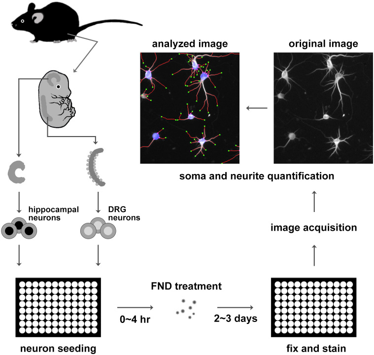
Figure 1
Schematic diagram of dissociated primary neurons preparation, fluorescent nanodiamond treatment, and imaging procedure.
Primary neurons from mouse hippocampi and the dorsal root ganglia were isolated from embryonic mice, dissociated with protease, and seeded into 96-well plates. Dissociated neurons were treated with FN...
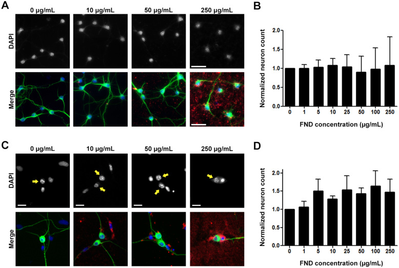
Figure 2
Fluorescent nanodiamonds did not cause cytotoxicity in dissociated hippocampal or dorsal root ganglion neuron cultures.
(A) Images of dissociated hippocampal neurons treated with various concentrations of FNDs 4u2005hours after seeding and incubated for 3 days in vitro . Images on the top row show DAPI-stained nuclei, ...
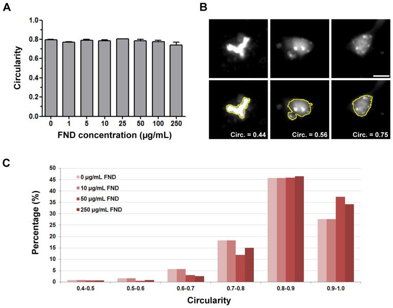
Figure 3
Fluorescent nanodiamonds did not induce apoptosis in dissociated hippocampal neuron cultures.
(A) Quantification of nucleus circularity in hippocampal neurons treated with various concentrations of FND. All quantification data were obtained from 3 independent repeats. No statistically signific...
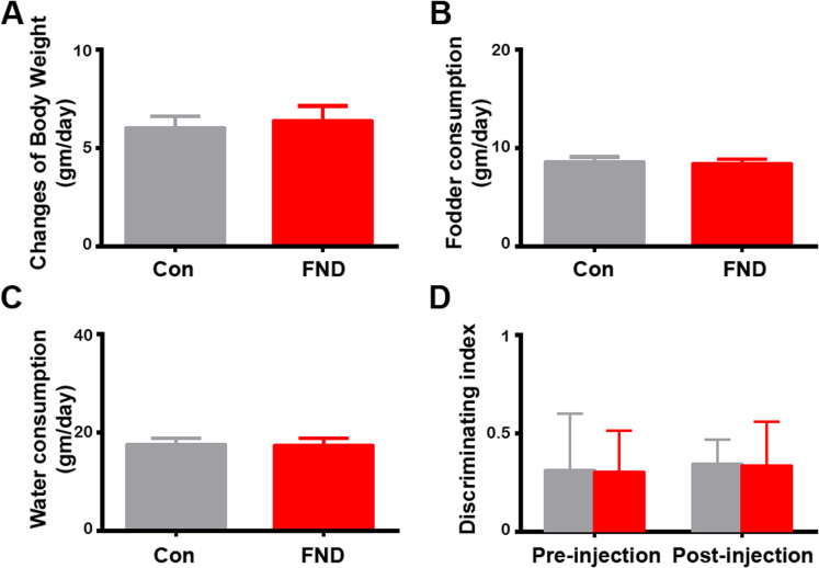
Figure 4
Intracranial injection of fluorescence nanodiamonds did not alter body weight change, fodder and water consumptions, nor the performance of novel object recognition test.
Quantification of daily changes of body weight (A), fodder (B), and water (C) consumption per day in control (saline-injected, n = 4) and FND-injected (n = 4) rats. No statistically significant differ...
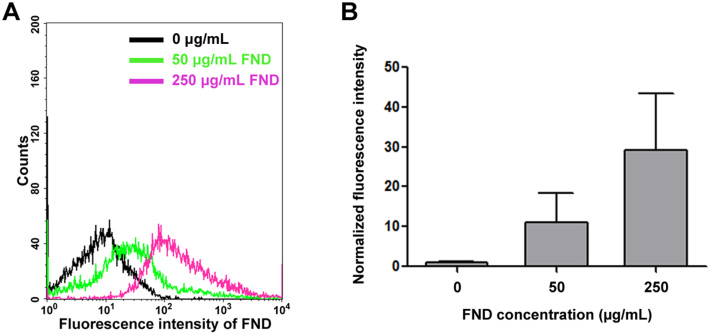
Figure 5
The uptake ability of fluorescent nanodiamonds in dissociated neurons by flow cytometry analysis.
(A) Dissociated mouse cortical neurons treated with 0, 50, or 250u2005u03bcg/mL FND for 3 days. FNDs were excited by with the 488u2005nm wavelength light, and the emitted light was collect in >650u200...
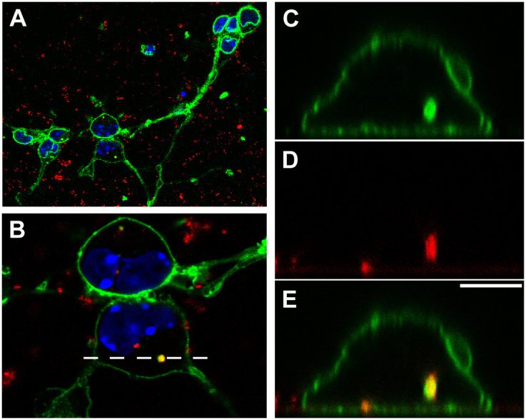
Figure 6
Fluorescent nanodiamonds can be internalized into dissociated neurons.
Confocal image of dissociated mouse cortical neurons cultured in the presence of 20u2005u03bcg/mL FND for 2 days, fixed, and stained with Alexa Fluor 488-conjugated concanavalin-A and DAPI. (A) Image ...
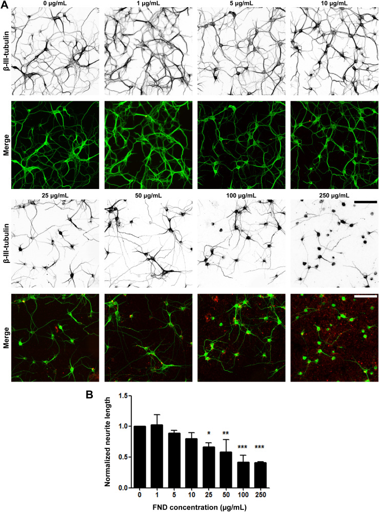
Figure 7
Fluorescent nanodiamonds reduced neurite outgrowth in a dosage-dependent manner in dissociated hippocampal neurons.
(A) Images of dissociated hippocampal neurons treated with various concentrations of FNDs 4u2005hours after seeding and incubated for 3 days in vitro . Images on the top row show u03b2-III-tubulin sta...
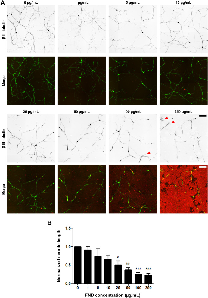
Figure 8
Fluorescent nanodiamonds reduced neurite outgrowth in a dosage-dependent manner in dissociated dorsal root ganglion neurons.
(A) Images of dissociated DRG neurons treated with various concentrations of FNDs after seeding and incubated for 2 days in vitro . Images on the top row show u03b2-III-tubulin staining (inverted to e...
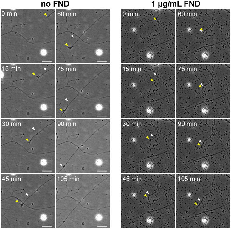
Figure 9
Fluorescent nanodiamond clusters acted as spatial hindrance on advancing neuronal growth cones.
The time-lapse phase contrast image sequences showing the advancing growth cones of DRG neurons under no (left panels) or 1u2005u03bcg/mL of FND (right panels) treatment for 1 day. The yellow arrowhea...
Figure images are served from the NIH/NLM PubMed Central Open Access Subset or Europe PMC; copyright remains with the publishers and authors.
💬 Discussion
0 commentsNo comments yet. Be the first to start a discussion!
Leave a Comment