Abstract
Chromosomes must be accurately partitioned to daughter cells to prevent aneuploidy, a hallmark of many tumors and birth defects. Kinetochores are the macromolecular machines that segregate chromosomes by maintaining load-bearing attachments to the dynamic tips of microtubules. Here, we present the structure of isolated budding-yeast kinetochore particles, as visualized by EM and electron tomography of negatively stained preparations. The kinetochore appears as an ~126-nm particle containing a large central hub surrounded by multiple outer globular domains. In the presence of microtubules, some particles also have a ring that encircles the microtubule. Our data, showing that kinetochores bind to microtubules via multivalent attachments, lay the foundation to uncover the key mechanical and regulatory mechanisms by which kinetochores control chromosome segregation and cell division.
🔬 Techniques
🏭 Microscope Brands
🧪 Reagent Suppliers
📷 Detectors
💻 Software Details
💾 Data Repositories
🏛️ Research Organizations (ROR)
Affiliated research institutions:
📋 Methods
Yeast strains, plasmids and microbial techniques Media and genetic and microbial techniques were essentially as described 41 . Mitotic cultures were prepared with benomyl as described 30 . For temperature sensitive mutants, cells were shifted to 37 °C for 3 hours. Yeast strains and plasmids used in this study are listed in Supplemental Table S4 . The 3xFlag epitope tag strains were made using a PCR-based integration system and confirmed by PCR 42 - 44 . The 6xHis-3xFlag epitope tagging of the endogenous DSN1 gene was performed using a PCR-based integration system using primers SB2434 and SB2435 and plasmid pSB1590 as a template 29 . All tagged strains we constructed are functional in vivo and do not cause any detectable growth defects or temperature sensitivity. Specific primer sequences are available upon request.
Isolation of kinetochore particles
Native kinetochore particles were isolated from budding yeast as described 29 . Briefly, 2 L of yeast cells (SBY8253 or relevant strain) expressing Dsn1-Flag or Dsn1-His-Flag were arrested in mitosis with 60 μg/ml of microtubule depolymerizing drug benomyl for 3 hours and harvested. Cells were lysed in Buffer H (25 mM HEPES pH 8.0, 2 mM MgCl 2 , 0.1 mM EDTA, 0.5 mM EGTA, 150 mM KCl, 15% glycerol and 0.1% NP-40) supplemented with protease and phosphatase inhibitors, and kinetochore particles were captured with anti-Flag antibodies and eluted with 40 μl of Buffer H containing 0.5 mg/ml Flag peptide. The eluted material was used directly for negative stain EM studies as described below (note that this buffer resulted in less compact structures as shown in Figure 1d ). Alternatively, the eluate was prepared for microtubule binding experiments as described below (note that these kinetochores appeared more compact as shown in Figure 1c ). All measurements in the paper were done on particles that had been incubated for microtubule binding experiments.
Show full methods section
Yeast strains, plasmids and microbial techniques Media and genetic and microbial techniques were essentially as described 41 . Mitotic cultures were prepared with benomyl as described 30 . For temperature sensitive mutants, cells were shifted to 37 °C for 3 hours. Yeast strains and plasmids used in this study are listed in Supplemental Table S4 . The 3xFlag epitope tag strains were made using a PCR-based integration system and confirmed by PCR 42 - 44 . The 6xHis-3xFlag epitope tagging of the endogenous DSN1 gene was performed using a PCR-based integration system using primers SB2434 and SB2435 and plasmid pSB1590 as a template 29 . All tagged strains we constructed are functional in vivo and do not cause any detectable growth defects or temperature sensitivity. Specific primer sequences are available upon request.
Isolation of kinetochore particles
Native kinetochore particles were isolated from budding yeast as described 29 . Briefly, 2 L of yeast cells (SBY8253 or relevant strain) expressing Dsn1-Flag or Dsn1-His-Flag were arrested in mitosis with 60 μg/ml of microtubule depolymerizing drug benomyl for 3 hours and harvested. Cells were lysed in Buffer H (25 mM HEPES pH 8.0, 2 mM MgCl 2 , 0.1 mM EDTA, 0.5 mM EGTA, 150 mM KCl, 15% glycerol and 0.1% NP-40) supplemented with protease and phosphatase inhibitors, and kinetochore particles were captured with anti-Flag antibodies and eluted with 40 μl of Buffer H containing 0.5 mg/ml Flag peptide. The eluted material was used directly for negative stain EM studies as described below (note that this buffer resulted in less compact structures as shown in Figure 1d ). Alternatively, the eluate was prepared for microtubule binding experiments as described below (note that these kinetochores appeared more compact as shown in Figure 1c ). All measurements in the paper were done on particles that had been incubated for microtubule binding experiments.
Electron microscopy and image processing
All samples were prepared for negative stain EM as previously described 45 with the following modifications. A 3 μl drop of Flag eluate was applied to a negatively charged carbon coated copper grid (Gilder 400 or 200 mesh) for 20 seconds and washed with a single drop of water, followed by 2 drops of freshly prepared 0.75% uranyl formate. Samples containing microtubules were treated similarly but were applied to positively charged copper grids. Specimens were screened on either a 100 kV transmission electron microscope (TEM) (Morgagni, FEI) or a 120 kV TEM (Spirit T12, FEI). Images were recorded using a Gatan slow scan bottom mount charge coupled device (CCD) camera at a nominal magnification of 18,000x at the specimen level. Measurements were taken either in the Digital Micrograph suite (Gatan, v 1.71.38) or ImageJ64. (v 1.43).
Electron tomography
Negatively stained samples prepared as above were coated with a second layer of carbon by evaporation and anti-mouse IgG-gold (5-10 nm) (Sigma-Aldrich) added as fiducial markers. Tilt series were collected using a Spirit T12 120 kV transmission electron microscope (FEI Company). Images were recorded using a Gatan slow scan 4K x 4K bottom mount CCD with a pixel size of 4.3Å at the sample level (52,000x). Tilt series were recorded from –70° to +70° with an increment of 2° at 2 μ m defocus. Three-dimensional reconstructions were calculated using Amira (v 5.3.1) 46 and IMOD (v. 4.1.9) 47 software. Microtubule binding experiments Taxol stabilized microtubules were prepared freshly as described 48 . A 200 μl aliquot was centrifuged at 58,000 r.p.m. (Beckman TLA 100.1 rotor), 37 °C for 10 minutes. The supernatant was decanted and the pellet was used for microtubule-kinetochore binding experiments as follows. 2 μl of Dsn1-Flag eluate was mixed with 7 μl of the microtubule pellet and incubated at RT for 10 minutes. The sample was then diluted with 200 μl warm BTAX (80 mM PIPES pH 6.9, 1 mM MgCl 2 , 1 mM EGTA and 10 μM Taxol, 37 °C). Grids for EM were prepared as described above. Note that excess tubulin dimers could be seen on the grids due to the microtubule polymerization buffer. Images were recorded on a CCD camera using either the 100 kV TEM or the 120 kV TEM at nominal magnification range of 14,000 – 36,000x at the specimen level. Measurements were taken either in the Digital Micrograph suite (Gatan, v 1.71.38) or ImageJ64. (v 1.43).
Quantification of MT binding
A total of 100 microtubules (for the dad1-1 mutant) or 200 microtubules (for WT and ndc80-1 ) ranging in size from 0.5 to 9 microns were assayed for the number of large particles bound in the presence or absence of a ring (Table 3). Eluates from SBY9047 or SBY7441 were used for all microtubule-binding experiments with WT kinetochore particles.
Supplementary Material 1
📊 Figures
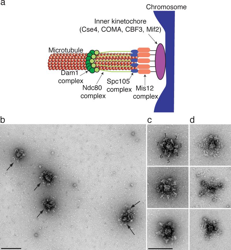
Figure 1
Kinetochore particles contain a central hub surrounded by a number of globular domains
(a) A model for the budding yeast kinetochore shows that multiple copies of the Dam1, Ndc80, KNL-1 (Spc105) and Mis12 kinetochore subcomplexes mediate binding of the chromosome (blue) to the microtubu...
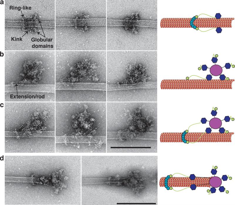
Figure 2
Kinetochore particles bound to taxol-stabilized microtubules
(a) Representative images of fragments of kinetochore particles (56 nm long) bound to taxol-stabilized microtubules reveal a rod with a kink (arrow) connected to a ring on one end and a globular domai...
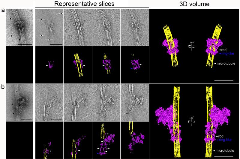
Figure 3
Three-dimensional structures of two types of kinetochore particles bound to a microtubule
a and b , projection images of the two types of kinetochore assemblies on taxol-stabilized microtubules selected for tomographic reconstruction. Representative slices through the each particle are pre...
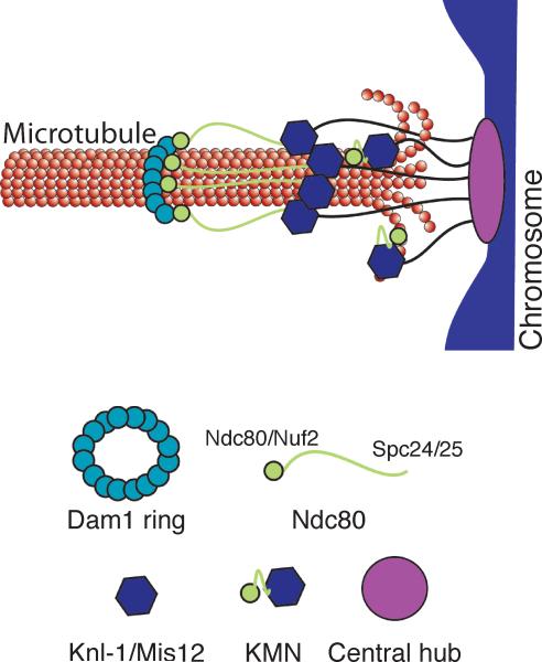
Figure 4
Schematic of the proposed model of kinetochore architecture
The central globular domain binds to the centromeric locus of the chromosome and globular domains containing the KMN complex extend to attach to the microtubule. The Ndc80 subcomplex makes an addition...
Figure images are served from the NIH/NLM PubMed Central Open Access Subset or Europe PMC; copyright remains with the publishers and authors.
💬 Discussion
0 commentsNo comments yet. Be the first to start a discussion!
Leave a Comment