Abstract
The cornea is the main refracting lens in the eye. As part of the outer tunic it has to be resilient, a property conferred by the organisation of the constituent collagen. It also has to be sufficiently elastic to regain its exact shape when deformed, in order not to distort the retinal image. The basis of this elasticity is not fully understood. The purpose of this study was to characterise in three dimensions the arrangement and distribution of elastic fibers in the human corneal stroma, using serial block face scanning electron microscopy. We have demonstrated that there exists a complex network of elastic fibers that appear to originate in the sclera or limbus. These appear as elastic sheets in the limbus and peripheral cornea immediately above the trabecular meshwork which itself appears to extend above Descemet's membrane in the peripheral stroma. From these sheets, elastic fibers extend into the cornea; moving centrally they bifurcate and trifurcate into narrower fibers and are concentrated in the posterior stroma immediately above Descemet's membrane. We contend that elastic sheets will play an important role in the biomechanical deformation and recovery of the peripheral cornea. The network may also have practical implications for understanding the structural basis behind a number of corneal surgeries.
🔬 Techniques
💻 Software
✨ Fluorophores
🧪 Sample Preparation
🏭 Microscope Brands
🧪 Reagent Suppliers
💻 Software Details
📋 Protocols
🏛️ Research Organizations (ROR)
Affiliated research institutions:
📋 Methods
Tissue specimens
Five human corneas were obtained from the CTS Eye Bank, Bristol, UK. Cornea 1 was from a 69-year–old female. The whole enucleated eye was fixed in 4% paraformaldehyde. The cornea was removed with a scleral rim and placed in modified Karnovsky's fixative (2.5% glutaraldehyde and 2% paraformaldehyde in 0.1M cacodylate buffer at pH 7.2) for 30 min, dissected and processed for serial block face scanning electron microscopy (SBF SEM) and TEM. Cornea 2 was from a 50-year-old male and was received from the Eye Bank in Eagle's minimum essential medium as a cornea with about 2 mm of the adjacent sclera. It was de-swelled with 8% dextran overnight, mounted in a Barron artificial anterior chamber to maintain a trans-corneal pressure and was fixed using the same modified Karnovsky's fixative for 3 h. It was then dissected and used for SBF SEM and TEM. Cornea 3 was from a 79-year-old male. It was collected and fixed in modified Karnovsky's fixative within two days of death and was processed for TEM using the orcein method (see below). Cornea 4 was from a 72-year old female. It was fixed in 4% paraformaldehyde then wax embedded for histology. Cornea 5 from a 67-year-old male was processed for non-linear microscopy as described below. Cornea 6 was from a 13-week old foetus obtained from the Human Developmental Biology Resource (HDBR). The whole globe was fixed in modified Karnovsky's fixative and was then processed for SBF SEM as below.
Institutional Ethics
Committee approval was obtained for this study and the research followed the Tenets of the Declaration of Helsinki.
Show full methods section
Tissue specimens
Five human corneas were obtained from the CTS Eye Bank, Bristol, UK. Cornea 1 was from a 69-year–old female. The whole enucleated eye was fixed in 4% paraformaldehyde. The cornea was removed with a scleral rim and placed in modified Karnovsky's fixative (2.5% glutaraldehyde and 2% paraformaldehyde in 0.1M cacodylate buffer at pH 7.2) for 30 min, dissected and processed for serial block face scanning electron microscopy (SBF SEM) and TEM. Cornea 2 was from a 50-year-old male and was received from the Eye Bank in Eagle's minimum essential medium as a cornea with about 2 mm of the adjacent sclera. It was de-swelled with 8% dextran overnight, mounted in a Barron artificial anterior chamber to maintain a trans-corneal pressure and was fixed using the same modified Karnovsky's fixative for 3 h. It was then dissected and used for SBF SEM and TEM. Cornea 3 was from a 79-year-old male. It was collected and fixed in modified Karnovsky's fixative within two days of death and was processed for TEM using the orcein method (see below). Cornea 4 was from a 72-year old female. It was fixed in 4% paraformaldehyde then wax embedded for histology. Cornea 5 from a 67-year-old male was processed for non-linear microscopy as described below. Cornea 6 was from a 13-week old foetus obtained from the Human Developmental Biology Resource (HDBR). The whole globe was fixed in modified Karnovsky's fixative and was then processed for SBF SEM as below.
Institutional Ethics
Committee approval was obtained for this study and the research followed the Tenets of the Declaration of Helsinki.
Serial block face scanning electron microscopy Human
Corneas were fixed for 3 h in 2.5% glutaraldehyde/2% paraformaldehyde in 100 mM sodium cacodylate buffer pH 7.2 at room temperature (RT). The cornea was cut into thin segments and post fixed with 1% osmium tetroxide for 1 h. After a brief wash with fresh buffer followed by a 20 min wash in distilled water the samples were incubated in 0.5% low molecular weight (di-Gallic) C 14 H 10 O 9 tannic acid (mw 322.22) (AR Mallinckrodt, Dublin, Ireland) in distilled water for 2 h. The samples were then washed in distilled water for 30 min and placed in 1% aqueous uranyl acetate for 1 h in the dark at RT. Corneal samples were then dehydrated in an ethanol series from 70% to 100% ethanol for 1 h after which they were placed in 2% uranyl acetate in 100% ethanol for 2 h in the dark at RT. After washing in 100% ethanol for 40 min, the corneal samples were placed in a 1:1 mixture of 100% ethanol: 100% acetone for 20 min. The samples were then incubated in a saturated solution of lead acetate in a mixture of 1:1 100% ethanol 100% acetone for 2 h ( Kushida, 1966 ). After staining, the corneal samples were washed in two changes of 1:1 mixture of 100% ethanol and 100% acetone for 15 min and placed in 100% solution of acetone for 20 min. After washing the samples 3 times for 20 min in 100% acetone, the samples were finally embedded in CY212 (TAAB) epoxy resin and polymerised for 24 h at 60° C. The surfaces of polymerised resin blocks were then trimmed and attached to Gatan (PEP6590) specimen pins. The pins were then gold coated and transferred to a Zeiss Sigma VP FEG SEM equipped with a Gatan 3View2 system, where data sets of up to 1000 images were acquired of the block surface every 50 nm through automated sectioning. Each image was acquired at 4K × 4K pixels, at a pixel resolution of 4 nm and a pixel dwell time of 8 μs, using an accelerating voltage of 3.4 keV in low vacuum variable pressure mode (28 Pa). Imaging data was acquired from a 16.19 μm × 16.19 μm region of interest. Selected serial image sequences were extracted from the image data and 3D reconstructions were generated with Amira 6.0 software (FEI, Mérignac, France) using both manual hand tracing for the larger structures and automated thresholding for the fibers. For the full thickness quantification analysis, the peripheral cornea was imaged every 50 nm en face at 5000 × magnification, starting from the epithelium and ending at Descemet's membrane, obtaining over 21,000 images with resolutions of 2000 × 2000 pixels. The data were split into sets of ∼1000 images, fibers were segmented using a mixture of manual and automatic thresholding, resulting in a fiber voxel count that was subsequently converted to percentage using the total volume voxel count. Cells were blocked out to background level in order to remove their contribution to the images. The more detailed quantification above Descemet's membrane was carried out by measuring fiber volume every 200 images. Both were done using Amira 6.0 software with XImagePAQ extension. When sectioning the 16 μm × 16 μm block face through a thickness of almost 1 mm, it was very difficult to ensure that the en face sections were exactly parallel to the surface of the cornea. When viewing our series of images from different depths, it was evident that the blocks were being cut at a slight angle to the surface of the cornea. To correct for this, the number of 50 nm sections between where Descemet's membrane was just visible down to the depth where the stroma was just not visible (which ideally would be zero if the block was being cut parallel to the corneal surface) was used to make a geometrical calculation of the offset angle and hence correct the section thickness and the depth measurements to allow for this when plotting the % fiber volume as a function of depth. This led to an uncertainty of ±5 μm in each depth measurement.
Transmission electron microscopy
Embedded samples from Corneas 1 and 2 that had been en bloc stained for SBF SEM, were also used for TEM. A Leica UC6 ultra-microtome was used to cut 90 nm-thick gold sections that were floated on distilled water before being mounted on copper grids. Additionally, separate sections were cut from Cornea 3 and stained with the elastic fiber-specific stain 0.2% orcein in acid alcohol (1% HCl in 70% EtOH), then rinsed with 70% EtOH, air-dried, and counterstained with uranyl acetate and lead citrate. Both normal and orcein stained ultra-thin sections were visualised using a Jeol 1010 TEM (JEOL, Tokyo, Japan).
Non-linear microscopy
Corneal buttons 8 mm in diameter were dissected from the centre of Cornea 5, placed into dialysis tubing (molecular cut-off 14 kDa), and brought to physiological hydration by immersion in 2.5% polyethylene glycol (PEG) overnight. The buttons were then mounted on Superfrost glass slides, in 1:1 phosphate buffered saline (PBS) – glycerol solution, protected by a 0.16 mm thick glass coverslip. A modified confocal microscope (Olympus FluoView IX71 and F300) was used to obtain TPF and SHG images (see Bell et al. (2014) for a complete description of the system configuration). TPF and SHG images from the same area of cornea were overlaid using ImageJ software ( Schneider et al., 2012 ), resulting in a composite image.
Histology
Paraffin wax sections were dewaxed in xylene and rehydrated in a descending industrial methylated spirit (IMS) gradient to water. Tissue sections were incubated in Miller's elastic stain at 60 °C for 1 h, destained in 95% IMS and washed in water before 10 min of nuclear staining in Mayer's haematoxylin, followed by counterstaining of collagen fibrils with Van Gieson's stain for 5 min. Sections were then washed in water, dehydrated through an ascending IMS gradient, cleared in xylene and mounted under coverslips with DPX mountant. Elastic fibers and collagen appeared purple/black and pinkish/red, respectively.
📊 Figures
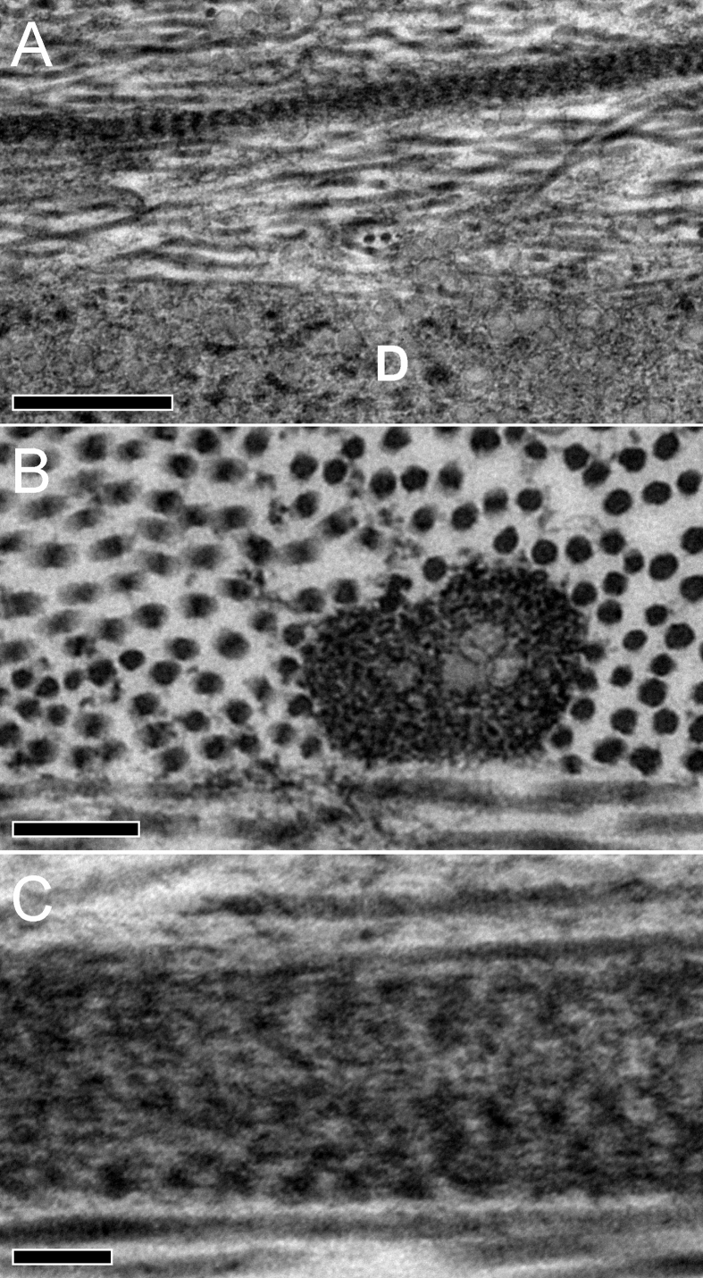
Fig.u00a01
TEM images of elastic fibers from the central cornea (Cornea 1) stained with tannic acid. Fig.u00a01 A: A banded fiber running longitudinally above Descemet's membrane (D) (baru00a0=u00a0500u00a0nm). ...
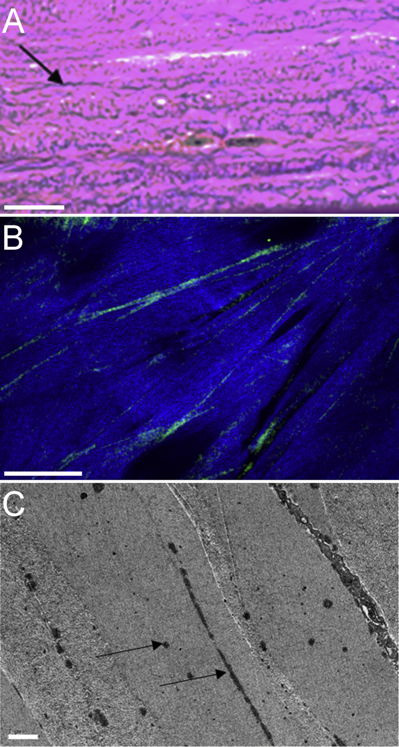
Fig.u00a02
Images of the peripheral stroma (4u20135u00a0mm from the optic axis) stained for elastic fibers. Fig.u00a02 A: Transverse histological section (Cornea 5) immediately above Descemet's membrane, stained...
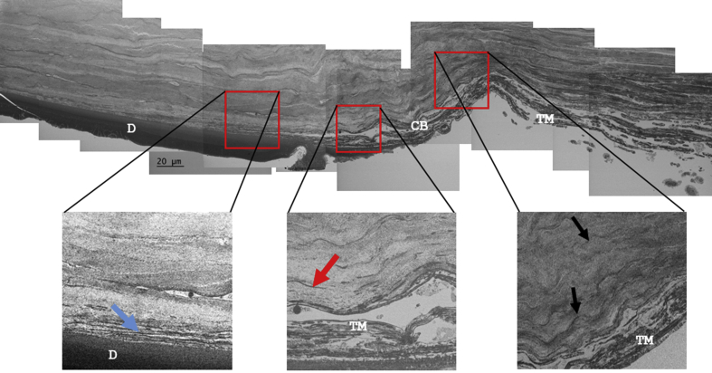
Fig.u00a03
Low magnification montage of SEM images showing a transverse section through the corneolimbal region from Cornea 2. Numerous elastic structures are evident within the corneolimbal region (black arrows...
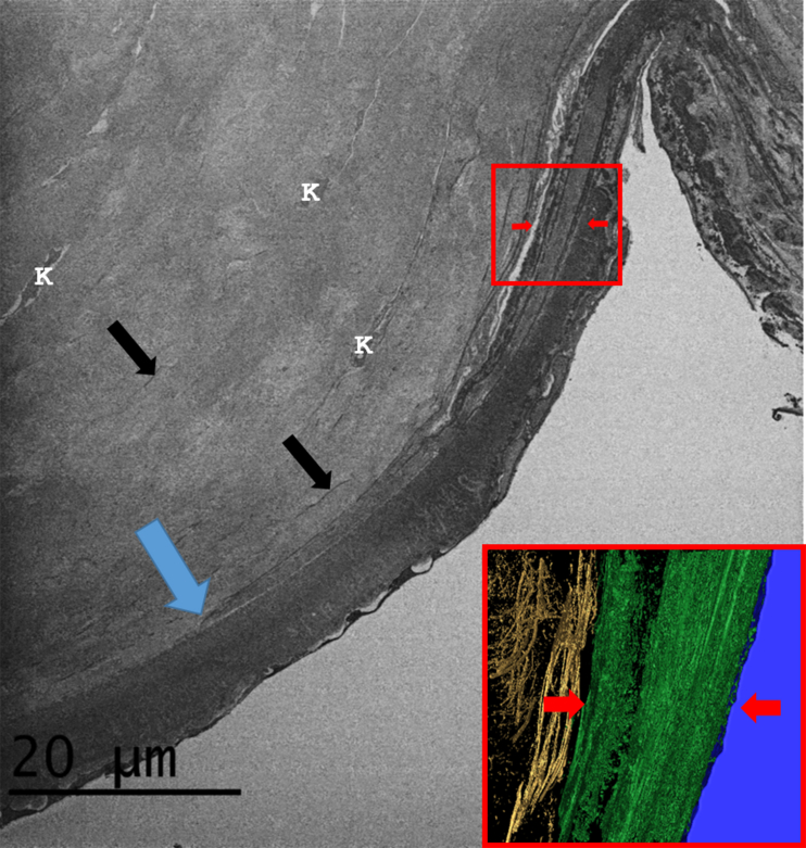
Fig.u00a04
The corneolimbal region (Cornea 1). The main SEM image shows the trabecular meshwork insertion between Descemet's membrane and the posterior corneal stroma. The trabecular meshwork insertion is separa...
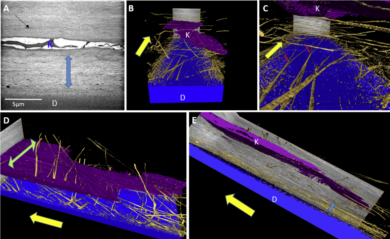
Fig.u00a05
The peripheral cornea (Cornea 1). The yellow arrows indicate the radial direction towards the limbus. Fig.u00a05 A: SBF SEM image of posterior peripheral cornea reveals the presence of numerous elasti...
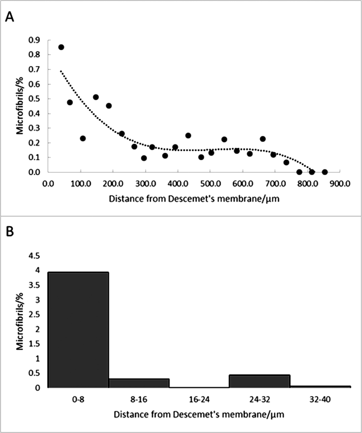
Fig.u00a06
Distribution of elastic fibers as a function of depth in the peripheral cornea measured from en face images through the cornea (Cornea 1). The top graph shows the percentage of the tissue volume occup...
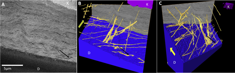
Fig.u00a07
The central cornea (Cornea 1). Fig.u00a07 A: SBF SEM image of posterior central cornea. The black arrow highlights a single elastic fiber running longitudinally within the stroma. (Du00a0=u00a0Desceme...
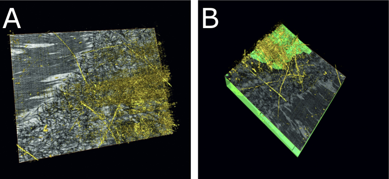
Fig.u00a08
SBF SEM from the central posterior cornea of a 13-week-old human foetus (Cornea 6). Fig.u00a08 A shows the presence of elastic fibers (gold) above a meshwork of dark filaments which will later become ...
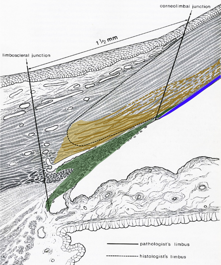
Fig.u00a09
Proposed elastic fiber organisation in the limbal region. Translucent green: Trabecular meshwork. Solid green: Trabecular meshwork insertion. Gold: Elastic limbal region with concentrated elastic fibe...
Figure images are served from the NIH/NLM PubMed Central Open Access Subset or Europe PMC; copyright remains with the publishers and authors.
💬 Discussion
0 commentsNo comments yet. Be the first to start a discussion!
Leave a Comment