Abstract
Podocytes are critical in the maintenance of a healthy glomerular filter; however, they have been difficult to study in the intact kidney because of technical limitations. Here we report the development of serial multiphoton microscopy (MPM) of the same glomeruli over several days to visualize the motility of podocytes and parietal epithelial cells (PECs) in vivo. In podocin-GFP mice, podocytes formed sporadic multicellular clusters after unilateral ureteral ligation and migrated into the parietal Bowman's capsule. The tracking of single cells in podocin-confetti mice featuring cell-specific expression of CFP, GFP, YFP or RFP revealed the simultaneous migration of multiple podocytes. In phosphoenolpyruvate carboxykinase (PEPCK)-GFP mice, serial MPM found PEC-to-podocyte migration and nanotubule connections. Our data support a highly dynamic rather than a static nature of the glomerular environment and cellular composition. Future application of this new approach should advance our understanding of the mechanisms of glomerular injury and regeneration.
🔬 Techniques
🔭 Microscopes
💻 Software
✨ Fluorophores
🧪 Sample Preparation
🔬 Cell Lines
🏭 Microscope Brands
🧪 Reagent Suppliers
🔴 Lasers
💻 Software Details
💾 Data Repositories
🏛️ Research Organizations (ROR)
Affiliated research institutions:
📋 Methods
Animals
Both male and female C57BL6/J mice at the age of 4–12 weeks were used randomly. Four new fluorescent reporter mouse models were generated: (i) Pod-GFP by crossing mice expressing Cre recombinase under the control of the podocin promoter 21 and animals with a Tomato floxSTOP EGFP sequence in the ROSA 26 locus (B6.129(Cg)- Gt(ROSA)26Sor tm4(ACTB-tdTomato,-EGFP)Luo ) 16 . This results in an expression of Tomato in all cells except in podocytes, which express GFP. To study the effect of different backgrounds those mice were used in a C57BL6 background, a FvB background or the F1 generation crossed between these backgrounds. (ii) iPod-GFP by crossing mice expressing tamoxifen-inducible improved Cre recombinase under the control of the podocin promoter (podocin-iCreER T2 mice 22 , kind gift from Dr. Farhad R. Danesh, Baylor College of Medicine) and animals with the Tomato floxSTOP EGFP construct mentioned above. (iii) PEPCK-GFP by crossing mice expressing Cre recombinase under the control of the phosphoenolpyruvate-carboxykinase promoter (PEPCK-Cre, kind gift from Dr. Volker Haase, Vanderbilt University) 31 and animals with the Tomato floxSTOP EGFP construct mentioned above. (iv) Pod-Confetti by crossing mice expressing the R26R-Confetti construct 17 and podocin-Cre mice 21 resulting in the expression of either membrane-targeted CFP, nuclear GFP, cytosolic YFP or cytosolic RFP in podocytes. All animals were purchased from the Jackson laboratory (Bar Harbor, ME) or otherwise noted. All animal protocols were approved by the Institutional Animal Care and Use Committee at the University of Southern California or by local government authorities in Germany (LANUV NRW) under the license 8.87–50.10.31.08.049. Tamoxifen induction 4 weeks-old mice received Tamoxifen chow (40 mg/kg body weight, Harlan Laboratories, Indianapolis, IN) for 2 weeks, followed by a 2 week washout period before additional procedures were performed. UUO Between 3 and 8 weeks of age the animals were anaesthetized with isoflurane, and after a midline laparotomy the left ureter was exposed and ligated three times. Successful ligation was confirmed by the hydronephrotic distension of the kidney at the time of imaging. Adriamycin nephropathy Pod-GFP animals received a single dose of 25mg/kg Adriamycin (Doxorubicin) via retroorbital injection and were imaged, sacrificed and their tissues collected 4–6 days later. Successful induction of albuminuria was confirmed by measuring the urinary albumin/creatinine ratio (Exocell, Philadelphia, PA) between days 0–6 after adriamycin treatment.
Show full methods section
Animals
Both male and female C57BL6/J mice at the age of 4–12 weeks were used randomly. Four new fluorescent reporter mouse models were generated: (i) Pod-GFP by crossing mice expressing Cre recombinase under the control of the podocin promoter 21 and animals with a Tomato floxSTOP EGFP sequence in the ROSA 26 locus (B6.129(Cg)- Gt(ROSA)26Sor tm4(ACTB-tdTomato,-EGFP)Luo ) 16 . This results in an expression of Tomato in all cells except in podocytes, which express GFP. To study the effect of different backgrounds those mice were used in a C57BL6 background, a FvB background or the F1 generation crossed between these backgrounds. (ii) iPod-GFP by crossing mice expressing tamoxifen-inducible improved Cre recombinase under the control of the podocin promoter (podocin-iCreER T2 mice 22 , kind gift from Dr. Farhad R. Danesh, Baylor College of Medicine) and animals with the Tomato floxSTOP EGFP construct mentioned above. (iii) PEPCK-GFP by crossing mice expressing Cre recombinase under the control of the phosphoenolpyruvate-carboxykinase promoter (PEPCK-Cre, kind gift from Dr. Volker Haase, Vanderbilt University) 31 and animals with the Tomato floxSTOP EGFP construct mentioned above. (iv) Pod-Confetti by crossing mice expressing the R26R-Confetti construct 17 and podocin-Cre mice 21 resulting in the expression of either membrane-targeted CFP, nuclear GFP, cytosolic YFP or cytosolic RFP in podocytes. All animals were purchased from the Jackson laboratory (Bar Harbor, ME) or otherwise noted. All animal protocols were approved by the Institutional Animal Care and Use Committee at the University of Southern California or by local government authorities in Germany (LANUV NRW) under the license 8.87–50.10.31.08.049. Tamoxifen induction 4 weeks-old mice received Tamoxifen chow (40 mg/kg body weight, Harlan Laboratories, Indianapolis, IN) for 2 weeks, followed by a 2 week washout period before additional procedures were performed. UUO Between 3 and 8 weeks of age the animals were anaesthetized with isoflurane, and after a midline laparotomy the left ureter was exposed and ligated three times. Successful ligation was confirmed by the hydronephrotic distension of the kidney at the time of imaging. Adriamycin nephropathy Pod-GFP animals received a single dose of 25mg/kg Adriamycin (Doxorubicin) via retroorbital injection and were imaged, sacrificed and their tissues collected 4–6 days later. Successful induction of albuminuria was confirmed by measuring the urinary albumin/creatinine ratio (Exocell, Philadelphia, PA) between days 0–6 after adriamycin treatment.
Multiphoton imaging
The animals were anaesthetized with a combination of ketamine (100 mg/kg) and xylazine (10 mg/kg). A tracheal tube was placed to facilitate breathing and the right carotid artery and/or jugular vein was cannulated for dye infusion. A 70kDa Texas red dextran or Alexa 594 bovine serum albumin was injected to label the vasculature. In some experiments Lucifer Yellow (Invitrogen) was injected as iv bolus to visualize glomerular filtration. The left kidney was exteriorized through a flank incision and the animal placed on the microscope stage as described before 34 . Body temperature was maintained with a homeothermic blanket system (Harvard Apparatus). The images were acquired using a Leica TCS SP5 multiphoton confocal fluorescence imaging system with a 63x Leica glycerine-immersion objective (NA 1.3) powered by a Chameleon Ultra-II MP laser at 860 nm (Coherent) and a DMI 6000 inverted microscope’s external nondescanned detectors. Short pass filters (680 nm for blue and red, 700 nm for green and yellow), dichroic mirrors (cut off at 515 nm for green and yellow, 560 nm for blue and red) and bandpass filters were specific for detecting CFP/GFP/YFP/RFP emission (473nm/514nm/545nm/585nm) (Chroma). The images of iPod-GFP animals were acquired using the external detectors of an inverted Zeiss LSM710 NLO multiphoton confocal fluorescence microscope powered by a Chameleon Ultra-II MP laser at 860 nm and a 40x Zeiss water-immersion objective (NA1.2). Serial survival imaging of the same glomerulus After anaesthesia the left kidney was exteriorized via a small cut in the left flank below the kidney to avoid sutures right above the kidney afterwards. To avoid invasive vascular access surgeries, the dyes were administered by retroorbital injections. The animal was transferred to the microscope stage and the exteriorized kidney was placed into a kidney cup. An area of the kidney suitable for imaging was identified and the position of the kidney was noted for identical placement on the following days. After acquiring z-stacks of the glomerulus a small distant area in the field of view was marked by shortly focusing the laser beam on this area with high power. This maneuver generated an easy to find highly fluorescent spot (reference point) which remained there for 3–5 days. The position of the mark relative to the glomerulus of interest was documented. After imaging, the kidney was placed back into the retroperitoneum and the flank cut was closed with two layers of sutures. This procedure was repeated 24, and 48 hours later by removing the sutures and exteriorizing the kidney again. With this technique we were able to subsequently find approximately 70% of the glomeruli that were marked in the first imaging session. Z-stacks of marked glomeruli were acquired with identical imaging settings as the day before. The potential toxicity of laser excitation and fluorescence to the cells were minimized by using a low laser power and high scan speeds to keep total laser exposure as small as possible. The usual image acquisition consisted of only one z-stack per glomerulus (
📊 Figures
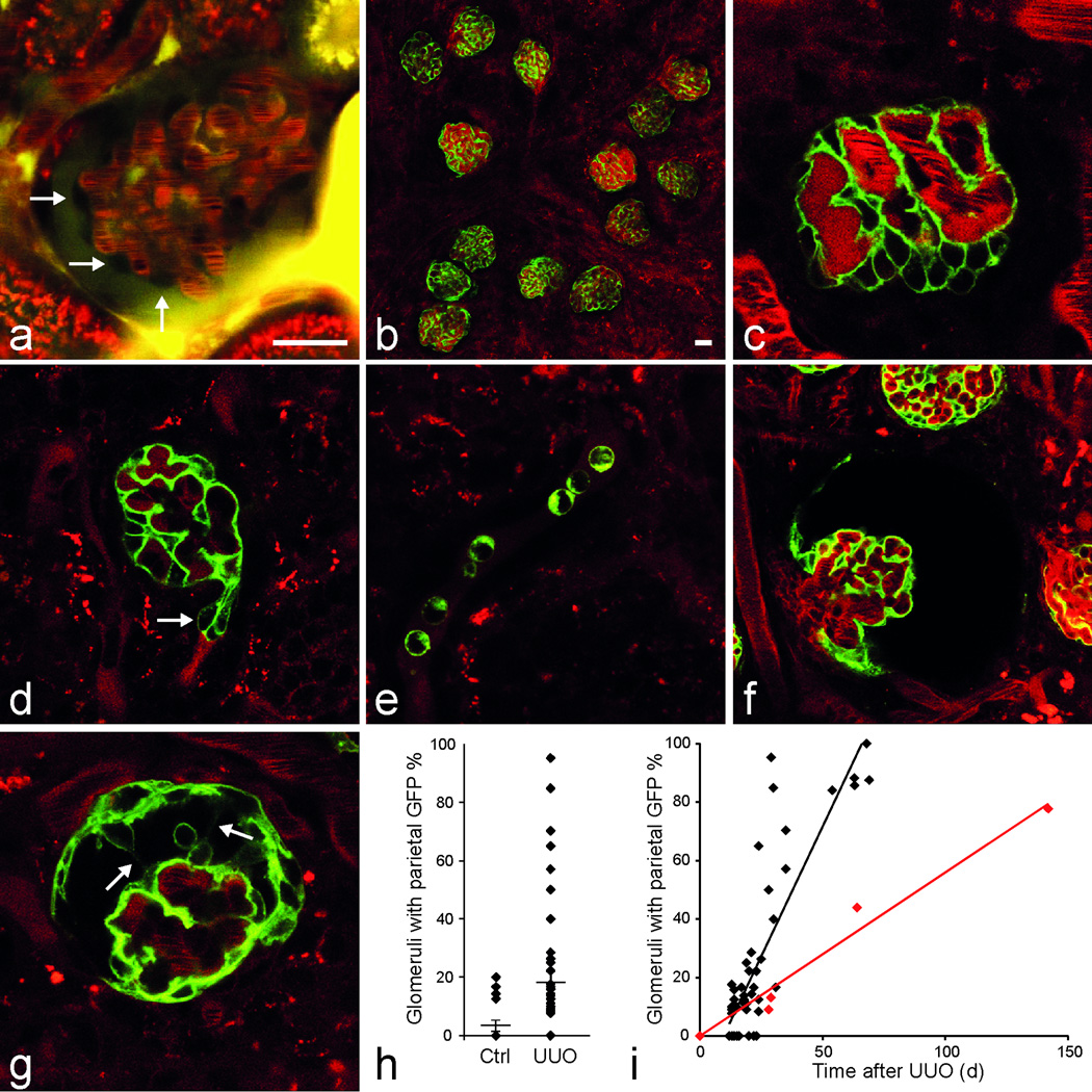
Figure 1
MPM imaging in vivo reveals signs of podocyte migration in the intact kidney in the model of unilateral ureteral obstruction (UUO) in Pod-GFP mice
(a) In a wild-type mouse kidney the glomerular podocytes appear as dark, unlabeled cells around capillaries (arrows). Plasma is labeled red with Alexa594-Albumin. Lucifer yellow (yellow) was injected ...
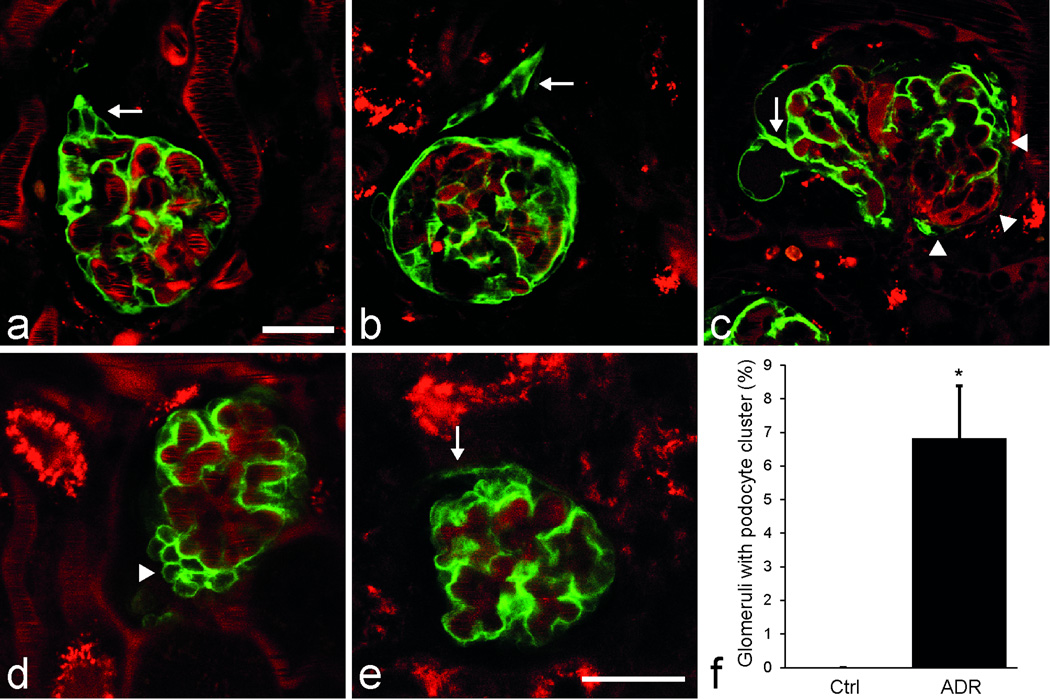
Figure 2
Confirmation of podocyte clustering and migration in iPod-GFP mice after UUO (au2013c) and podocyte clustering in the model of adriamycin nephropathy in Pod-GFP mice (du2013f)
In mice with tamoxifen-induced podocyte GFP expression (iPod-GFP) podocyte clustering was observed 4 weeks following UUO, podocyte projections to the parietal Bowmanu2019s capsule developed either at ...
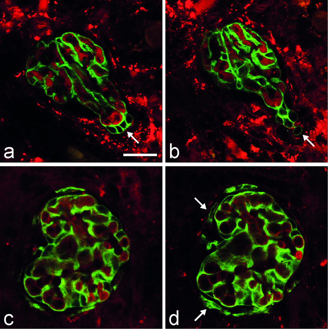
Figure 3
Serial in vivo MPM imaging of the same glomerulus in Pod-GFP mice after UUO over time, once in 24 hours
(au2013b) Podocytes from a hypercellular area at the urinary pole of a collapsed, non-filtering glomerulus (a, arrow) appear to migrate away from the capillary tuft to form a projection into the remai...
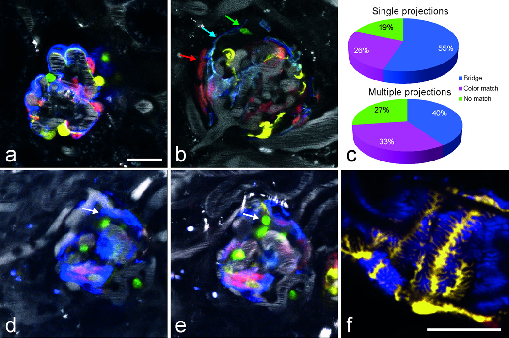
Figure 4
Identification and tracking of single podocytes in the multi-color Pod-Confetti mouse model using in vivo MPM imaging
(a) Podocytes are labeled in one of the four colors, either by membrane-targeted CFP, nuclear GFP, cytosolic YFP or cytosolic RFP. Labeled dextran (plasma dye) is shown in grayscale. (b) A CFP-labeled...
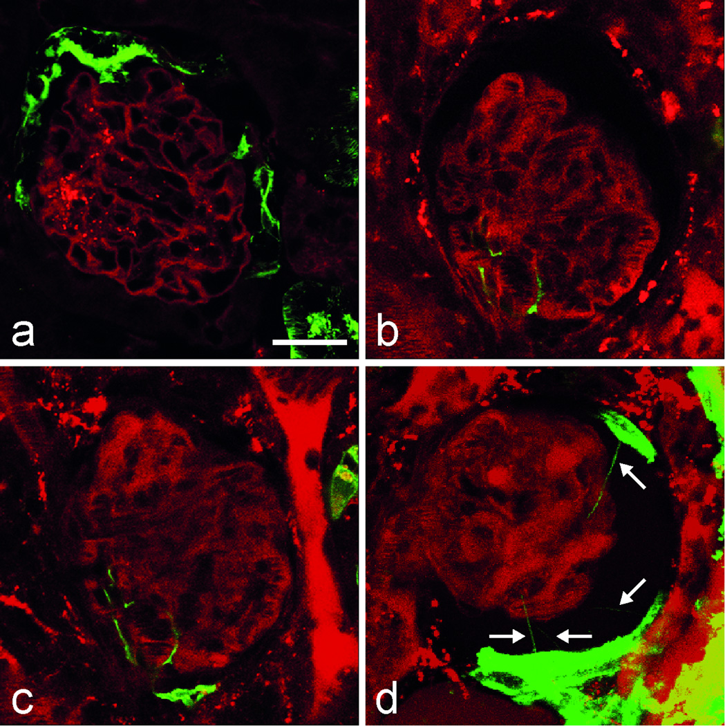
Figure 5
MPM imaging of PEC migration in vivo in the PEPCK-GFP mouse model after UUO
(a) Cell-specific GFP expression in the proximal tubule and in some, but not all PECs. All other cells are labeled with Tomato. (bu2013c) Serial MPM imaging of the same glomerulus shows a GFP expressi...
Figure images are served from the NIH/NLM PubMed Central Open Access Subset or Europe PMC; copyright remains with the publishers and authors.
💬 Discussion
0 commentsNo comments yet. Be the first to start a discussion!
Leave a Comment