Abstract
Abstract Tissue ischemia adversely affects the function of mitochondria, which results in impairment of oxidative phosphorylation and compromised recovery of the affected organ. The impact of ischemia on mitochondrial function has been extensively studied in the heart because of the morbidity and mortality associated with injury to this organ. As conventional methods to preserve cardiac cell viability and contractile function following ischemia are limited in their efficacy, we developed a unique approach to protect the heart by transplanting respiration-competent mitochondria to the injured region. Our previous animal experiments showed that transplantation of isolated mitochondria to ischemic heart tissue leads to decreases in cell death, increases in energy production, and improvements in contractile function. We also discovered that exogenously-derived mitochondria injected or perfused into ischemic hearts were rapidly internalised by cardiac cells. Here, we used three-dimensional super-resolution microscopy and transmission electron microscopy to determine the intracellular fate of endocytosed exogenous mitochondria in human iPS-derived cardiomyocytes and primary cardiac fibroblasts. We found isolated mitochondria are incorporated into cardiac cells within minutes and then transported to endosomes and lysosomes. The majority of exogenous mitochondria escape from these compartments and fuse with the endogenous mitochondrial network, while some of these organelles are degraded through hydrolysis.
🔬 Techniques
🔭 Microscopes
💻 Software
✨ Fluorophores
🧪 Sample Preparation
🔬 Cell Lines
🏭 Microscope Brands
🧪 Reagent Suppliers
💻 Software Details
💾 Data Repositories
🏛️ Research Organizations (ROR)
Affiliated research institutions:
📋 Methods
Reagents
The following antibodies were purchase from Abcam: anti-α-actinin (ab68167), anti-vimentin (ab92547), anti-TOMM20 (ab56783), anti-mitochondria [MTC02] (ab3298), anti-mitochondria [113-1] (ab92824), anti-Mitofusin 1 [MFN1] (ab57602 and ab104274), anti-Mitofusin 2 [MFN2] (ab56889 and ab101055), anti-OPA1 (ab90857), anti-Parkin (ab15954), anti-DRP1 [ EPR19275 ] (ab184248), anti-DRP1 [phospho S637] (ab193216) and anti-Rab7 (ab50533). The anti-phosphoDRP1 Ser616 [D9A1] antibody (CST-4494S) was purchased from Cell Signaling. The mouse IgG isotype control (10400 C) and rabbit IgG isotype control (10500 C) antibodies were purchased from ThermoFisher Scientific. The following CellLight reagents were purchased from ThermoFisher Scientific: Early endosomes ( C10586 and C10587 ), Endoplasmic Reticulum ( C10590 and C10591 ), Golgi Complex ( C10592 and C10593 ), Late endosomes ( C10588 and C10589 ), Lysosomes ( C10596 and C10597 ), Mitochondria ( C10600 and C10601 ) and Null Virus Negative Control ( C10615 ). MitoTracker Red CMXRos (M7512) was purchased from ThermoFisher Scientific and used according to the manufacturer’s directions at 100 nM. Cells iCell-Cardiomyocytes 2 (iPS-CMs) (CMC-100-012-001) were purchased from Cellular Dynamics and Human Cardiac Fibroblasts (HCFs) (6300) were purchased from Sciencell Research Laboratories. Cells were cultured according to the manufacturer’s directions. Baculovirus infections were performed on dividing HCFs by adding 400 μL of CellLight reagent to a 100-mm confluent plate of cells for 16 h. Spontaneously contracting syncytial monolayers of iPS-CMs (≥96 hours after plating) were infected for 16 h by using 10% CellLight reagent in iCell Cardiomyocyte Maintenance Medium (CMM-100-120-001) (Cellular Dynamics) containing 0.2 μM BacMam Enhancer (ThermoFisher Scientific). Mitochondria HCF media was replaced with 4 mL of 4 °C homogenisation buffer (300 mM sucrose, 10 mM HEPES-KOH, 1 mM EGTA-KOH, pH 7.4) containing 2 mg Subtilisin A protease from Bacillus licheniformis (Sigma-Aldrich) and incubated at room temperature for 5 min. Digested cells were transferred to a 15-mL tube on ice and digested for an additional 15 min before filtration through a 10 μm mesh filter (PluriSelect) saturated with cold homogenisation buffer. Mitochondria were collected by centrifugation at 9,500 RCF at 4 °C for 5 minutes and washed 3 times in cold homogenisation buffer before resuspension in culture media. Mitochondrial number was determined by using a Multisizer 4e Coulter Counter (Beckman-Coulter) and corroborated by hemocytometry. Isolated mitochondria were incubated with HCFs or iPS-CMs for 0 to 4 h.
Show full methods section
Reagents
The following antibodies were purchase from Abcam: anti-α-actinin (ab68167), anti-vimentin (ab92547), anti-TOMM20 (ab56783), anti-mitochondria [MTC02] (ab3298), anti-mitochondria [113-1] (ab92824), anti-Mitofusin 1 [MFN1] (ab57602 and ab104274), anti-Mitofusin 2 [MFN2] (ab56889 and ab101055), anti-OPA1 (ab90857), anti-Parkin (ab15954), anti-DRP1 [ EPR19275 ] (ab184248), anti-DRP1 [phospho S637] (ab193216) and anti-Rab7 (ab50533). The anti-phosphoDRP1 Ser616 [D9A1] antibody (CST-4494S) was purchased from Cell Signaling. The mouse IgG isotype control (10400 C) and rabbit IgG isotype control (10500 C) antibodies were purchased from ThermoFisher Scientific. The following CellLight reagents were purchased from ThermoFisher Scientific: Early endosomes ( C10586 and C10587 ), Endoplasmic Reticulum ( C10590 and C10591 ), Golgi Complex ( C10592 and C10593 ), Late endosomes ( C10588 and C10589 ), Lysosomes ( C10596 and C10597 ), Mitochondria ( C10600 and C10601 ) and Null Virus Negative Control ( C10615 ). MitoTracker Red CMXRos (M7512) was purchased from ThermoFisher Scientific and used according to the manufacturer’s directions at 100 nM. Cells iCell-Cardiomyocytes 2 (iPS-CMs) (CMC-100-012-001) were purchased from Cellular Dynamics and Human Cardiac Fibroblasts (HCFs) (6300) were purchased from Sciencell Research Laboratories. Cells were cultured according to the manufacturer’s directions. Baculovirus infections were performed on dividing HCFs by adding 400 μL of CellLight reagent to a 100-mm confluent plate of cells for 16 h. Spontaneously contracting syncytial monolayers of iPS-CMs (≥96 hours after plating) were infected for 16 h by using 10% CellLight reagent in iCell Cardiomyocyte Maintenance Medium (CMM-100-120-001) (Cellular Dynamics) containing 0.2 μM BacMam Enhancer (ThermoFisher Scientific). Mitochondria HCF media was replaced with 4 mL of 4 °C homogenisation buffer (300 mM sucrose, 10 mM HEPES-KOH, 1 mM EGTA-KOH, pH 7.4) containing 2 mg Subtilisin A protease from Bacillus licheniformis (Sigma-Aldrich) and incubated at room temperature for 5 min. Digested cells were transferred to a 15-mL tube on ice and digested for an additional 15 min before filtration through a 10 μm mesh filter (PluriSelect) saturated with cold homogenisation buffer. Mitochondria were collected by centrifugation at 9,500 RCF at 4 °C for 5 minutes and washed 3 times in cold homogenisation buffer before resuspension in culture media. Mitochondrial number was determined by using a Multisizer 4e Coulter Counter (Beckman-Coulter) and corroborated by hemocytometry. Isolated mitochondria were incubated with HCFs or iPS-CMs for 0 to 4 h.
Labelling
Some isolated mitochondria (1 × 10 8 ) were conjugated to 10 nm NHS-activated gold nanoparticles (CytoDiagnostics) according to the manufacturer’s directions. Labeled mitochondria were washed 5 times with homogenisation buffer containing 1 mg/mL fraction V bovine serum albumin (BSA) to remove unreacted nanoparticles. Gold-labeled mitochondria were incubated with iPS-CMs for 0, 2.5, 5 and 10 minutes prior to fixation and preparation for transmission electron microscopy. Immunoblots Whole cell proteins were isolated by rinsing cells with ice-cold PBS (pH 7.4) and lifting them from the plates with a rubber policeman. Mitochondrial proteins were isolated by rinsing the mitochondrial pellet with ice-cold PBS (pH 7.4) five times. Pelleted cells or isolated mitochondria were then resuspended in a small volume of freshly-prepared ice-cold lysis solution (150 mM NaCl, 20 mM Tris-HCl (pH 7.6), 1 mM EDTA, 0.5% sodium deoxycholate, 70 mM NaF, 1% Nonidet P-40, Complete protease inhibitor cocktail (Sigma-Aldrich), 200 μM sodium orthovanadate, and 2 μM phenylmethylsulfonyl fluoride) 49 . After a 10-min incubation on ice with intermittent, brief agitation, debris was pelleted in a microcentrifuge, and the supernatants were stored at −80 °C. Protein concentrations were determined using the bicinchoninic acid (BCA) protein determination kit (Pierce). SDS-PAGE and transfer to nitrocellulose was performed using the XCell SureLock Mini-Cell System and XCell II Blot Module (ThermoFisher), respectively. Identical 4–20% Tris-Glycine gels (ThermoFisher) were stained with Coomassie brilliant blue R250 to confirm equal protein loading. The ColorBurst Electrophoresis Marker (Sigma-Aldrich) and MagicMark XP Western Protein Standard (ThermoFisher) were used according to the manufacturer’s directions. Nitrocellulose membranes were rinsed in Tris-buffered saline (pH 7.4) containing 0.1% Tween 20 (TBS-T) and blocked in 5% non-fat milk in TBS-T for 1 h at 22 °C on a rocking platform. Membranes were incubated overnight at 4 °C on an orbital shaker with primary antibodies diluted according to the manufacturer’s directions in TBS-T containing 1% non-fat milk (Sigma). Primary antibodies were detected with the Amersham ECL Western Blotting Analysis System (GE Healthcare) using HRP-conjugated secondary antibodies (GE-Healthcare).
Staining
Cells cultured on No 1.5 H (170 μm ± 5 μm) coverslips (Marienfeld-Superior) were fixed in 4% freshly-prepared formaldehyde in phosphate buffered saline (PBS). Cells were permeabilised in 0.1% Triton X-100 in PBS for 3 minutes and incubated with primary antibodies diluted 1:1000 in 10% fetal bovine serum (FBS) in PBS for 1 h. Primary antibodies were detected with species-appropriate Alexa Fluor 488-, 568-, or 633-conjugated secondary antibodies (ThermoFisher) diluted in PBS and cells were simultaneously stained using 4′,6-diamidino-2-phenylindole (DAPI) (ThermoFisher). Coverslips were mounted to slides using ProLong Diamond Mountant (ThermoFisher).
Microscopy
Cells were visualised on a ELYRA PS.1 (Zeiss) attached to a LSM 710 inverted microscope (Zeiss) with a 100x oil objective (Zeiss) and a DAPI/GFP/mRFP/Alexa 633 fluorescence filter set. Optical sections (84 nm) were collected through entire cell volumes using 5 grid patterns for structured illumination microscopy. Image stacks were processed using ZEN Black software (Zeiss) and displayed as transparent three-dimensional (3-D) volumetric renderings. Widefield fluorescence microscopy was performed using a FSX100 (Olympus). For transmission electron microscopy, iPS-CMs cultured on Thermanox coverslips (ThermoFisher) were fixed in 2% formaldehyde, 2.5% grade I glutaraldehyde, and 0.03% picric acid in 0.1 M cacodylate buffer, pH 7.4 at 4 °C prior to incubation in 1% osmium tetroxide and 1.5% potassium ferrocyanide dissolved in cacodylate buffer. After contrast staining with 1% uranyl acetate in cacodylate buffer, cells were dehydrated and infiltrated with a 1:1 mixture of Epon-Araldite (Electron Microscopy Sciences) and propylene oxide. Embedding resins were polymerised for 24 to 48 h at 60 °C and 60 nm-thick sections on were placed on copper grids and imaged on a Jeol 1200EX (80 kV). Biochemistry ATP content was determined in the presence or absence of 1 μM adenosine diphosphate (ADP) using the ATPlite luminescence assay (PerkinElmer).
Statistics Control and experimental
ATP data was expressed as concentration ± standard error of the mean (SEM). Statistical analysis was performed by Student’s t-test and a p < 0.05 was considered significant. The number of internalised GFP-labeled mitochondria associated with RFP-labeled early endosomes, late endosomes, lysosomes, or mitochondria was enumerated at 0.5, 1, 2, and 4 h by fluorescence microscopy. Randomly-selected, high-powered (400x) fields were acquired at each time point and in each of the 4 cell compartments. Between 1,852 to 12,007 mitochondria were counted per compartment by assessing anywhere from 100 to 473 individual cells at any given time (0.5, 1, 2, and 4 h). The raw data for these analyses is presented in Extended Data Fig. 6 . For early endosomes, late endosomes and lysosomes, GFP-labeled mitochondria were quantified as fully encapsulated, partially encapsulated (escaping) or not associated with each compartment. GFP-mitochondria were determined to be either fusing with the RFP-labeled endogenous mitochondria or not associated with these organelles. Analysis of the cell count data was subsequently performed using a generalised linear modeling approach with the gamma distribution and a log link function to evaluate the escaping mitochondria as a percentage of total cells between the 4 time points (0.5, 1, 2, and 4 h) within each compartment (early endosome, late endosome, and lysosome) 50 . Fit of the model was assessed by the Akaike information criterion. Summary data are expressed in terms of the mean percentage with the standard error and two-tailed Bonferroni adjusted. p < 0.05 are considered statistically significant based on the Wald test. Statistical analysis was conducted using SPSS Statistics version 23.0 (IBM).
Data Availability
The datasets generated during and/or analysed during the current study are available from the corresponding author on reasonable request.
Electronic supplementary material Supplementary Information Movie S1 Movie S2 Movie S3 Movie S4
📊 Figures
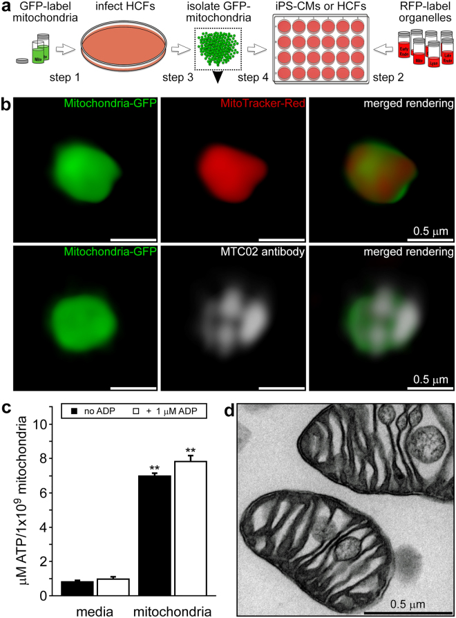
Figure 1
Experimental strategy and characterisation of isolated human fibroblast mitochondria. ( a) HCFs infected with BacMam CellLight Mitochondria-GFP were used for mitochondrial isolations and iPS-CMs or HC...
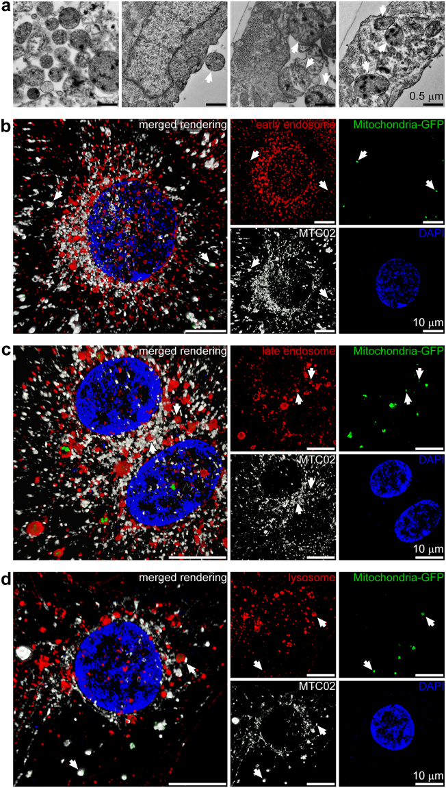
Figure 2
Uptake and endocytic transport of extracellular mitochondria in cardiomyocytes. ( a) iPS-CMs were exposed to gold-labelled HCF mitochondria for 2.5, 5, and 10u2009min and imaged by TEM. Labelled mitoc...
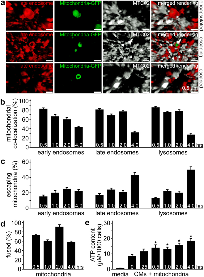
Figure 3
Encapsulation and escape of internalised mitochondria from cardiomyocyte cell compartments. ( a) Late endosomes (red) associated with GFP-labelled exogenous mitochondria (green) were imaged by 3-D SR-...
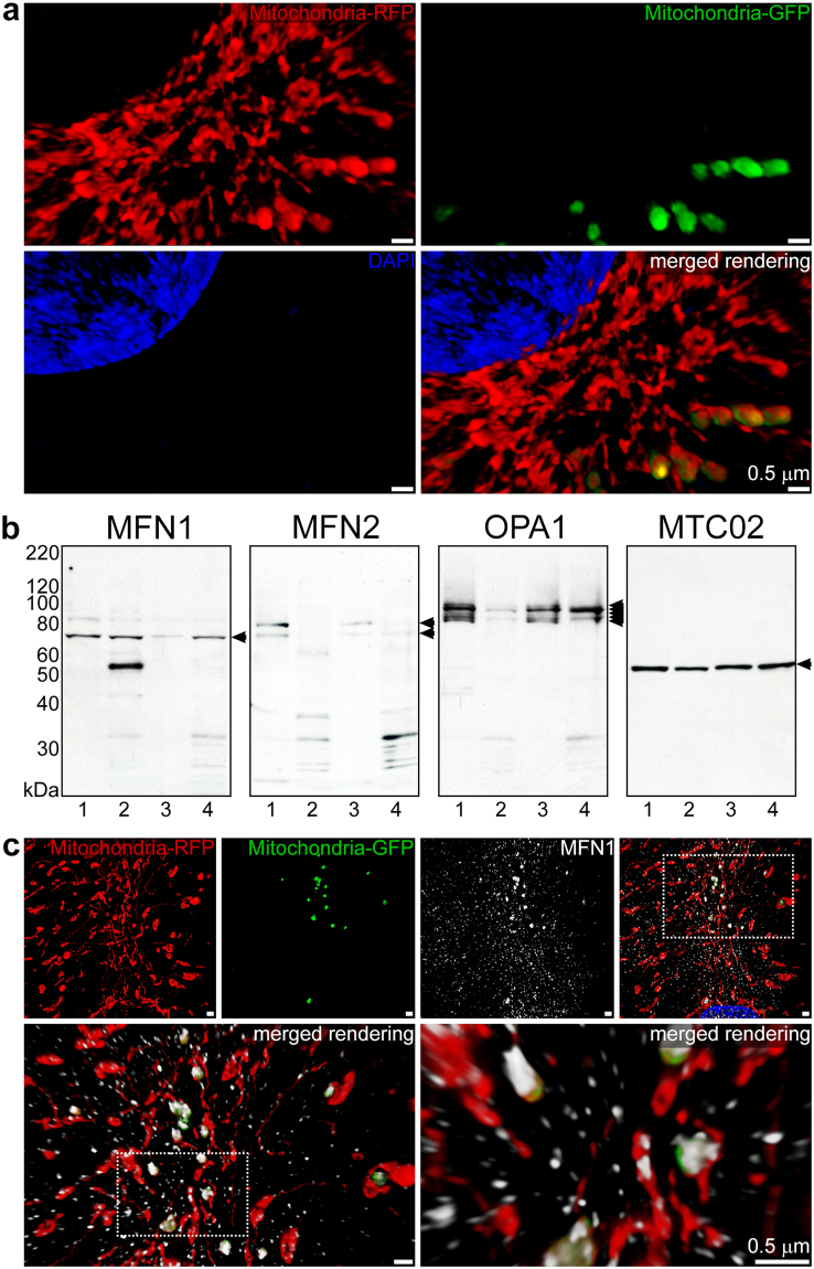
Figure 4
Fusion of exogenous mitochondria with the cardiomyocyte mitochondrial network. ( a ) iPS-CMs with RFP-labelled mitochondria (red) treated for 4u2009h with isolated GFP-labelled HCF mitochondria (green...
Figure images are served from the NIH/NLM PubMed Central Open Access Subset or Europe PMC; copyright remains with the publishers and authors.
💬 Discussion
0 commentsNo comments yet. Be the first to start a discussion!
Leave a Comment