Abstract
DNA-PAINT's imaging speed has recently been significantly enhanced by optimized sequence design and buffer conditions. However, this implementation has not reached an ultimate speed limit and is only applicable to imaging of single targets. To further improve acquisition speed, we introduce concatenated, periodic DNA sequence motifs, yielding up to 100-fold-faster sampling in comparison to traditional DNA-PAINT. We extend this approach to six orthogonal sequence motifs, now enabling speed-optimized multiplexed imaging.
🔬 Techniques
🔭 Microscopes
✨ Fluorophores
🧪 Sample Preparation
🔬 Cell Lines
🏭 Microscope Brands
🧪 Reagent Suppliers
📷 Detectors
🎨 Filters
💻 Code & Software
💾 Data Repositories
🏛️ Research Organizations (ROR)
Affiliated research institutions:
📋 Methods
Materials Unmodified DNA oligonucleotides were purchased from MWG Eurofins and Metabion. C3-azide, Cy3B and ATTO 655 modified DNA oligonucleotides were ordered from Metabion and MWG Eurofins. M13mp18 scaffold was obtained from Tilibit. Magnesium 1 M (cat: AM9530G), sodium chloride 5 M (cat: AM9759), ultrapure water (cat: 10977-035), Tris 1 M, pH 8 (cat: AM9855G), EDTA 0.5 M, pH 8.0 (cat: AM9260G) and 10×PBS (cat: 70011051) was purchased from Thermo Fisher Scientific. Bovine serum albumin (cat: A4503-10G) was ordered from Sigma-Aldrich. Triton X-100 (cat: 6683.1), Sodium borohydride > 97 % (cat: 4051.1), ammonium chloride (cat: K298.1), and potassium chloride (cat: 6781.1) was purchased from Carl Roth. Sodium hydroxide (cat: 31627.290) was purchased from VWR. Paraformaldehyde (cat: 15710) and glutaraldehyde (cat: 16220) were obtained from Electron Microscopy Sciences. Tween 20 (cat: P9416-50ML), glycerol (cat: 65516-500ml), methanol (cat: 32213-2.5L), protocatechuate 3,4-dioxygenase pseudomonas (PCD) (cat: P8279), 3,4-dihydroxybenzoic acid (PCA) (cat: 37580-25G-F) and (+−)-6-hydroxy-2,5,7,8- tetra-methylchromane-2-carboxylic acid (Trolox) (cat: 238813-5 G) were ordered from Sigma Aldrich. Streptavidin (cat: S-888) was purchased from Thermo Fisher. BSA-Biotin (cat: A8549) was obtained from Sigma-Aldrich. Coverslips (cat: 0107032) and glass slides (cat: 10756991) were purchased from Marienfeld and Thermo Fisher Scientific. Double-sided tape (cat: 665D) was ordered from Scotch. Two component silica twinsil speed 22 (cat. 1300 1002) was purchased from Picodent. Fetal Bovine Serum (FBS) (cat: 10500-064), 1× Phosphate Buffered Saline (PBS) pH 7.2 (cat: 20012-019), 0.05 % Trypsin–EDTA (cat: 25300-054) and were purchased from Thermo Fisher Scientific. 90 nm diameter Gold Nanoparticles (cat: G-90-100) were ordered from cytodiagnostics. Buffers The following buffers were used for sample preparation and imaging Buffer A: 10 mM Tris pH 8, 100 mM NaCl, 0.05 % Tween 20 Buffer B: 10 mM MgCl 2, 5 mM Tris-HCl pH 8, 1 mM EDTA, 0.05 % Tween 20, pH 8 Buffer C (Imaging buffer): 1× PBS, 1mM EDTA, 500 mM NaCl, pH 7.4. Optionally supplemented with: 1× Trolox, 1× PCA and 1× PCD Blocking buffer: 1× PBS pH 7.4, 1 mM EDTA, 3% BSA, 0.02% Tween-20. Trolox, PCA and PCD 100× Trolox: 100 mg Trolox, 430 μl 100 % Methanol, 345 μl 1M NaOH in 3.2 ml H 2 O. 40× PCA: 154 mg PCA, 10 ml water and NaOH were mixed and pH was adjusted 9.0. 100× PCD: 9.3 mg PCD, 13.3 ml of buffer (100 mM Tris-HCl pH 8, 50 mM KCl, 1 mM EDTA, 50 % Glycerol). DNA origami self-assembly All DNA origami structures were designed with the Picasso design tool 4 . Self-assembly of DNA origami was accomplished in a one-pot reaction mix with 40 μl total volume, consisting of 10 nM scaffold strand (sequence see Supplementary Spreadsheet 1 ), 100 nM folding staples ( Supplementary Spreadsheet 2 – 4 ), 500 nM biotinylated staples ( Supplementary Table 3 ), and 1 μM of staple strands with docking site extensions (for respective sequences see Supplementary Table 4 and 5 and Supplementary Data 2 – 4 ) in folding buffer (5 mM Tris pH 8, 1 mM EDTA 12.5 mM MgCl 2 ). The reaction mix was then subjected to a thermal annealing ramp using a thermocycler. The reaction mix was first incubated at 80 °C for 5 min, then cooled down using a temperature gradient from 60 to 4 °C in steps of 1 °C per 3.21 min and finally held at 4 °C.
Show full methods section
Materials Unmodified DNA oligonucleotides were purchased from MWG Eurofins and Metabion. C3-azide, Cy3B and ATTO 655 modified DNA oligonucleotides were ordered from Metabion and MWG Eurofins. M13mp18 scaffold was obtained from Tilibit. Magnesium 1 M (cat: AM9530G), sodium chloride 5 M (cat: AM9759), ultrapure water (cat: 10977-035), Tris 1 M, pH 8 (cat: AM9855G), EDTA 0.5 M, pH 8.0 (cat: AM9260G) and 10×PBS (cat: 70011051) was purchased from Thermo Fisher Scientific. Bovine serum albumin (cat: A4503-10G) was ordered from Sigma-Aldrich. Triton X-100 (cat: 6683.1), Sodium borohydride > 97 % (cat: 4051.1), ammonium chloride (cat: K298.1), and potassium chloride (cat: 6781.1) was purchased from Carl Roth. Sodium hydroxide (cat: 31627.290) was purchased from VWR. Paraformaldehyde (cat: 15710) and glutaraldehyde (cat: 16220) were obtained from Electron Microscopy Sciences. Tween 20 (cat: P9416-50ML), glycerol (cat: 65516-500ml), methanol (cat: 32213-2.5L), protocatechuate 3,4-dioxygenase pseudomonas (PCD) (cat: P8279), 3,4-dihydroxybenzoic acid (PCA) (cat: 37580-25G-F) and (+−)-6-hydroxy-2,5,7,8- tetra-methylchromane-2-carboxylic acid (Trolox) (cat: 238813-5 G) were ordered from Sigma Aldrich. Streptavidin (cat: S-888) was purchased from Thermo Fisher. BSA-Biotin (cat: A8549) was obtained from Sigma-Aldrich. Coverslips (cat: 0107032) and glass slides (cat: 10756991) were purchased from Marienfeld and Thermo Fisher Scientific. Double-sided tape (cat: 665D) was ordered from Scotch. Two component silica twinsil speed 22 (cat. 1300 1002) was purchased from Picodent. Fetal Bovine Serum (FBS) (cat: 10500-064), 1× Phosphate Buffered Saline (PBS) pH 7.2 (cat: 20012-019), 0.05 % Trypsin–EDTA (cat: 25300-054) and were purchased from Thermo Fisher Scientific. 90 nm diameter Gold Nanoparticles (cat: G-90-100) were ordered from cytodiagnostics. Buffers The following buffers were used for sample preparation and imaging Buffer A: 10 mM Tris pH 8, 100 mM NaCl, 0.05 % Tween 20 Buffer B: 10 mM MgCl 2, 5 mM Tris-HCl pH 8, 1 mM EDTA, 0.05 % Tween 20, pH 8 Buffer C (Imaging buffer): 1× PBS, 1mM EDTA, 500 mM NaCl, pH 7.4. Optionally supplemented with: 1× Trolox, 1× PCA and 1× PCD Blocking buffer: 1× PBS pH 7.4, 1 mM EDTA, 3% BSA, 0.02% Tween-20. Trolox, PCA and PCD 100× Trolox: 100 mg Trolox, 430 μl 100 % Methanol, 345 μl 1M NaOH in 3.2 ml H 2 O. 40× PCA: 154 mg PCA, 10 ml water and NaOH were mixed and pH was adjusted 9.0. 100× PCD: 9.3 mg PCD, 13.3 ml of buffer (100 mM Tris-HCl pH 8, 50 mM KCl, 1 mM EDTA, 50 % Glycerol). DNA origami self-assembly All DNA origami structures were designed with the Picasso design tool 4 . Self-assembly of DNA origami was accomplished in a one-pot reaction mix with 40 μl total volume, consisting of 10 nM scaffold strand (sequence see Supplementary Spreadsheet 1 ), 100 nM folding staples ( Supplementary Spreadsheet 2 – 4 ), 500 nM biotinylated staples ( Supplementary Table 3 ), and 1 μM of staple strands with docking site extensions (for respective sequences see Supplementary Table 4 and 5 and Supplementary Data 2 – 4 ) in folding buffer (5 mM Tris pH 8, 1 mM EDTA 12.5 mM MgCl 2 ). The reaction mix was then subjected to a thermal annealing ramp using a thermocycler. The reaction mix was first incubated at 80 °C for 5 min, then cooled down using a temperature gradient from 60 to 4 °C in steps of 1 °C per 3.21 min and finally held at 4 °C.
DNA origami purification
DNA origami structures were purified via ultrafiltration using Amicon Ultra Centrifugal Filters with a 50 kDa MWCO (Merck Millipore, UFC505096) as previously described 23 . In brief, folded origami was filled up to 500 μl with FoB5 buffer (5 mM Tris, 1 mM EDTA, 5 mM NaCl, 5 mM MgCl2, pH 8) and spun for 6 min at 5000 g. This process was repeated twice. Purified DNA origami structures were recovered into a new tube by centrifugation for 5 min at 5000 g.
DNA origami sample preparation
For sample preparation, a piece of coverslip and a glass slide were sandwiched together by two strips of double-sided tape to form a flow chamber with inner volume of ~20 μl. First, 20 μl of biotin labeled bovine albumin (1 mg/ml, dissolved in buffer A) was flushed into the chamber and incubated for 2 min. The chamber was then washed with 40 μl of buffer A. A volume of 20 μl of streptavidin (0.1 mg/ml, dissolved in buffer A) was then flushed through the chamber and allowed to bind for 2 min. After washing with 20 μl of buffer A and subsequently with 20 μl of buffer B, 20 μl of biotin labeled DNA structures (~200 pM) in buffer B were flushed into the chamber and incubated for 2 min. The chamber was washed with 40 μl of buffer B. Finally, 20 μl of the imager solution in imaging buffer (see Supplementary Table 8 ) was flushed into the chamber, which was subsequently sealed with two component silica before imaging. For multiplexing experiments a bottomless 6 channel slide (ibidi, cat: 80608) was attached to a coverslip. The same sample preparation was performed as described above with adjusted volumes of 120 μl for each washing and incubation step. In between imaging rounds the sample was washed 4–5 times with 120 μl PBS until no residual signal from the previous imager solution was detected (total washing time of approx. 3–5 min). Then, the next imager solution was introduced. Antibody-DNA conjugation Antibodies were conjugated to DNA-PAINT docking sites (see Supplementary Table 7 ) via DBCO-sulfo-NHS ester chemistry as previously reported 4 . In brief, antibodies were reacted with 20-fold excess of a bifunctional DBCO-sulfo-NHS ester (Jena Biosciences, cat: CLK-A124-10). Unreacted linker was removed using Zeba Spin Desalting columns (0.5 ml, 40k MWCO, Thermo Fisher Scientific, cat: 89882). Azide-DNA was added to the DBCO-antibodies with a 10-fold molar excess and reacted overnight at 4°C. Afterwards, buffer was exchange to PBS using Amicon centrifugal filters (100k MWCO). Nanobody-DNA conjugation Nanobodies against GFP, tagFP, rabbit and mouse IgG were purchased from Nanotag with a single ectopic cysteine at the C-terminus for site specific and quantitative conjugation. The conjugation to DNA-PAINT docking sites (see Supplementary Table 7 ) was performed similar as described previously 20 . First, buffer was exchanged to 1× PBS + 5 mM EDTA, pH 7.0 using Amicon centrifugal filters (10k MWCO) and free cysteines were reacted with 20-fold molar excess of bifunctional maleimide-DBCO linker (Sigma Aldrich, cat: 760668) for 2-3 hours on ice. Unreacted linker was removed by buffer exchange to PBS using Amicon centrifugal filters. Azide-functionalized DNA was added with 5-10 molar excess to the DBCO-nanobody and reacted overnight at 4°C. Unconjugated nanobody and free azide-DNA was removed by anion exchange using an ÄKTA Pure liquid chromatography system equipped with a Resource Q 1 ml column.
Cell culture U-2
OS-CRISPR-Nup96-mEGFP (gift from Ries and Ellenberg lab) and SKOV3 GFP-Her2 RFP-tagEGFR cells (purchased from Sigma Aldrich, cat: CLL1143-1VL) were cultured in McCoy’s 5A medium (Thermo Fisher Scientific, cat: 16600082) supplemented with 10% fetal bovine serum. For imaging, cells were seeded 1-2 days prior to fixation into Glass-bottomed 8-well μ-slides (ibidi, cat: 80827).
Cell fixation
All fixatives were pre-heated to 37°C before adding to the cells. Microtubules and vimentin U-2 OS-CRISPR-Nup96-mEGFP cells were first pre-extracted with 0.3 % glutaraldehyde and 0.25 % Triton X-100 for 90 s, followed by fixation with 3 % glutaraldehyde for 10 min. Afterwards, samples were rinsed twice with PBS and free aldehyde groups were reduced with 0.1 % NaBH4 for 5 min. After rinsing four times with PBS, cells were blocked and permeabilized in blocking buffer with 0.25 % Triton X-100 for 2 h. Primary antibodies against anti-vimentin and anti-α-tubulin were incubated overnight at 4°C. After washing unbound primary antibodies for four times with PBS, secondary antibodies conjugated to DNA-PAINT docking sites were diluted in blocking buffer and incubated with the cells for 1 hour at room temperature. Finally, unbound antibodies were removed by washing three times with PBS for 5 min. Nup96-EGFP imaging U-2 OS-CRISPR-Nup96-mEGFP cells were fixed with 2.4% PFA in PBS for 30 min at room temperature. After fixation cells were washed three times with PBS and incubated with 0.1 M NH 4 Cl in PBS for 5 min. Then, cells were permeabilized with 0.25% Triton-X-100 for 5 min and afterwards blocked in blocking buffer for 1 hour. Anti-GFP nanobodies were incubated at a concentration of appr. 50 nM in blocking buffer supplemented with 0.05 mg/ml sheared salmon sperm DNA overnight at 4°C. Unbound nanobodies were removed by washing three times with PBS for 5 min. Receptor tyrosine kinases SKOV3 GFP-Her2 tagRFP-EGFR cells were fixed with 4% PFA. After fixation, cells were washed three times with PBS and incubated with 0.1 M NH 4 Cl in PBS for 5 min. Then, cells were permeabilized with 0.25% Triton-X-100 for 15 min and afterwards blocked in blocking buffer for 1 hour. Nanobodies and primary antibodies (see Supplementary Table 6 ) were diluted in blocking buffer supplemented with 0.05 mg/ml sheared salmon sperm DNA and incubated with the cells overnight at 4°C. Unbound nanobodies and antibodies were removed by washing three times with PBS for 5 min.
Microscope setup
Fluorescence imaging was carried out on an inverted microscope (Nikon Instruments, Eclipse Ti2) with the Perfect Focus System, applying an objective-type TIRF configuration equipped with an oil-immersion objective (Nikon Instruments, Apo SR TIRF 100×, NA 1.49, Oil). A 561 nm and 642 nm laser (MPB Communications Inc., 2 W, DPSS-system) were used for excitation. The laser beams were passed through cleanup filters (Chroma Technology, ZET561/10, ZET 640/10) and coupled into the microscope objective using a beam splitter (Chroma Technology, ZT561rdc, ZT640rdc). Fluorescence light was spectrally filtered with an emission filter (Chroma Technology, ET600/50m and ET700/75m) and imaged on a sCMOS camera (Andor, Zyla 4.2 Plus) without further magnification, resulting in an effective pixel size of 130 nm (after 2×2 binning). Images were acquired choosing a region of interest with the size of 512×512 pixels. More detailed imaging conditions for the respective experiments are shown in Supplementary Table 8 .
Image analysis
Raw fluorescence data was subjected super-resolution reconstruction using the ‘Picasso’ software package 4 (Latest version available on https://github.com/jungmannlab/picasso ). Drift correction was performed with a redundant cross-correlation and gold particles as fiducials. Gold particles were also used to align multiple rounds for Exchange-PAINT experiments. For quantification of binding kinetics, localizations were linked allowing a gap size of 3 frames and a maximum distance of 130 nm. Origami structures were automatically selected using Picasso’s ‘Pick similar’ function. Kinetic information of detected picks was extracted with the ‘save pick properties’ command. Further quantification such as histogram analysis and fitting were performed with Origin Pro (Version 2019b). Kinetic barcoding analysis ( Extended Data Figure 10 ) was performed as previously described 22 . The data was segmented using the HDBSCAN clustering algorithm 24 with input parameter ‘Min_cluster size’ set to 10.
Materials Unmodified DNA oligonucleotides were purchased from MWG Eurofins and Metabion. C3-azide, Cy3B and ATTO 655 modified DNA oligonucleotides were ordered from Metabion and MWG Eurofins. M13mp18 scaffold was obtained from Tilibit. Magnesium 1 M (cat: AM9530G), sodium chloride 5 M (cat: AM9759), ultrapure water (cat: 10977-035), Tris 1 M, pH 8 (cat: AM9855G), EDTA 0.5 M, pH 8.0 (cat: AM9260G) and 10×PBS (cat: 70011051) was purchased from Thermo Fisher Scientific. Bovine serum albumin (cat: A4503-10G) was ordered from Sigma-Aldrich. Triton X-100 (cat: 6683.1), Sodium borohydride > 97 % (cat: 4051.1), ammonium chloride (cat: K298.1), and potassium chloride (cat: 6781.1) was purchased from Carl Roth. Sodium hydroxide (cat: 31627.290) was purchased from VWR. Paraformaldehyde (cat: 15710) and glutaraldehyde (cat: 16220) were obtained from Electron Microscopy Sciences. Tween 20 (cat: P9416-50ML), glycerol (cat: 65516-500ml), methanol (cat: 32213-2.5L), protocatechuate 3,4-dioxygenase pseudomonas (PCD) (cat: P8279), 3,4-dihydroxybenzoic acid (PCA) (cat: 37580-25G-F) and (+−)-6-hydroxy-2,5,7,8- tetra-methylchromane-2-carboxylic acid (Trolox) (cat: 238813-5 G) were ordered from Sigma Aldrich. Streptavidin (cat: S-888) was purchased from Thermo Fisher. BSA-Biotin (cat: A8549) was obtained from Sigma-Aldrich. Coverslips (cat: 0107032) and glass slides (cat: 10756991) were purchased from Marienfeld and Thermo Fisher Scientific. Double-sided tape (cat: 665D) was ordered from Scotch. Two component silica twinsil speed 22 (cat. 1300 1002) was purchased from Picodent. Fetal Bovine Serum (FBS) (cat: 10500-064), 1× Phosphate Buffered Saline (PBS) pH 7.2 (cat: 20012-019), 0.05 % Trypsin–EDTA (cat: 25300-054) and were purchased from Thermo Fisher Scientific. 90 nm diameter Gold Nanoparticles (cat: G-90-100) were ordered from cytodiagnostics.
Supplementary Material Supplementary Data 1 Supplementary Data 2 Supplementary Data 3 Supplementary Data 4 Supplementary Information
📊 Figures
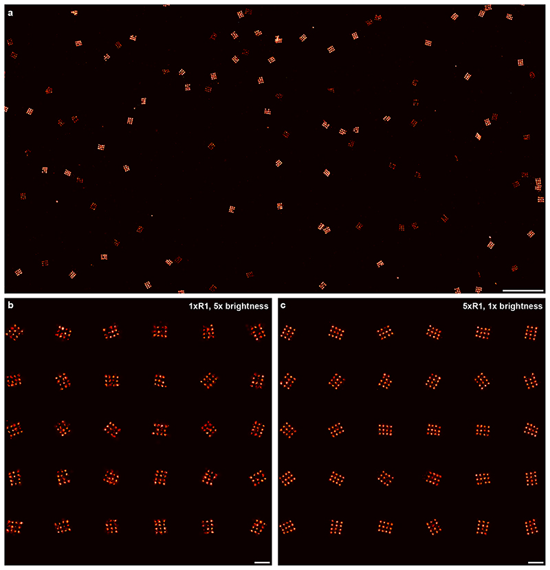
Extended Data Fig. 1
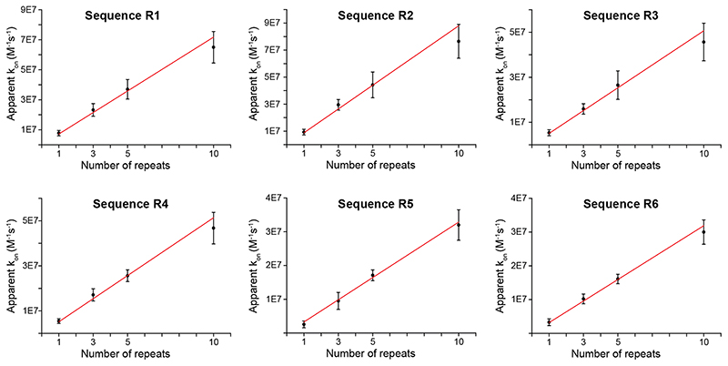
Extended Data Fig. 2
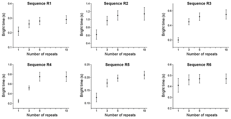
Extended Data Fig. 3
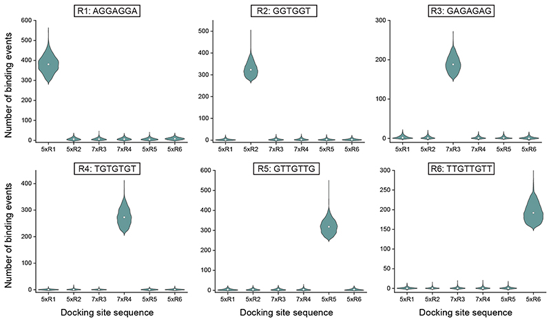
Extended Data Fig. 4
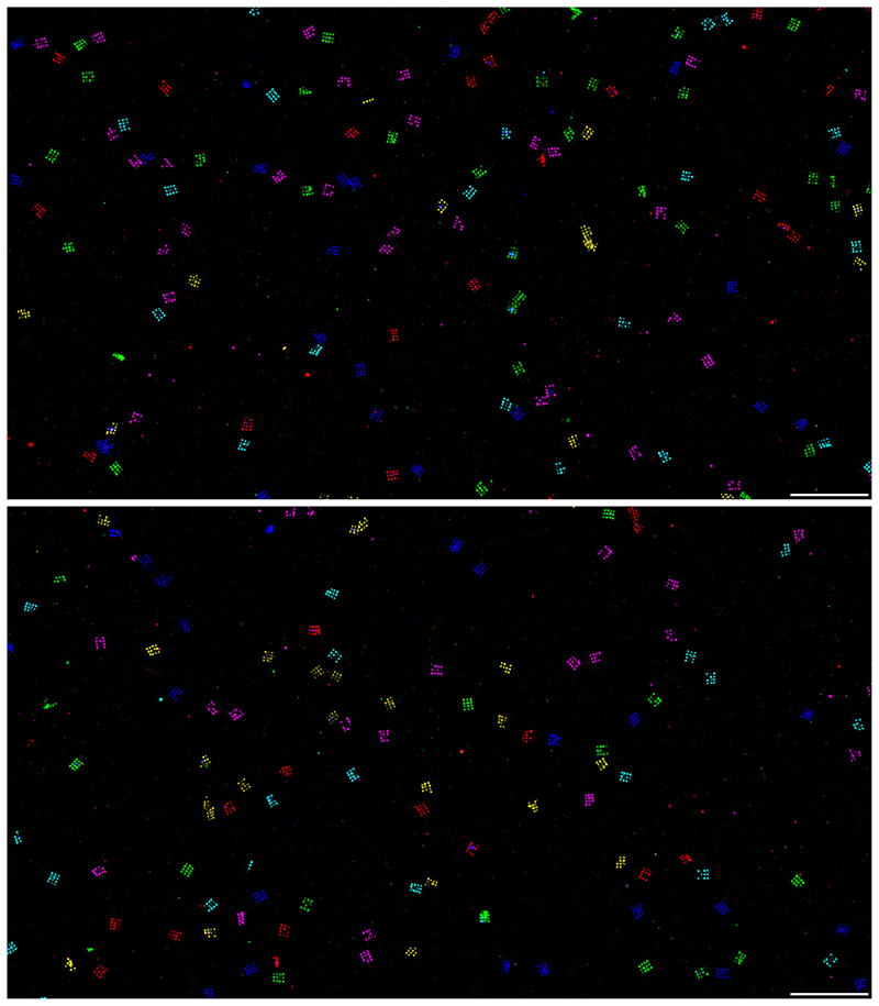
Extended Data Fig. 5
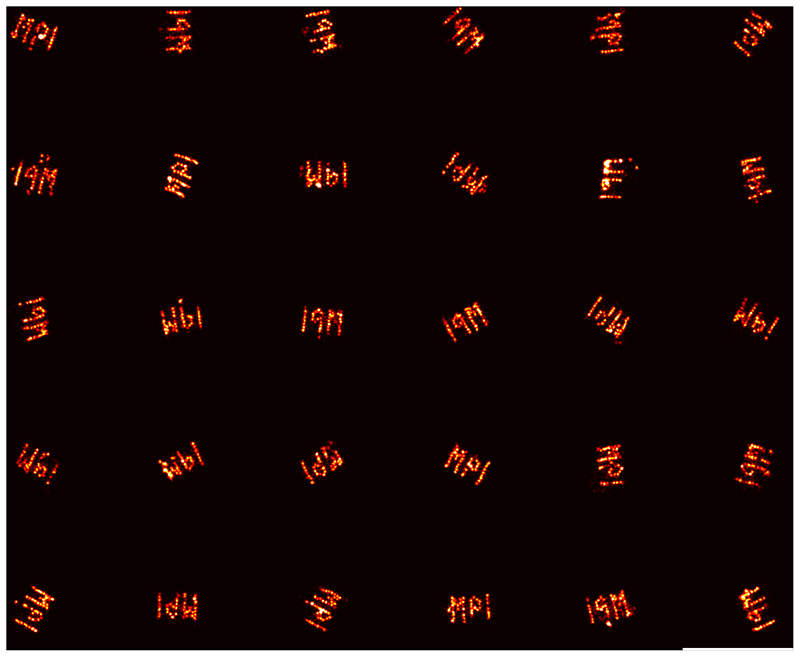
Extended Data Fig. 6
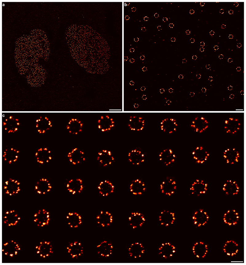
Extended Data Fig. 7
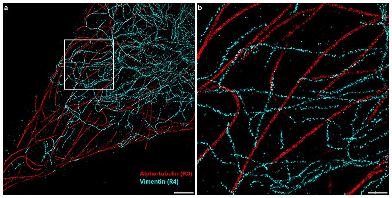
Extended Data Fig. 8
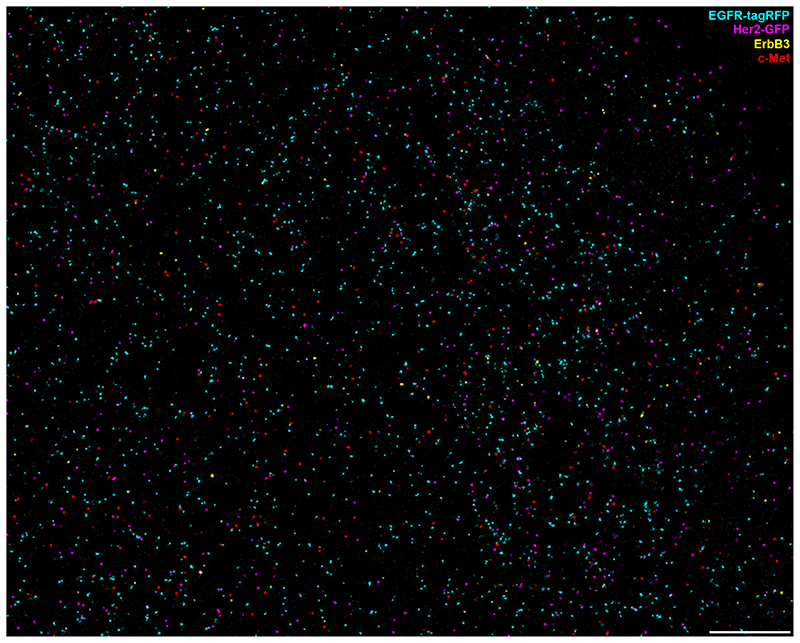
Extended Data Fig. 9
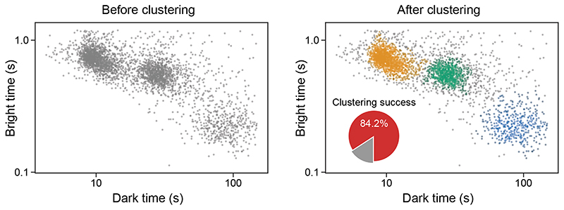
Extended Data Fig. 10
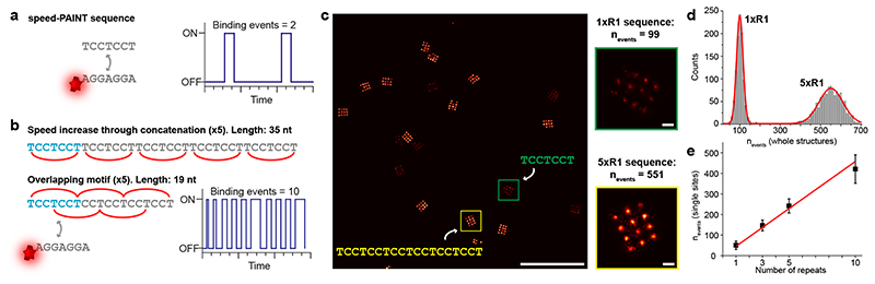
Fig. 1
Faster DNA-PAINT through overlapping sequence motifs.
( a ) A single speed-optimized DNA-PAINT sequence exhibits a certain number of binding events (e.g. two per unit time). ( b ) Concatenation leads to a linear increase in binding frequency and thus ima...
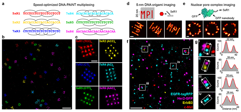
Fig. 2
Multiplexing with concatenated speed-optimized motifs.
( a ) Designing six orthogonal binding motifs enables speed-optimized multiplexing in Exchange-PAINT experiments. ( b ) Proof-of-concept experiments using 20-nm-grid DNA origami with six orthogonal se...
Figure images are served from the NIH/NLM PubMed Central Open Access Subset or Europe PMC; copyright remains with the publishers and authors.
💬 Discussion
0 commentsNo comments yet. Be the first to start a discussion!
Leave a Comment