Abstract
BACKGROUND: Myofibroblasts in the cancer microenvironment have recently been implicated in tumour growth and metastasis of gastric cancer. However, the mechanisms responsible for the regulation of myofibroblasts in cancer-associated fibroblasts (CAFs) remain unclear. This study was performed to clarify the mechanisms for regulation of myofibroblasts in gastric cancer microenvironment. METHODS: Two CAFs (CaF-29 and CaF-33) from the tumoural gastric wall and a normal fibroblast (NF-29) from the nontumoural gastric wall, 4 human gastric cancer cell lines from scirrhous gastric cancer (OCUM-2MD3 and OCUM-12), and non-scirrhous gastric cancer (MKN-45 and MKN-74) were used. Immunofluorescence microscopy by triple-immunofluorescence labelling (α-SMA, vimentin, and DAPI) was performed to determine the presence of α-SMA-positive myofibroblasts. Real-time RT-PCR was performed to examine α-SMA mRNA expression. RESULTS: Immunofluorescence microscopy showed that the frequency of myofibroblasts in CaF-29 was greater than that in NF-29. The number of myofibroblasts in gastric fibroblasts gradually decreased with serial passages. Transforming growth factor-β (TGF-β) significantly increased the α-SMA expression level of CAFs. Conditioned medium from OCUM-2MD3 or OCUM-12 cells upregulated the α-SMA expression level of CAFs, but that from MKN-45 or MKN-74 cells did not. The α-SMA upregulation effect of conditioned medium from OCUM-2MD3 or OCUM-12 cells was significantly decreased by an anti-TGF-β antibody or Smad2 siRNA. CONCLUSION: Transforming growth factor-β from scirrhous gastric carcinoma cells upregulates the number of myofibroblasts in CAFs.
🔬 Techniques
🔭 Microscopes
✨ Fluorophores
🧪 Sample Preparation
🔬 Cell Lines
🏭 Microscope Brands
🧪 Reagent Suppliers
🏛️ Research Organizations (ROR)
Affiliated research institutions:
📋 Methods
Two CAFs (CaF-29 and CaF-33) from the tumoural gastric wall and a normal fibroblast (NF-29) from the nontumoural gastric wall, 4 human gastric cancer cell lines from scirrhous gastric cancer (OCUM-2MD3 and OCUM-12), and non-scirrhous gastric cancer (MKN-45 and MKN-74) were used. Immunofluorescence microscopy by triple-immunofluorescence labelling ( α -SMA, vimentin, and DAPI) was performed to determine the presence of α -SMA-positive myofibroblasts. Real-time RT–PCR was performed to examine α-SMA mRNA expression.
Materials and methods Cell culture and cell lines
We used three human gastric fibroblast cell lines and four human gastric cancer cell lines in this study. Fibroblasts cell lines were established at our department. The NF-29 and CAF-29 were established from a 68-year-old male patient with poorly differentiated gastric carcinoma who had a total gastrectomy. The NF-29 was from nontumoural gastric wall, and CaF-29 was from tumoural gastric wall. The CaF-33 was established from a 65-year-old male patient with poorly differentiated gastric carcinoma who had a distal gastrectomy. The primary culture was initiated as follows: the primary tumour was excised under aseptic conditions, and minced with forceps and scissors. The tumour pieces were cultivated in Dulbecco's modified Eagle medium (DMEM; Nikken, Kyoto, Japan) with 10% heat-inactivated fetal calf serum (FCS; Life Technologies, Inc., Grand Island, NY, USA), 100 IU ml –1 penicillin (ICN Biomedical, Costa Mesa, CA, USA), 100 μ g ml –1 streptomycin (ICN Biomedical), and 0.5 m M sodium pyruvate (Cambrex, Walkersville, MD, USA), and incubated in humidified incubators at 37 °C in an atmosphere of 5% CO 2 in air. The fibroblasts initially grew in a monolayer. After ∼2 weeks, fibroblasts were collected and transferred to another culture dish. Serial passages were then carried out every 4–7 days. The fibroblasts were used 3–12th passage in culture. Four human gastric cancer cell lines, including OCUM-2MD3 (poorly differentiated adenocarcinoma) ( Yashiro et al , 1996 ), OCUM-12 (poorly differentiated adenocarcinoma) ( Kato et al , 2010 ), MKN-45 (poorly differentiated adenocarcinoma) ( Motoyama et al , 1986 ), and MKN-74 (well-differentiated adenocarcinoma) ( Motoyama et al , 1986 ) were seeded in a 100-mm dish (Falcon, Lincoln Park, NJ, USA) and cultured. OCUM-2MD3 and OCUM-12 were derived from scirrhous gastric carcinoma.
Show full methods section
Two CAFs (CaF-29 and CaF-33) from the tumoural gastric wall and a normal fibroblast (NF-29) from the nontumoural gastric wall, 4 human gastric cancer cell lines from scirrhous gastric cancer (OCUM-2MD3 and OCUM-12), and non-scirrhous gastric cancer (MKN-45 and MKN-74) were used. Immunofluorescence microscopy by triple-immunofluorescence labelling ( α -SMA, vimentin, and DAPI) was performed to determine the presence of α -SMA-positive myofibroblasts. Real-time RT–PCR was performed to examine α-SMA mRNA expression.
Materials and methods Cell culture and cell lines
We used three human gastric fibroblast cell lines and four human gastric cancer cell lines in this study. Fibroblasts cell lines were established at our department. The NF-29 and CAF-29 were established from a 68-year-old male patient with poorly differentiated gastric carcinoma who had a total gastrectomy. The NF-29 was from nontumoural gastric wall, and CaF-29 was from tumoural gastric wall. The CaF-33 was established from a 65-year-old male patient with poorly differentiated gastric carcinoma who had a distal gastrectomy. The primary culture was initiated as follows: the primary tumour was excised under aseptic conditions, and minced with forceps and scissors. The tumour pieces were cultivated in Dulbecco's modified Eagle medium (DMEM; Nikken, Kyoto, Japan) with 10% heat-inactivated fetal calf serum (FCS; Life Technologies, Inc., Grand Island, NY, USA), 100 IU ml –1 penicillin (ICN Biomedical, Costa Mesa, CA, USA), 100 μ g ml –1 streptomycin (ICN Biomedical), and 0.5 m M sodium pyruvate (Cambrex, Walkersville, MD, USA), and incubated in humidified incubators at 37 °C in an atmosphere of 5% CO 2 in air. The fibroblasts initially grew in a monolayer. After ∼2 weeks, fibroblasts were collected and transferred to another culture dish. Serial passages were then carried out every 4–7 days. The fibroblasts were used 3–12th passage in culture. Four human gastric cancer cell lines, including OCUM-2MD3 (poorly differentiated adenocarcinoma) ( Yashiro et al , 1996 ), OCUM-12 (poorly differentiated adenocarcinoma) ( Kato et al , 2010 ), MKN-45 (poorly differentiated adenocarcinoma) ( Motoyama et al , 1986 ), and MKN-74 (well-differentiated adenocarcinoma) ( Motoyama et al , 1986 ) were seeded in a 100-mm dish (Falcon, Lincoln Park, NJ, USA) and cultured. OCUM-2MD3 and OCUM-12 were derived from scirrhous gastric carcinoma.
Immunofluorescence microscopy
To examine incubating myofibroblast content of fibroblast, immunofluorescence microscopy was performed. Triple-immunofluorescence labelling was performed to examine the presence of α -SMA-positive myofibroblasts. Fibroblasts were washed twice with Dulbecco’s PBS and fixed with acetone for 5 min, and then blocked with 3% BSA (diluted in PBS) for 30 min at room temperature. Fibroblasts were further incubated with anti-human α -SMA antibody (R&D Systems, Minneapolis, MN, USA; 1 : 100) and vimentin (Santa Cruz, Santa Cruz, CA, USA; 1 : 50) and DAPI (Wako, Osaka, Japan; 1 : 10 000) for 60 min at room temperature. Fibroblasts were viewed under a fluorescence microscope Leica Digital Microscopy DMI 6000 (Leica Microsystems, Heidelberg, Germany) with a DAPI filter (365 nm excitation), α -SMA fluorescence with a PE filter (546 nm excitation), and vimentin with a FITC filter (450–490 nm excitation). Cells that were α -SMA positive were determined as myofibroblasts. The percentage of binding cells was calculated as follows: (number of myofibroblasts/number of total cells) × 100. The percentage of α -SMA-positive myofibroblast cells was determined in 10 random fields. At least, three independent experiments were performed.
Western blot analysis
Fibroblasts were rinsed with PBS and were lysed in a lysis buffer. Aliquots containing 30 μ g of total protein were subjected to SDS–PAGE, and the protein bands were transferred to a polyvinylidene difluoride membrane (Amersham, Aylesbury, UK). The membrane was placed in the TBS-T solution containing the primary antibody, α -SMA (Dako, Glostrup, Denmark; 1 : 1000) or β -Actin (Cell Signaling, Danvers, MA, USA; 1 : 1000), and allowed to react at 4 °C overnight for western blotting. The bands were detected using an enhanced chemiluminescence system (Amersham). An immunoblot analysis was performed twice.
Preparation of conditioned medium
Conditioned medium from gastric cancer cells was prepared as follows. Gastric cancer cells (5 × 10 4 cells ml –1 ) were seeded into 100-mm plastic dishes with 10 ml of DMEM containing 2% FCS and incubated for 3 days. The number of fibroblasts and gastric cancer cells in each dish was ∼2.5 × 10 6 cells after 3 days of incubation. To obtain conditioned medium, fibroblasts and gastric cancer cells were washed twice with PBS and then incubated for 3 days in 3 ml of DMEM. Conditioned medium was collected from each dish and centrifuged at 1000 g for 5 min. The supernatant was stored as conditioned medium at −20 °C until use. As a control, DMEM was used instead of conditioned medium. Quantitative real-time reverse transcriptase-polymerase chain reaction (RT–PCR) Real-time RT–PCR was performed to examine α-SMA mRNA expression. Gastric cancer cells and fibroblasts were incubated in 3 ml DMEM containing 2% FCS with 50% each conditioned medium. After 3 days of incubation, the total cellular RNA was extracted using Trizol reagent (Invitrogen, Carlsbad, CA, USA). After removal of genomic DNA by DNAse, cDNA was prepared from 20 μ g RNA using random primers (Invitrogen). To determine fold changes in each gene, real-time RT–PCR was performed on the ABI Prism 7000 (Applied Biosystems, Foster City, CA, USA), using commercially available gene expression assays for α-SMA (Hs00426835). Glyceraldehyde-3-phosphate dehydrogenase ( GAPDH ) was used as an internal standard to normalise mRNA levels. The threshold cycle ( C t ) values were used to calculate the relative expression ratios between control and treated cells using the formula described by Pfaffl ( Pfaffl, 2001 ). The α-SMA expression level was calculated relative to that of NF-29 at third passage (1.0-fold as the control). Quantitative RT–PCR reactions were performed in triplicate. Effect of conditioned medium, TGF- β , or anti-TGF- β neutralising antibody on α-SMA expression of fibroblasts Fibroblasts were incubated in 3 ml DMEM containing 2% FCS with 50% of each conditioned medium, 10 ng ml –1 TGF- β (R&D Systems), and 10 μ M anti-TGF- β neutralising antibody. After 3 days of incubation, α-SMA expression of fibroblasts was examined by RT–PCR as described above. The effect of Smad2 siRNA on α-SMA expression of fibroblasts The sequences for Smad2 small interfering RNA (siRNA) are designed as: Smad2 siRNA sense, 5′-GUCCCAUGAAAAGACUUAATT-3′ antisense, 5′-UUAAGUCUUUUCAUGGGACTT-3′. Control non-targeting siRNA was purchased from Ambion (Austin, TX, USA). The transfection mixture was prepared by incubating 5 μ l of siPORT Neo-Fx (Ambion) and 295 μ l of Opti-MEMI. The CAF-33 cells were prepared at 50–60% confluence in six-well dishes. The transfection mixture (final siRNA concentration was 30 n M ) was added to six-well dish containing 2 ml of DMEM with 10% FBS. At 24 h after transfection, CAF-33 cells were incubated in addition of conditioned medium from gastric cancer cells. After 3 days of incubation, the total cellular RNA was extracted, and RT–PCR was performed.
Statistical analysis
Data are expressed as the means±s.d. from at least three independent determinations. Significance of difference was analysed using unpaired Student’s t- tests. Values of P
📊 Figures
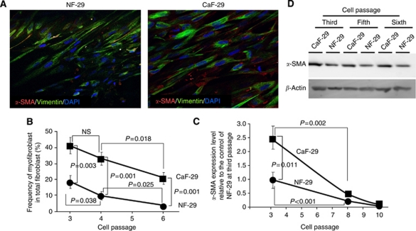
Figure 1
The u03b1 -smooth muscle actin ( u03b1 -SMA) expression in fibroblasts. ( A ) Immunofluorescence of NF-29 fibroblasts and CaF-29 fibroblasts. Fibroblasts were stained with u03b1 -SMA (red), vimentin (...
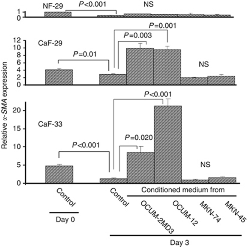
Figure 2
Effect of conditioned medium from gastric cancer cells on the u03b1-SMA expression of fibroblast. The u03b1-SMA expression levels of cancer-associated fibroblasts CaF-29 and CaF-33 were significantly ...
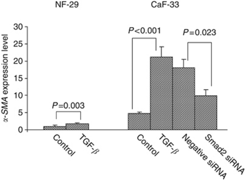
Figure 3
Effect of TGF- u03b2 or Smad2 siRNA on u03b1-SMA expression of fibroblasts. The TGF- u03b2 (10u2009ngu2009ml u20131 ) increased the u03b1-SMA expression level of gastric fibroblasts. The upregulation ...
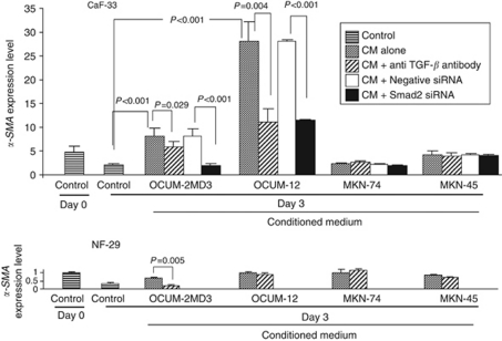
Figure 4
Effect of anti-TGF- u03b2 antibody or Smad2 siRNA on u03b1-SMA expression of fibroblasts. The conditioned medium (CM) from OCUM-2MD3 and OCUM-12 cells significantly increased the u03b1-SMA expression ...
Figure images are served from the NIH/NLM PubMed Central Open Access Subset or Europe PMC; copyright remains with the publishers and authors.
💬 Discussion
0 commentsNo comments yet. Be the first to start a discussion!
Leave a Comment