Abstract
Background: Oxaliplatin chemotherapy induced neuropathy is a dose related cumulative toxicity that manifests as tingling, numbness, and chronic pain, compromising the quality of life and leading to discontinued chemotherapy. Patients report marked hypersensitivity to cold stimuli at early stages of treatment, when sensory testing reveals cold and heat hyperalgesia. This study examined the morphological and functional effects of oxaliplatin treatment in cultured adult rat DRG neurons. Results: 48 hour exposure to oxaliplatin resulted in dose related reduction in neurite length, density, and number of neurons compared to vehicle treated controls, using Gap43 immunostaining. Neurons treated acutely with 20 μg/ml oxaliplatin showed significantly higher signal intensity for cyclic AMP immunofluorescence (160.5 ± 13 a.u., n = 3, P < 0.05), compared to controls (120.3 ± 4 a.u.). Calcium imaging showed significantly enhanced capsaicin (TRPV1 agonist), responses after acute 20 μg/ml oxaliplatin treatment where the second of paired capsaicin responses increased from 80.7 ± 0.6% without oxaliplatin, to 171.26 ± 29% with oxaliplatin, (n = 6 paired t test, P < 0.05); this was reduced to 81.42 ± 8.1% (P < 0.05), by pretretreatment with the cannabinoid CB2 receptor agonist GW 833972. Chronic oxaliplatin treatment also resulted in dose related increases in capsaicin responses. Similarly, second responses to icilin (TRPA1/TRPM8 agonist), were enhanced after acute (143.85 ± 7%, P = 0.004, unpaired t test, n = 3), and chronic (119.7 ± 11.8%, P < 0.05, n = 3) oxaliplatin treatment, compared to control (85.3 ± 1.7%). Responses to the selective TRPM8 agonist WS-12 were not affected. Conclusions: Oxaliplatin treatment induces TRP sensitization mediated by increased intracellular cAMP, which may cause neuronal damage. These effects may be mitigated by co-treatment with adenylyl cyclase inhibitors, like CB2 agonists, to alleviate the neurotoxic effects of oxaliplatin.
🔬 Techniques
💻 Software
✨ Fluorophores
🧪 Sample Preparation
🏭 Microscope Brands
🧪 Reagent Suppliers
💻 Software Details
🏛️ Research Organizations (ROR)
Affiliated research institutions:
📋 Methods
Bilateral DRG from all spinal levels were aseptically harvested from 19 freshly sacrificed adult female Wistar rats (250 grams), in Ham's F12 medium, enzyme digested in 0.2% collagenase (Worthington type IV)/0.5% dispase for 3 hours at 37°C, and dissociated in modified BSF2 medium [ 77 ], containing 2% fetal calf serum and soy-bean trypsin inhibitor (1 mg/ml), to obtain a single cell suspension. 100 μl neuronal suspension was plated on collagen (type I, 50 μg/ml), and laminin (20 μg/ml), coated MatTek dishes (glass bottomed plastic petri dishes), in BSF2 medium and incubated at 37°C in a humid environment, for 30 minutes before adding 2 mls BSF2 containing 50 ng/ml each NT3 and GDNF, 100 ng/ml NGF, to each dish. 24 hours later, oxaliplatin, was added to some of the cultures at 5 μg/ml (12 μM), 20 μg/ml (48 μM), 50 μg/ml (120 μM), or vehicle (0.4% distilled water). Immunostaining Cultures were fixed 48 hours after oxaliplatin treatment, with 4% paraformaldehyde (PFA), for 15 minutes, washed in PBS, permeabilised with methanol (-20°C, 3 minutes), washed in PBS and incubated with primary antibody, mouse monoclonal anti Gap43 (1: 200). This was followed by 3 PBS washes and secondary antibody Alexa Fluor 488 (Molecular probes, 1: 200), for 45 minutes at room temperature, washed with PBS, and mounted in Citifluor containing Dabco (antifade agent). Cyclic AMP (cAMP) immuostaining was performed in vehicle treated neurones, and neurons treated with 20 μg/ml oxaliplatin or 500 μM 8-bromo cAMP (Calbiochem, positive controls) for 15 minutes. Following PFA fixation, immunostaining with primary rabbit polyclonal antibody to cAMP (AB306, Millipore, USA, 1:1000), and Alexa 546 secondary antibody, was performed as above. Images were acquired with Smartcapture 3 (Digital Scientific, Cambridge UK), connected to an upright Olympus BX61 microscope, after confirming the absence of immunostaining in negative controls where the primary antibody had been omitted. Adobe Photoshop C53 software was used to measure the maximum neurite lengths in Gap43 immunostained neurons, and cells with and without neurites were counted in each group (n = 3 for each group). Images of cAMP immunostaining were captured using 0.2 seconds exposure time, and fluorescence intensity was measured (arbitrary units a.u.) with Metamorph software (Molecular Devices), imported and analysed with Excel software.
Show full methods section
Bilateral DRG from all spinal levels were aseptically harvested from 19 freshly sacrificed adult female Wistar rats (250 grams), in Ham's F12 medium, enzyme digested in 0.2% collagenase (Worthington type IV)/0.5% dispase for 3 hours at 37°C, and dissociated in modified BSF2 medium [ 77 ], containing 2% fetal calf serum and soy-bean trypsin inhibitor (1 mg/ml), to obtain a single cell suspension. 100 μl neuronal suspension was plated on collagen (type I, 50 μg/ml), and laminin (20 μg/ml), coated MatTek dishes (glass bottomed plastic petri dishes), in BSF2 medium and incubated at 37°C in a humid environment, for 30 minutes before adding 2 mls BSF2 containing 50 ng/ml each NT3 and GDNF, 100 ng/ml NGF, to each dish. 24 hours later, oxaliplatin, was added to some of the cultures at 5 μg/ml (12 μM), 20 μg/ml (48 μM), 50 μg/ml (120 μM), or vehicle (0.4% distilled water). Immunostaining Cultures were fixed 48 hours after oxaliplatin treatment, with 4% paraformaldehyde (PFA), for 15 minutes, washed in PBS, permeabilised with methanol (-20°C, 3 minutes), washed in PBS and incubated with primary antibody, mouse monoclonal anti Gap43 (1: 200). This was followed by 3 PBS washes and secondary antibody Alexa Fluor 488 (Molecular probes, 1: 200), for 45 minutes at room temperature, washed with PBS, and mounted in Citifluor containing Dabco (antifade agent). Cyclic AMP (cAMP) immuostaining was performed in vehicle treated neurones, and neurons treated with 20 μg/ml oxaliplatin or 500 μM 8-bromo cAMP (Calbiochem, positive controls) for 15 minutes. Following PFA fixation, immunostaining with primary rabbit polyclonal antibody to cAMP (AB306, Millipore, USA, 1:1000), and Alexa 546 secondary antibody, was performed as above. Images were acquired with Smartcapture 3 (Digital Scientific, Cambridge UK), connected to an upright Olympus BX61 microscope, after confirming the absence of immunostaining in negative controls where the primary antibody had been omitted. Adobe Photoshop C53 software was used to measure the maximum neurite lengths in Gap43 immunostained neurons, and cells with and without neurites were counted in each group (n = 3 for each group). Images of cAMP immunostaining were captured using 0.2 seconds exposure time, and fluorescence intensity was measured (arbitrary units a.u.) with Metamorph software (Molecular Devices), imported and analysed with Excel software.
Calcium imaging - capsaicin responses
Neuronal cultures were loaded with the calcium indicator dye Fura 2 AM, (Molecular Probes, 2 μM, 45 minutes at 37°C), in phenol-red free Hank's balanced salt solution (HBSS), containing 0.1% BSA, washed and incubated in HBSS containing 0.5% BSA for 20 minutes, and baseline changes in bound/unbound calcium ratio (340/380 λem nm), in response to capsaicin stimulation were monitored as previously described [ 63 ]. Acute effects of oxaliplatin were determined in normal neurons stimulated with a test dose of 200 nM capsaicin to identify capsaicin sensitivity, followed by washout and a second capsaicin stimulus of 1 μM after 30 minutes; this was compared with the responses to 1 μM capsaicin in a separate group of neurons, preincubated with 20 μg/ml oxaliplatin, or the cannabinoid receptor subtype 2 (CB2), agonist GW 833972 and oxaliplatin, for 10 minutes following the 200 nM test dose of capsaicin and washout. Chronic effects of oxaliplatin were determined by measuring the first response to 200 nM capsaicin in neurons pretreated with oxaliplatin for 24-48 hours and compared with vehicle treated controls. Icilin responses Fura 2 loaded normal untreated neurons were stimulated with a pair of stimuli of 1 μM icilin, separated by washout of medium and a 10 minute rest period. The responses were compared with those obtained after acute application of 20 μg/ml oxaliplatin before the second icilin stimulus. Chronic effects of oxaliplatin treatment were determined in neurons treated with 20 μg/ml oxaliplatin for 24-48 hours, with repeat stimulation using 1 μM icilin. WS-12 responses The TRPM8 specific ligand WS-12 was used in an identical protocol as icilin, with paired stimuli of 20 μM, separated by washout of medium and a 10 minute rest period between the two stimuli. The responses were compared with those from neurons acutely treated with 20 μg/ml oxaliplatin before the second WS-12 stimulus. Students t-test was used to compare groups and results are given as Mean ± SEM, n = number of culture preparations and n.s. indicates 'not significant'. P < 0.05 was set as the level for statistical significance. Capsaicin was freshly prepared in ethanol at 400× final concentration from 20 mM stock solution (in DMSO aliquoted and stored at -20°C). Oxaliplatin was dissolved in sterile distilled water at 5 mg/ml concentration, and stored as aliquots at -20°C, which were freshly thawed prior to use. Icilin and WS-12 stocks were prepared at 400× final concentration in ethanol; all chemicals were purchased from Sigma, U.K. unless stated otherwise. GW833972 is 2-[(2,4-dichlorophenyl)amino]-N-(4- pyridinylmethyl)-4-(trifluoromethyl)-5 pyrimidine carboxamide, CB2 EC50 = 60 nM [kind gift of GlaxoSmithKline, UK]. WS-12 is 2 S,5R-2-Isopropyl-N-(4-methoxyphenyl)-5-methylcyclohexanecarboxyamide. Rats were housed and tissues were harvested according to UK Home Office guidelines.
📊 Figures
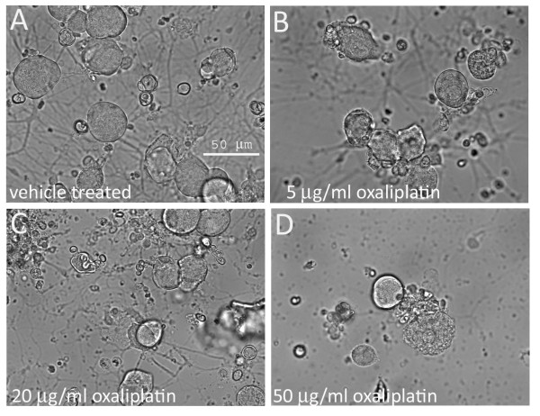
Figure 1
Phase contrast photomicrographs of cultured adult rat DRG neurons. A . without oxaliplatin; B . after 48 hour treatment with 5 u03bcg/ml oxaliplatin, C . 20 u03bcg/ml oxaliplatin and D . 50 u03bcg/ml ...
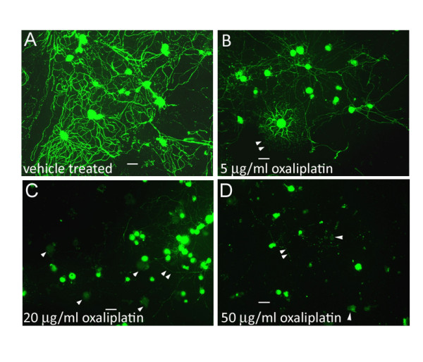
Figure 2
Gap 43 immunostaining in adult rat DRG neurons in vitro . A . without oxaliplatin treatment, DRG neurons are densely distributed and the cell bodies and neurites are intensely positive for Gap43 immun...
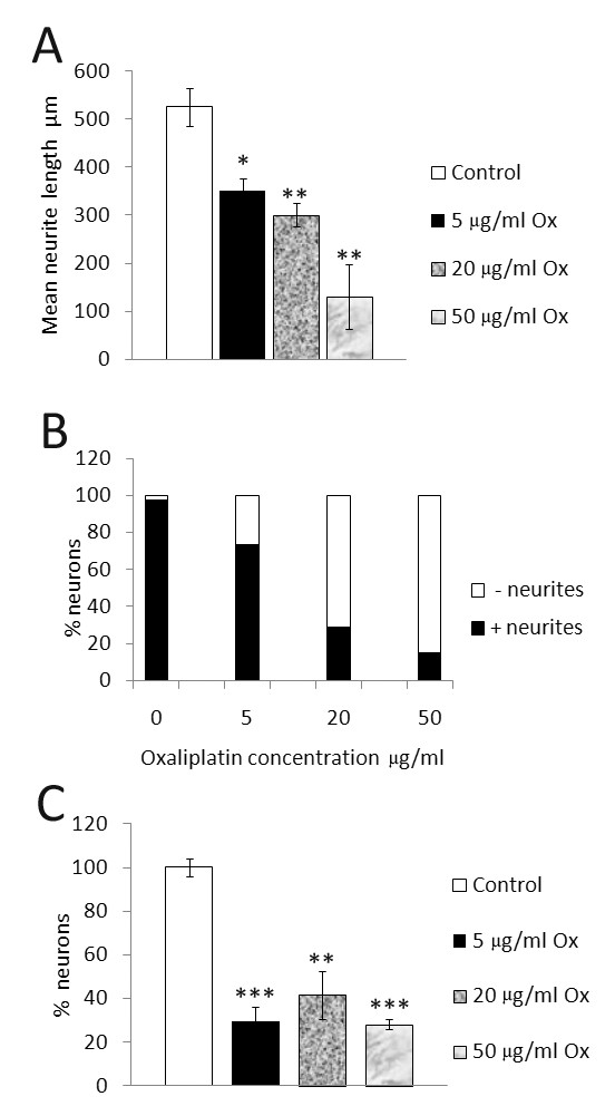
Figure 3
Morphological effects of oxaliplatin treated neurons . A . Dose-related decrease in maximum neurite length in oxaliplatin treated adult rat DRG neurons in vitro; 33% reduction after 5 u03bcg/ml oxalip...
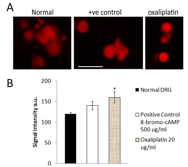
Figure 4
Increased cAMP signal intensity in oxaliplatin treated neurons. A . Immunofluorescence images of cAMP positive adult rat DRG neurons acutely treated with 8-bromo-cAMP (positive control), or oxaliplati...
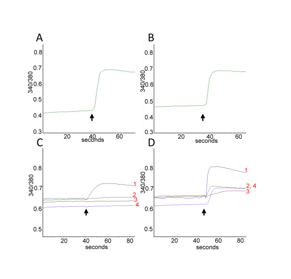
Figure 5
Sensitization of capsaicin responses by acute oxaliplatin treatment: A. Representative trace showing baseline 340/380 intracellular ratio in a DRG neuron and its increase in response to addition of 20...
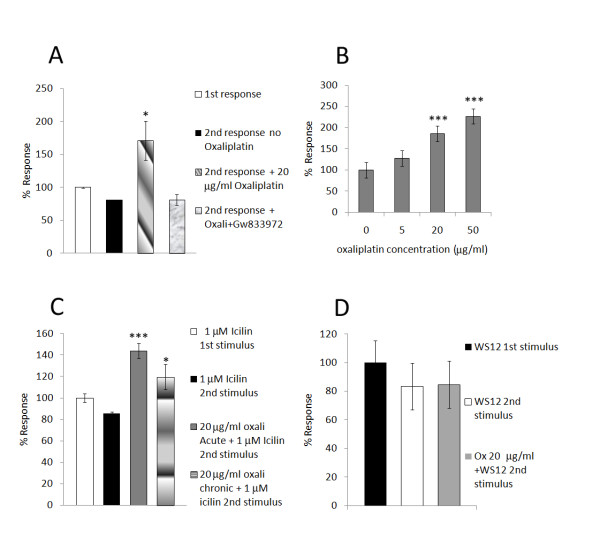
Figure 6
Sensitization of capsaicin responses after acute (A) and chronic (B) oxaliplatin treatment; Sensitization of responses to icilin (C), but not WS12 (D), after oxaliplatin treatment . A . Graph showing ...
Figure images are served from the NIH/NLM PubMed Central Open Access Subset or Europe PMC; copyright remains with the publishers and authors.
💬 Discussion
0 commentsNo comments yet. Be the first to start a discussion!
Leave a Comment