Abstract
Essentials Dimeric high-affinity collagen receptor glycoprotein VI (GPVI) is present on resting platelets. Spatio-temporal organization of platelet GPVI-dimers was evaluated using advanced microscopy. Upon platelet adhesion to collagenous substrates, GPVI-dimers coalesce to form clusters. Clustering of GPVI-dimers may increase avidity and facilitate platelet activation SUMMARY: Background Platelet glycoprotein VI (GPVI) binding to subendothelial collagen exposed upon blood vessel injury initiates thrombus formation. Dimeric GPVI has high affinity for collagen, and occurs constitutively on resting platelets. Objective To identify higher-order oligomerization (clustering) of pre-existing GPVI dimers upon interaction with collagen as a mechanism to initiate GPVI-mediated signaling. Methods GPVI was located by use of fluorophore-conjugated GPVI dimer-specific Fab (antigen-binding fragment). The tested substrates include Horm collagen I fibers, soluble collagen III, GPVI-specific collagen peptides, and fibrinogen. GPVI dimer clusters on the platelet surface interacting with these substrates were visualized with complementary imaging techniques: total internal reflection fluorescence microscopy to monitor real-time interactions, and direct stochastic optical reconstruction microscopy (dSTORM), providing relative quantification of GPVI cluster size and density. Confocal microscopy was used to locate GPVI dimer clusters, glycoprotein Ib, integrin α2 β1 , and phosphotyrosine. Results Upon platelet adhesion to all collagenous substrates, GPVI dimers coalesced to form clusters; notably clusters formed along the fibers of Horm collagen. dSTORM revealed that GPVI density within clusters depended on the substrate, collagen III being the most effective. Clusters on fibrinogen-adhered platelets were much smaller and more numerous; whether these are pre-existing oligomers of GPVI dimers or fibrinogen-induced is not clear. Some GPVI dimer clusters colocalized with areas of phosphotyrosine, indicative of signaling activity. Integrin α2 β1 was localized to collagen fibers close to GPVI dimer clusters. GPVI clustering depends on a dynamic actin cytoskeleton. Conclusions Platelet adhesion to collagen induces GPVI dimer clustering. GPVI clustering increases both avidity for collagen and the proximity of GPVI-associated signaling molecules, which may be crucial for the initiation and persistence of signaling.
🔬 Techniques
💻 Software
✨ Fluorophores
🧪 Sample Preparation
🏭 Microscope Brands
🧪 Reagent Suppliers
📷 Detectors
💻 Software Details
🏛️ Research Organizations (ROR)
Affiliated research institutions:
📋 Methods
GPVI was located by use of fluorophore‐conjugated GPVI dimer‐specific Fab (antigen‐binding fragment). The tested substrates include Horm collagen I fibers, soluble collagen III , GPVI ‐specific collagen peptides, and fibrinogen. GPVI dimer clusters on the platelet surface interacting with these substrates were visualized with complementary imaging techniques: total internal reflection fluorescence microscopy to monitor real‐time interactions, and direct stochastic optical reconstruction microscopy ( dSTORM ), providing relative quantification of GPVI cluster size and density. Confocal microscopy was used to locate GPVI dimer clusters, glycoprotein I b, integrin α 2 β 1 , and phosphotyrosine.
Materials and methods
Non‐inhibitory, recombinant dimer‐specific 204‐11 Fab 10 was derived from clone 204‐11 21 by Kaketsuken (Kumamoto, Japan). Antibodies were fluorescently labeled by use of a Microscale Protein Labelling kit (Molecular Probes, Eugene, OR, USA); degree of fluorescent labeling = 2–5 dye molecules per protein molecule. The antibodies used were: 4G10 (anti‐phosphotyrosine; Millipore Merck, Billerica, MA, USA); anti‐human CD42b/GPIb, clone 486805 (R&D Systems, Abingdon, UK); 16B4 (mouse anti‐human CD49b/integrin α 2 chain; Bio‐Rad, Hercules, CA, USA); Alexa Fluor 647‐conjugated AffiniPure F(ab′)2 fragment goat anti‐mouse IgG, Fcγ‐specific (Jackson ImmunoResearch Laboratories, Inc., West Grove, PA, USA); Gi9 (anti‐integrin α 2 ; Abcam, Cambridge, UK); and Alexa Fluor 647‐conjugated anti‐human CD62P (Bio‐Rad). Other materials, of reagent grade or better, were obtained from commercial sources.
Show full methods section
GPVI was located by use of fluorophore‐conjugated GPVI dimer‐specific Fab (antigen‐binding fragment). The tested substrates include Horm collagen I fibers, soluble collagen III , GPVI ‐specific collagen peptides, and fibrinogen. GPVI dimer clusters on the platelet surface interacting with these substrates were visualized with complementary imaging techniques: total internal reflection fluorescence microscopy to monitor real‐time interactions, and direct stochastic optical reconstruction microscopy ( dSTORM ), providing relative quantification of GPVI cluster size and density. Confocal microscopy was used to locate GPVI dimer clusters, glycoprotein I b, integrin α 2 β 1 , and phosphotyrosine.
Materials and methods
Non‐inhibitory, recombinant dimer‐specific 204‐11 Fab 10 was derived from clone 204‐11 21 by Kaketsuken (Kumamoto, Japan). Antibodies were fluorescently labeled by use of a Microscale Protein Labelling kit (Molecular Probes, Eugene, OR, USA); degree of fluorescent labeling = 2–5 dye molecules per protein molecule. The antibodies used were: 4G10 (anti‐phosphotyrosine; Millipore Merck, Billerica, MA, USA); anti‐human CD42b/GPIb, clone 486805 (R&D Systems, Abingdon, UK); 16B4 (mouse anti‐human CD49b/integrin α 2 chain; Bio‐Rad, Hercules, CA, USA); Alexa Fluor 647‐conjugated AffiniPure F(ab′)2 fragment goat anti‐mouse IgG, Fcγ‐specific (Jackson ImmunoResearch Laboratories, Inc., West Grove, PA, USA); Gi9 (anti‐integrin α 2 ; Abcam, Cambridge, UK); and Alexa Fluor 647‐conjugated anti‐human CD62P (Bio‐Rad). Other materials, of reagent grade or better, were obtained from commercial sources.
Platelet preparation
Washed platelets were prepared from acid–citrate–dextrose‐anticoagulated blood from healthy volunteers 22 , and resuspended at 3–5 × 10 7 platelets mL −1 in HEPES–Tyrodes buffer (HT) (134 m m NaCl, 0.34 m m Na 2 HPO 4 , 2.9 m m KCl, 12 m m NaHCO 3 , 20 m m HEPES, 5 m m glucose, pH 7.3).
Preparation of collagenous substrate‐coated glass dishes for imaging
Thirty‐five‐millimeter glass (0.7 mm)‐bottomed MatTek dishes (MatTek, Ashland, MA, USA) were coated with 10 μg mL −1 cross‐linked CRP (CRP‐XL), III‐30, or collagen III (Col III), in phosphate‐buffered saline (PBS) (0.01 m phosphate buffer, 0.0027 m KCl, 0.137 m NaCl, pH 7.4) and Horm collagen (Horm), in manufacturer‐supplied diluent, overnight at 4 °C. The dishes were washed with PBS, blocked with 1% bovine serum albumin/PBS (heat‐denatured, filtered) for 1 h, and then washed with PBS to make them ready for platelet spreading. TIRFM Adhesion of Alexa Fluor 488–204‐11 Fab‐labeled washed platelets (3 × 10 7 platelets mL −1 in HT containing 2 m m MgCl 2 ) to immobilized collagenous substrate was imaged with TIRFM (Nikon TIRF system mounted on a Nikon Eclipse Ti inverted microscope, with a Nikon × 60 numerical aperture [NA] 1.49 TIRF objective; Nikon UK, Ltd., Surrey, UK). Images were obtained at 5‐s intervals for 20–30 min at 37 °C, and this was followed by fixation in formalin and confocal imaging. Integrin α 2 β 1 blockade was achieved by preincubating platelets with 10 μg mL −1 Gi9 blocking antibody. Platelet morphology was followed with differential interference contrast (DIC) microscopy. dSTORM Alexa Fluor 647‐204‐11 Fab‐labeled washed platelets were allowed to adhere to collagenous substrate‐coated MatTek dishes. Adherent platelets were fixed, permeabilized, and stained with phalloidin–Alexa Fluor 488. Samples were imaged in switching buffer as described previously 22 on a Nikon Eclipse Ti‐E N‐STORM system in dSTORM mode using Perfect Focus, with a CFl SR Apochromat TIRF × 100 oil, 1.49‐NA objective lens and an N‐STORM filter cube with excitation from the Agilent Ultra High Power Dual Output Laser bed (170‐mW, 647‐nm laser), and image capture with an Andor IXON Ultra 897 EMCCD camera. Thirty thousand frames were captured with Nikon nis elements v4.2, and reconstructed by the use of storm analysis module v3.2, with drift correction and Gaussian rendering of data points. Detected points with a photon count of < 500 were discarded from reconstructed data before further processing. Points in the reconstructed images represent individually identified fluorescent blinking events, which are referred to as molecules.
Confocal imaging
Platelets were preincubated with Alexa Fluor 488‐conjugated or Alexa Fluor 647‐conjugated 204‐11 Fab (4 μg mL −1 ) alone or with another fluorescently labeled antibody. In some experiments, platelets were also treated with an inhibitor or inhibitory antibody. The platelets were allowed to adhere to a collagenous substrate, fixed, and imaged with an FV300 IX81 laser‐scanning confocal microscope with a × 60 oil immersion objective (Olympus UK, Southend‐on‐Sea, UK). Where indicated, platelets were permeabilized (0.1% Triton/PBS) following fixation, and stained for actin (Alexa Fluor 647–phalloidin) or phosphotyrosine (4G10, 5 μg mL −1 ).
Flow cytometry to measure GPVI dimer formation
Washed platelets were preincubated with dimethylsulfoxide (DMSO) (0.25% final concentration) or actin antagonist (cytochalasin D, latrunculin A, or jasplakinolide; 10 μ m ; 0.25% DMSO, final concentration), and GPVI dimer formation was measured by flow cytometry 10 .
Data analyses
Data analyses, performed with prism v7 (GraphPad, San Diego, CA, USA), are described in the figure legends. Differences among treatment groups in the flow cytometry experiments were calculated with paired t ‐tests. dSTORM cluster analysis was performed within matlab for each 3 × 3‐μm region of interest (ROI), as described by Owen et al . 23 , with modifications described in Pollitt et al . 18 . Platelets were identified by phalloidin staining of the actin cytoskeleton. ROIs were positioned within the platelet, ensuring that the edges of the ROI fell within the platelet boundary. Clusters of < 3 points were discounted. The numerical data, processed with Microsoft Excel, were analyzed as detailed in the figure legends. The degree of colocalization between the GPVI and phophotyrosine confocal images was quantified with image‐pro premiere 9.2 (Media Cybernetics, Rockville, MD, USA).
Supporting information Movie S1. GPVI forms clusters when platelets spread on immobilized collagenous substrates. Click here for additional data file. Fig. S1. DIC images corresponding to the fluorescence images in Fig. 6 . Click here for additional data file. Click here for additional data file.
📊 Figures
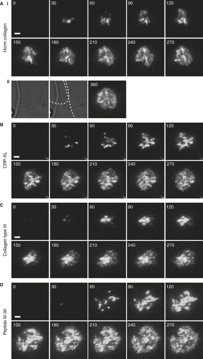
Figure 1
Glycoprotein VI forms clusters when platelets spread on immobilized collagenous substrates. Total internal reflection fluorescence microscopy time courses of washed human platelets, labeled with Alexa...
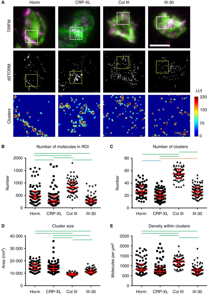
Figure 2
Direct stochastic optical reconstruction microscopy ( dSTORM ) analysis of glycoprotein VI ( GPVI ) clustering on collagenous substrates. (A) Platelets spread on the collagenous substrates indicated w...
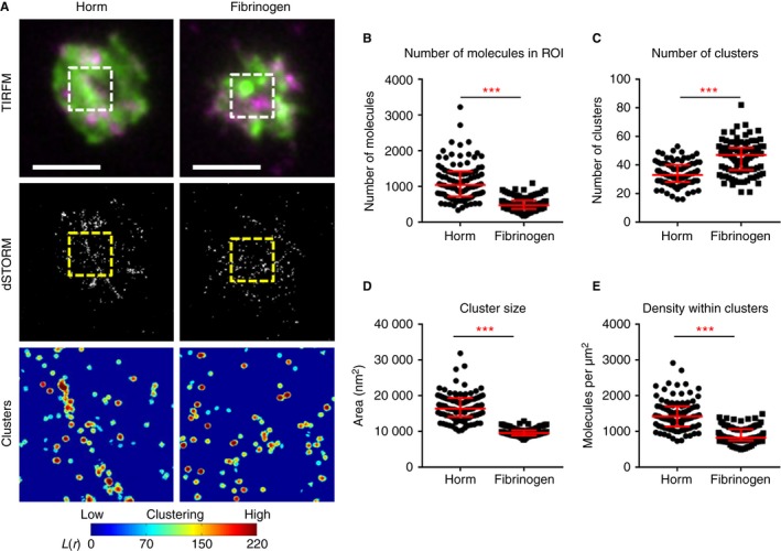
Figure 3
Comparison of glycoprotein VI ( GPVI ) clustering on Horm collagen (Horm) and fibrinogen. (A) Platelets spread on the Horm or fibrinogen as indicated at the top were labeled for dimeric GPVI with the ...
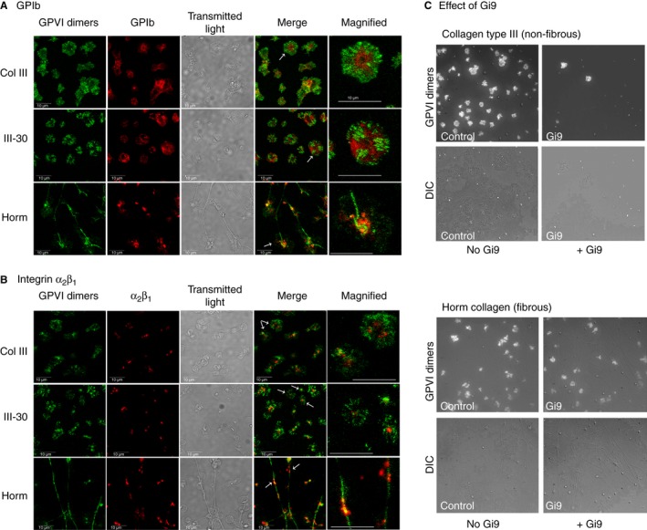
Figure 4
Comparison of distributions of glycoprotein VI ( GPVI ) dimer clusters, glycoprotein I b ( GPI b) and integrin u03b1 2 u03b2 1 on adhered platelets, and the effect of inhibiting u03b1 2 u03b2 1 on clu...
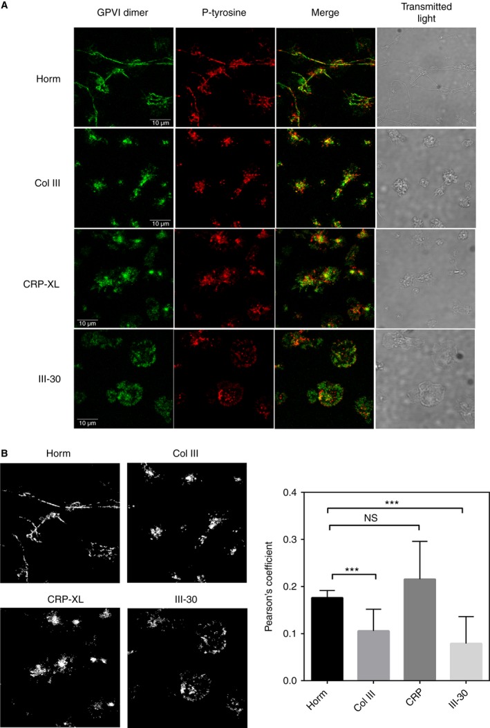
Figure 5
Phosphou2010tyrosine (Pu2010tyrosine) staining and glycoprotein VI ( GPVI ) dimer clustering. To determine whether signaling reactions may be occurring in the vicinity of GPVI dimer clusters, washed p...
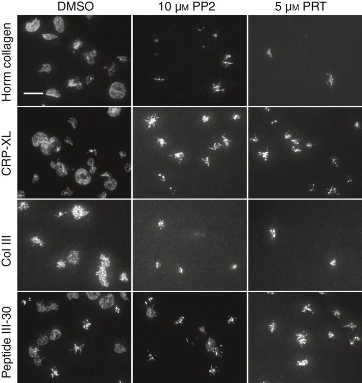
Figure 6
Effect of signaling inhibitors on glycoprotein VI ( GPVI ) dimer cluster formation in live platelets adhering to immobilized collagenous substrate. Alexau00a0Fluoru00a0488u2010conjugated 204u201011u20...
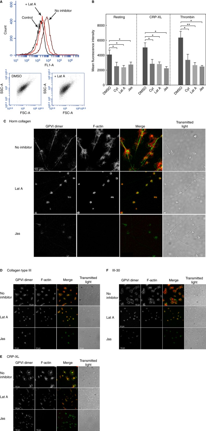
Figure 7
Effect of actin antagonists on glycoprotein VI ( GPVI ) dimerization and GPVI dimer clustering. (A) Raw flow cytometry data: effects of the actin antagonists latrunculinu00a0A (Latu00a0A) and jasplaki...
Figure images are served from the NIH/NLM PubMed Central Open Access Subset or Europe PMC; copyright remains with the publishers and authors.
💬 Discussion
0 commentsNo comments yet. Be the first to start a discussion!
Leave a Comment