Abstract
Neuronal inclusions composed of α-synuclein (α-syn) characterize Parkinson's Disease (PD) and Dementia with Lewy bodies (DLB). Cognitive dysfunction defines DLB, and up to 80% of PD patients develop dementia. α-Syn inclusions are abundant in the hippocampus, yet functional consequences are unclear. To determine if pathologic α-syn causes neuronal defects, we induced endogenous α-syn to form inclusions resembling those found in diseased brains by treating hippocampal neurons with α-syn fibrils. At seven days after adding fibrils, α-syn inclusions are abundant in axons, but there is no cell death at this time point, allowing us to assess for potential alterations in neuronal function that are not caused by neuron death. We found that exposure of neurons to fibrils caused a significant reduction in mushroom spine densities, adding to the growing body of literature showing that altered spine morphology is a major pathologic phenotype in synucleinopathies. The reduction in spine densities occurred only in wild type neurons and not in neurons from α-syn knockout mice, suggesting that the changes in spine morphology result from fibril-induced corruption of endogenously expressed α-syn. Paradoxically, reduced postsynaptic spine density was accompanied by increased frequency of miniature excitatory postsynaptic currents (EPSCs) and presynaptic docked vesicles, suggesting enhanced presynaptic function. Action-potential dependent activity was unchanged, suggesting compensatory mechanisms responding to synaptic defects. Although activity at the level of the synapse was unchanged, neurons exposed to α-syn fibrils, showed reduced frequency and amplitudes of spontaneous Ca2+ transients. These findings open areas of research to determine the mechanisms that alter neuronal function in brain regions critical for cognition at time points before neuron death.
🔬 Techniques
🔭 Microscopes
💻 Software
✨ Fluorophores
🧪 Sample Preparation
🔬 Cell Lines
🏭 Microscope Brands
🧪 Reagent Suppliers
💻 Software Details
🏛️ Research Organizations (ROR)
Affiliated research institutions:
📊 Figures
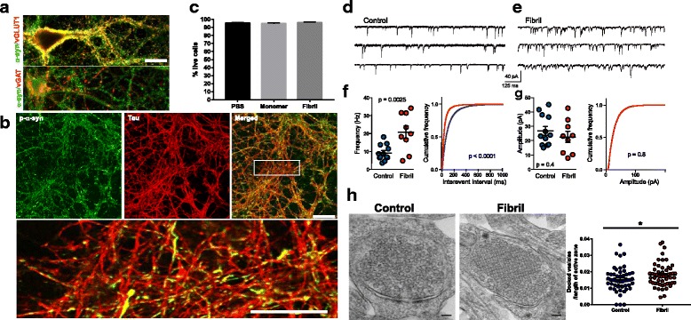
Fig. 1
Seven days after exposure to u03b1-syn fibrils, u03b1-syn inclusions localize to axons, increase mEPSC frequency and increase the number of docked presynaptic vesicles. a Untreated primary hippocampal...
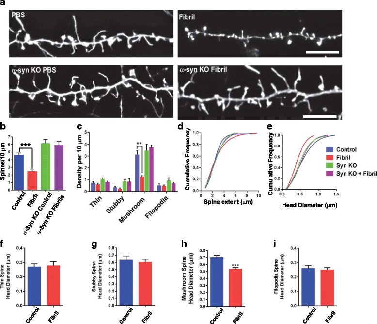
Fig. 2
Formation of u03b1-syn inclusions in primary hippocampal neurons reduces density and head diameter of mushroom-shaped dendritic spines. Primary hippocampal neurons from wild type mice or u03b1-syn kno...
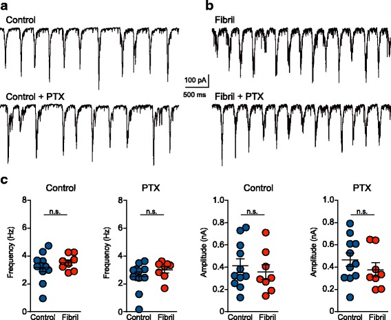
Fig. 3
Spontaneous synaptic activity driven by action potentials is normal in neurons with u03b1-syn inclusions. EPSCs were analyzed over 10u00a0min in neurons 7 days following exposure to fibrils (DIV14) or...
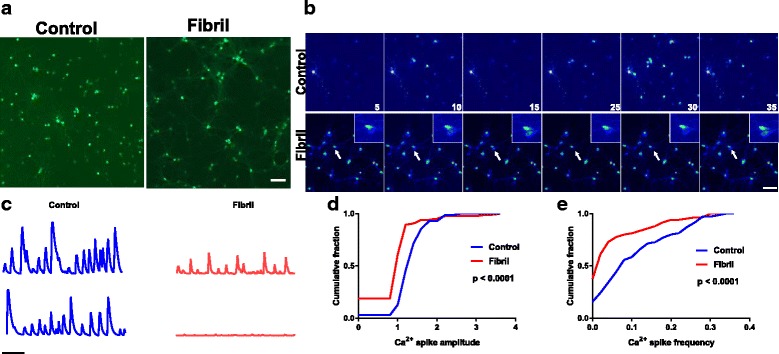
Fig. 4
Formation of u03b1-syn inclusions impair spontaneous Ca 2+ transients. Primary hippocampal neurons from wild type mice were exposed to fibrils (or PBS) at DIV 7. On DIV 8, primary neurons were transdu...
Figure images are served from the NIH/NLM PubMed Central Open Access Subset or Europe PMC; copyright remains with the publishers and authors.
💬 Discussion
0 commentsNo comments yet. Be the first to start a discussion!
Leave a Comment