Abstract
Current imaging approaches limit the ability to perform multi-scale characterization of three-dimensional (3D) organotypic cultures (organoids) in large numbers. Here, we present an automated multi-scale 3D imaging platform synergizing high-density organoid cultures with rapid and live 3D single-objective light-sheet imaging. It is composed of disposable microfabricated organoid culture chips, termed JeWells, with embedded optical components and a laser beam-steering unit coupled to a commercial inverted microscope. It permits streamlining organoid culture and high-content 3D imaging on a single user-friendly instrument with minimal manipulations and a throughput of 300 organoids per hour. We demonstrate that the large number of 3D stacks that can be collected via our platform allows training deep learning-based algorithms to quantify morphogenetic organizations of organoids at multi-scales, ranging from the subcellular scale to the whole organoid level. We validated the versatility and robustness of our approach on intestine, hepatic, neuroectoderm organoids and oncospheres.
🔬 Techniques
🧬 Organisms
✨ Fluorophores
🧪 Sample Preparation
🔬 Cell Lines
🏭 Microscope Brands
🏛️ Research Organizations (ROR)
Affiliated research institutions:
📊 Figures
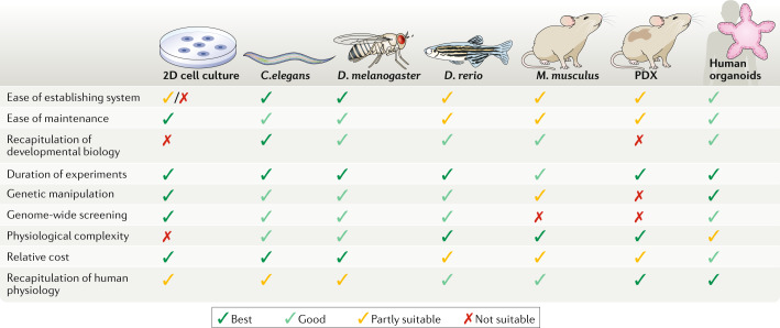
Fig. 1
Comparison of organoids with other model systems.
The most common model organisms that are used in biomedical research are Caenorhabditis elegans , Drosophila melanogaster , Danio rerio and Mus musculus , along with patient-derived xenografts (PDX). ...
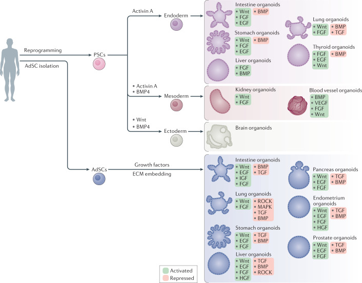
Fig. 2
Process for the establishment of human PSC-derived and AdSC-derived organoids.
Pluripotent stem cell (PSC)-derived organoids are established following directed differentiation of PSCs, which requires a first step that involves germ-layer specification (endoderm, mesoderm or ecto...
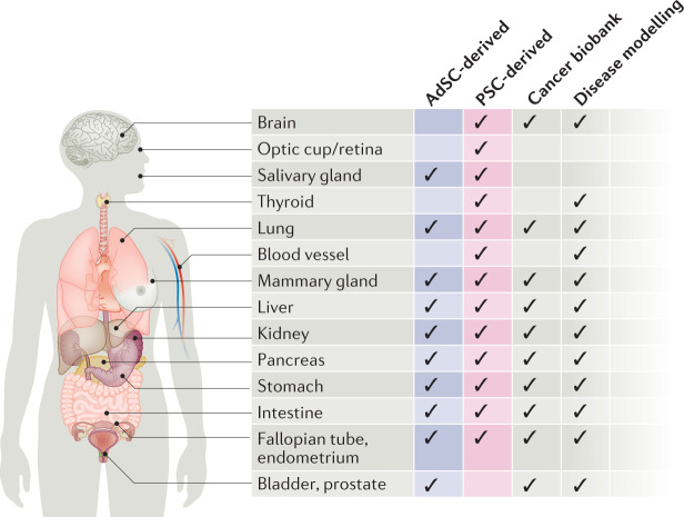
Fig. 3
Reported human organoid systems by tissue of origin.
The chart provides information on the type (either pluripotent stem cell (PSC)-derived or adult stem cell (AdSC)-derived), biobanking status and use in disease modelling of the human organoid systems ...
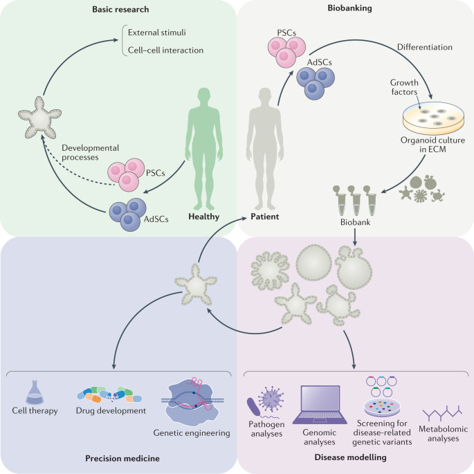
Fig. 4
Potential applications of human organoids.
Two types of organoids are widely used, derived from either pluripotent stem cells (PSCs) or adult stem cells (AdSCs). Organoids can be used for (1) basic research, including studies of human biology ...
Figure images are served from the NIH/NLM PubMed Central Open Access Subset or Europe PMC; copyright remains with the publishers and authors.
💬 Discussion
0 commentsNo comments yet. Be the first to start a discussion!
Leave a Comment