Abstract
Broadly neutralizing antibodies (bNAbs) against HIV-1 envelope (Env) inform vaccine design and are potential therapeutic agents. We identified SF12 and related bNAbs with up to 62% neutralization breadth from an HIV-infected donor. SF12 recognized a glycan-dominated epitope on Env's silent face and was potent against clade AE viruses, which are poorly covered by V3-glycan bNAbs. A 3.3Å cryo-EM structure of a SF12-Env trimer complex showed additional contacts to Env protein residues by SF12 compared with VRC-PG05, the only other known donor-derived silentface antibody, explaining SF12's increased neutralization breadth, potency, and resistance to Env mutation routes. Asymmetric binding of SF12 was associated with distinct N-glycan conformations across Env protomers, demonstrating intra-Env glycan heterogeneity. Administrating SF12 to HIV-1-infected humanized mice suppressed viremia and selected for viruses lacking the N448gp120 glycan. Effective bNAbs can therefore be raised against HIV-1 Env's silent face, suggesting their potential for HIV-1 prevention, therapy, and vaccine development.
🔬 Techniques
🔭 Microscopes
🧬 Organisms
💻 Software
✨ Fluorophores
🧪 Sample Preparation
🔬 Cell Lines
🏭 Microscope Brands
🧪 Reagent Suppliers
📷 Detectors
💻 Software Details
💾 Data Repositories
🏷️ Research Resource Identifiers (RRIDs)
Verified research resources used in this paper:
🏛️ Research Organizations (ROR)
Affiliated research institutions:
📋 Methods
Key Resources Table REAGENT or RESOURCE SOURCE IDENTIFIER Antibodies
Monoclonal anti-HIV-1 Env SF5 Michel C. Nussenzweig, The Rockefeller University (This Paper) N/A Monoclonal anti-HIV-1 Env SF12 Michel C. Nussenzweig, The Rockefeller University (This Paper) N/A Monoclonal anti-HIV-1 Env 3BNC117 NIH AIDS Reagent Program Cat# 12474 Monoclonal anti-HIV-1 Env 10-1074 NIH AIDS Reagent Program Cat# 12477 Monoclonal anti-HIV-1 Env PGDM1400 Dennis R. Burton, Scripps; Sok et al., 2014 N/A Monoclonal anti-HIV-1 Env 8ANC131 Michel C. Nussenzweig, The Rockefeller University; Scheid et al., 2011 N/A Monoclonal anti-HIV-1 Env 8ANC195 Michel C. Nussenzweig, The Rockefeller University; Scheid et al., 2011 N/A Monoclonal anti-HIV-1 Env PGT151 Dennis R. Burton, Scripps; Falkowska et al., 2014 N/A Monoclonal anti-HIV-1 Env 35O22 NIH AIDS Reagent Program Cat# 12586 Monoclonal anti-human IgM-PE-Cy5, Clone G20-127 BD Biosciences Cat# 551079; RRID: AB_394036 Monoclonal anti-human IgD-FITC, Clone IA6-2 BD Biosciences Cat# 555778; RRID: AB_396113 Monoclonal anti-human CD3-APC-Cy7, Clone SK7 BD Biosciences Cat# 557832; RRID: AB_396890 Monoclonal anti-human CD19-PE-Cy7, Clone HIB19 BD Biosciences Cat# 560728; RRID: AB_1727438 Monoclonal anti-human CD16-PB, Clone N/A Mario Roederer, NIH N/A Monoclonal anti-human CD19-BV421, Clone HIB19 Biolegend Cat# 302233; RRID: AB_10897802 Monoclonal anti-human CD20-BV421, Clone 2H7 Biolegend Cat# 302329; RRID: AB_10933088 Monoclonal anti-human CD3-PerCP-Cy5.5, Clone OKT3 Biolegend Cat# 317336; RRID: AB_2561628 Monoclonal anti-human CD14-PerCP-Cy5.5, Clone HCD14 Biolegend Cat# 325622; RRID: AB_893250 Monoclonal anti-human CD335-PerCP-Cy5.5, Clone 9E2 Biolegend Cat# 331920; RRID: AB_2561665 Monoclonal anti-human CD66b-PerCP-Cy5.5, Clone G10F5 Biolegend Cat# 305108; RRID: AB_2077855 Monoclonal anti-human anti-human-IgM-BV605, Clone MHM-88 Biolegend Cat# 314523; RRID: AB_2562373 Monoclonal anti-human anti-human-IgG-APC, Clone G18-145 BD Biosciences Cat# 550931; RRID: AB_398478 Anti-6X His tag antibody Abcam Cat# ab9108; RRID: AB_307016 Goat Anti-Human IgG Fc, Multi-Species SP ads-HRP Southern Biotech Cat# 2014-05; RRID: AB_2795580 Peroxidase AffiniPure Goat Anti-Human IgG, Fcγ fragment specific Jackson ImmunoResearch Cat# 109-035-098; RRID: AB_2337586 Bacterial and Virus Strains f61 Panel of 20 HIV-1 Env-pseudotyped viruses for neutralization fingerprinting Nicole Doria-Rose, NIH; Doria-Rose et al., 2017 N/A Global Panel of 12 HIV-1 Env-pseudotyped viruses NIH AIDS Reagent Program; deCamp et al., 2014 Cat# 12670 119 HIV-1 Env-pseudotyped viruses cross-clade panel Michael S. Seaman, BIDMC; Freund et al., 2017 , Mouquet et al., 2012 N/A YU2 HIV-1 Env-pseudotyped viruses carrying mutations in common anti-HIV-1-mAb binding sites Florian Klein, University of Cologne N/A Replication-competent HIV YU2 (YU2-envelope in pNL/HXB) for mouse experiment Paul D. Bieniasz, The Rockefeller University; Zhang et al., 2002 N/A Biological Samples PBMCs from Donor 27845 M. Juliana McElrath, Fred Hutchinson Cancer Research Center N/A Plasma from Donor 27845 M. Juliana McElrath, Fred Hutchinson Cancer Research Center N/A Human cord blood/placental tissue (for isolation of Human CD34+ cells) Department of Gynecology and Obstetrics, University Hospital of Cologne N/A Chemicals, Peptides, and Recombinant Proteins Dulbecco’s Modified Eagle Medium (DMEM) GIBCO Cat# 11960-044 Fetal bovine serum (FBS) Sigma-Aldrich Cat# F9665 Penicillin/Streptomycin GIBCO Cat# 15140-122 Sodium Pyruvate GIBCO Cat# 11360-070 L-Glutamine Thermo Fisher Scientific Cat# 25030024 Gentamicin Sigma-Aldrich Cat# G1397-10ML HEPES Biochrom Cat# L1613 Freestyle 293 Expression Medium Thermo Fisher Scientific Cat# 12338001 Interleukin 2 (IL-2) Roche Cat# 11147528001 Interleukin 21 (IL-21) Life Technologies Cat# PHC0211 Streptavidin-PE BioLegend Cat# 405203 BG505-SOSIP.664.Avi John P. Moore, Weill Cornell Medical College; Sok et al., 2014 N/A Superscript III Reverse Transcriptase Thermo Fisher Scientific Cat# 18080044 RNasin Plus RNase inhibitor Promega Cat# N2615 Random primers Invitrogen Cat# 48190-011 HotStarTaq DNA Polymerase QIAGEN Cat# 203203 Polyethylenimine (PEI), branched 25 kDa Sigma Cat# 408727 Protein G Sepharose 4 Fast Flow GE Healthcare Cat# 17-0618-05 BG505-SOSIP.664.His John P. Moore, Weill Cornell Medical College; de Taeye et al., 2015 , Sanders et al., 2013 N/A YU2 gp120 monomer John R. Mascola, NIH N/A YU2 gp120 monomer mutants (N160K, N332A, D368R) Michel C. Nussenzweig, The Rockefeller University N/A YU2 gp140 foldon trimer Richard Wyatt, The Scripps Research Institute; Yang et al., 2000 N/A YU2 gp140 foldon trimer mutants (A281T+368K, N160K+A281T+368K) Michel C. Nussenzweig, The Rockefeller University N/A ABTS 1-Step Solution Thermo Fisher Scientific Cat# 002024 Peroxidase Streptavidin Jackson Immuno Cat# 016-030-084 T4 DNA Polymerase New England Biolabs Cat# M0203L Platinum Taq Green Hot Start Thermo Fisher Cat# 11966034 Fugene 6 Transfection Reagent Promega Cat# E2691 B41 SOSIP.664 v4.2 Pugach et al., 2015 N/A SF12 Fab Michel C. Nussenzweig, The Rockefeller University (This Paper) N/A 10-1074 Fab Mouquet et al., 2012 N/A Critical Commercial Assays LIVE/DEAD Fixable Aqua Dead Cell Stain Kit Invitrogen Cat# L34957 TOPO TA cloning Kit Thermo Fisher Scientific Cat# K457501 BirA-500: BirA biotin-protein ligase standard reaction kit Avidity Cat# BirA500 FluoReporter Mini-Biotin-XX Protein Labeling Kit Thermo Fisher Scientific Cat# F6347 QuikChange II XL Site-Directed Mutagenesis Kit Agilent Cat# 200521 Q5-Site-Directed Mutagenesis Kit New England BioLabs Cat# E0554S NOVA Lite® HEp-2 ANA IgG (H&L) Immunoglobulin (External Evan’s Blue) Inova Diagnostics Cat# 704230 Deposited Data Silent face antibody family nucleotide sequences GenBank GenBank: MK722158–MK722171 SF12–B41 SOSIP.664–10-1074 coordinates PDB PDB: 6OKP SF12 Fab coordinates PDB PDB: 6OKQ SF12–B41 SOSIP.664–10-1074 complex cryoEM maps (class 1 and class 2) EMDB EMDB: 20100, 20101 Experimental Models: Cell Lines Mouse: 3T3-msCD40L Cells NIH AIDS Reagent Program Cat# 12535 Human: HEK293EBNA1-6E (293-6E) National Research Council Canada NRC File 11565 Human: HEK293T ATCC Cat# CRL-11268 Human: HeLa-derived TZM-bl NIH AIDS Reagent Program Cat# 8129 CHO Flp-In™ cells Invitrogen Cat# R75807 Experimental Models: Organisms/Strains NOD-Rag1 null IL2rg null (NRG) mice The Jackson Laboratory Stock No. 007799 Oligonucleotides Human immunoglobulin variable region amplification primers for VH-, Vκ−, and Vλ Michel C. Nussenzweig, The Rockefeller University; Scheid et al., 2011 , Doria-Rose et al., 2015 N/A HIV-1 qPCR Primer and Probe Set Michel C. Nussenzweig, The Rockefeller University; Horwitz et al., 2017 N/A HIV-1 YU2 env single genome sequencing primers Michel C. Nussenzweig, The Rockefeller University; Horwitz et al., 2017 N/A Recombinant DNA Human Expression vectors Igγ1, Igκ, Igλ, Ig-Fab heavy chain Michel C. Nussenzweig, The Rockefeller University; Tiller et al., 2008 N/A HIV-1 BG505.T332N gp160 env expression plasmid Rogier W. Sanders, Academic Medical Center, Netherlands N/A HIV-1 BG505.T332N gp160 env expression plasmids carrying silent face antibody binding mutations This Paper N/A HIV-1 YU2 Env gp160 env expression plasmid Joseph Sodroski, Dana-Farber Cancer Institute N/A HIV-1 Env YU2 expression plasmid carrying silent face antibody binding mutations This Paper N/A HIV-1 SG3 ΔEnv Non-infectious Molecular Clone (pSG3ΔEnv) NIH AIDS Reagent Program Cat# 11051 Software and Algorithms IgBLAST National Library of Medicine; Ye et al., 2013 https://www.ncbi.nlm.nih.gov/igblast/ IMGT International ImMunoGeneTics Information System; Lefranc et al., 2009 http://www.imgt.org Geneious v8.1.9 Biomatters Ltd. N/A Prism 7 GraphPad N/A Pymol Schrodinger, LLC 2015 RRID: SCR_000305 UCSF Chimera Pettersen et al., 2004 https://www.cgl.ucsf.edu/chimera/ Phenix Adams et al., 2010 https://www.phenix-online.org Coot Emsley and Cowtan, 2004 http://www2.mrc-lmb.cam.ac.uk/personal/pemsley/coot/ Relion Scheres, 2012 https://www2.mrc-lmb.cam.ac.uk/relion/index.php?title=Main_Page CCP4 suite Winn et al., 2011 http://www.ccp4.ac.uk/index.php XDS Kabsch, 2010 http://xds.mpimf-heidelberg.mpg.de/ Antibody Database v2.0 Pamela J. Bjorkman, California Institute of Technology; West et al., 2013 N/A Other HiLoad 16/600 Superdex 200 pg column GE Healthcare Cat# 28989335 2G12 5 ml column made in-house using using NHS-activated HP resin and 2G12 IgG GE Healthcare Cat# 17071601 Protein A column GE Healthcare Cat# 17040301 300 Mesh Quantifoil R2/2 copper grids EM Resolutions QR22300Cu25 Contact for Reagent and Resource Sharing Further information and requests for resources and reagents should be directed to and will be fulfilled by the Lead Contact, Pamela J. Bjorkman ( bjorkman@caltech.edu ). The Bjorkman laboratory cannot lawfully distribute clones in the pTT5 vector. Those wishing to obtain these clones must first obtain a license from the National Research Council of Canada.
Show full methods section
Key Resources Table REAGENT or RESOURCE SOURCE IDENTIFIER Antibodies
Monoclonal anti-HIV-1 Env SF5 Michel C. Nussenzweig, The Rockefeller University (This Paper) N/A Monoclonal anti-HIV-1 Env SF12 Michel C. Nussenzweig, The Rockefeller University (This Paper) N/A Monoclonal anti-HIV-1 Env 3BNC117 NIH AIDS Reagent Program Cat# 12474 Monoclonal anti-HIV-1 Env 10-1074 NIH AIDS Reagent Program Cat# 12477 Monoclonal anti-HIV-1 Env PGDM1400 Dennis R. Burton, Scripps; Sok et al., 2014 N/A Monoclonal anti-HIV-1 Env 8ANC131 Michel C. Nussenzweig, The Rockefeller University; Scheid et al., 2011 N/A Monoclonal anti-HIV-1 Env 8ANC195 Michel C. Nussenzweig, The Rockefeller University; Scheid et al., 2011 N/A Monoclonal anti-HIV-1 Env PGT151 Dennis R. Burton, Scripps; Falkowska et al., 2014 N/A Monoclonal anti-HIV-1 Env 35O22 NIH AIDS Reagent Program Cat# 12586 Monoclonal anti-human IgM-PE-Cy5, Clone G20-127 BD Biosciences Cat# 551079; RRID: AB_394036 Monoclonal anti-human IgD-FITC, Clone IA6-2 BD Biosciences Cat# 555778; RRID: AB_396113 Monoclonal anti-human CD3-APC-Cy7, Clone SK7 BD Biosciences Cat# 557832; RRID: AB_396890 Monoclonal anti-human CD19-PE-Cy7, Clone HIB19 BD Biosciences Cat# 560728; RRID: AB_1727438 Monoclonal anti-human CD16-PB, Clone N/A Mario Roederer, NIH N/A Monoclonal anti-human CD19-BV421, Clone HIB19 Biolegend Cat# 302233; RRID: AB_10897802 Monoclonal anti-human CD20-BV421, Clone 2H7 Biolegend Cat# 302329; RRID: AB_10933088 Monoclonal anti-human CD3-PerCP-Cy5.5, Clone OKT3 Biolegend Cat# 317336; RRID: AB_2561628 Monoclonal anti-human CD14-PerCP-Cy5.5, Clone HCD14 Biolegend Cat# 325622; RRID: AB_893250 Monoclonal anti-human CD335-PerCP-Cy5.5, Clone 9E2 Biolegend Cat# 331920; RRID: AB_2561665 Monoclonal anti-human CD66b-PerCP-Cy5.5, Clone G10F5 Biolegend Cat# 305108; RRID: AB_2077855 Monoclonal anti-human anti-human-IgM-BV605, Clone MHM-88 Biolegend Cat# 314523; RRID: AB_2562373 Monoclonal anti-human anti-human-IgG-APC, Clone G18-145 BD Biosciences Cat# 550931; RRID: AB_398478 Anti-6X His tag antibody Abcam Cat# ab9108; RRID: AB_307016 Goat Anti-Human IgG Fc, Multi-Species SP ads-HRP Southern Biotech Cat# 2014-05; RRID: AB_2795580 Peroxidase AffiniPure Goat Anti-Human IgG, Fcγ fragment specific Jackson ImmunoResearch Cat# 109-035-098; RRID: AB_2337586 Bacterial and Virus Strains f61 Panel of 20 HIV-1 Env-pseudotyped viruses for neutralization fingerprinting Nicole Doria-Rose, NIH; Doria-Rose et al., 2017 N/A Global Panel of 12 HIV-1 Env-pseudotyped viruses NIH AIDS Reagent Program; deCamp et al., 2014 Cat# 12670 119 HIV-1 Env-pseudotyped viruses cross-clade panel Michael S. Seaman, BIDMC; Freund et al., 2017 , Mouquet et al., 2012 N/A YU2 HIV-1 Env-pseudotyped viruses carrying mutations in common anti-HIV-1-mAb binding sites Florian Klein, University of Cologne N/A Replication-competent HIV YU2 (YU2-envelope in pNL/HXB) for mouse experiment Paul D. Bieniasz, The Rockefeller University; Zhang et al., 2002 N/A Biological Samples PBMCs from Donor 27845 M. Juliana McElrath, Fred Hutchinson Cancer Research Center N/A Plasma from Donor 27845 M. Juliana McElrath, Fred Hutchinson Cancer Research Center N/A Human cord blood/placental tissue (for isolation of Human CD34+ cells) Department of Gynecology and Obstetrics, University Hospital of Cologne N/A Chemicals, Peptides, and Recombinant Proteins Dulbecco’s Modified Eagle Medium (DMEM) GIBCO Cat# 11960-044 Fetal bovine serum (FBS) Sigma-Aldrich Cat# F9665 Penicillin/Streptomycin GIBCO Cat# 15140-122 Sodium Pyruvate GIBCO Cat# 11360-070 L-Glutamine Thermo Fisher Scientific Cat# 25030024 Gentamicin Sigma-Aldrich Cat# G1397-10ML HEPES Biochrom Cat# L1613 Freestyle 293 Expression Medium Thermo Fisher Scientific Cat# 12338001 Interleukin 2 (IL-2) Roche Cat# 11147528001 Interleukin 21 (IL-21) Life Technologies Cat# PHC0211 Streptavidin-PE BioLegend Cat# 405203 BG505-SOSIP.664.Avi John P. Moore, Weill Cornell Medical College; Sok et al., 2014 N/A Superscript III Reverse Transcriptase Thermo Fisher Scientific Cat# 18080044 RNasin Plus RNase inhibitor Promega Cat# N2615 Random primers Invitrogen Cat# 48190-011 HotStarTaq DNA Polymerase QIAGEN Cat# 203203 Polyethylenimine (PEI), branched 25 kDa Sigma Cat# 408727 Protein G Sepharose 4 Fast Flow GE Healthcare Cat# 17-0618-05 BG505-SOSIP.664.His John P. Moore, Weill Cornell Medical College; de Taeye et al., 2015 , Sanders et al., 2013 N/A YU2 gp120 monomer John R. Mascola, NIH N/A YU2 gp120 monomer mutants (N160K, N332A, D368R) Michel C. Nussenzweig, The Rockefeller University N/A YU2 gp140 foldon trimer Richard Wyatt, The Scripps Research Institute; Yang et al., 2000 N/A YU2 gp140 foldon trimer mutants (A281T+368K, N160K+A281T+368K) Michel C. Nussenzweig, The Rockefeller University N/A ABTS 1-Step Solution Thermo Fisher Scientific Cat# 002024 Peroxidase Streptavidin Jackson Immuno Cat# 016-030-084 T4 DNA Polymerase New England Biolabs Cat# M0203L Platinum Taq Green Hot Start Thermo Fisher Cat# 11966034 Fugene 6 Transfection Reagent Promega Cat# E2691 B41 SOSIP.664 v4.2 Pugach et al., 2015 N/A SF12 Fab Michel C. Nussenzweig, The Rockefeller University (This Paper) N/A 10-1074 Fab Mouquet et al., 2012 N/A Critical Commercial Assays LIVE/DEAD Fixable Aqua Dead Cell Stain Kit Invitrogen Cat# L34957 TOPO TA cloning Kit Thermo Fisher Scientific Cat# K457501 BirA-500: BirA biotin-protein ligase standard reaction kit Avidity Cat# BirA500 FluoReporter Mini-Biotin-XX Protein Labeling Kit Thermo Fisher Scientific Cat# F6347 QuikChange II XL Site-Directed Mutagenesis Kit Agilent Cat# 200521 Q5-Site-Directed Mutagenesis Kit New England BioLabs Cat# E0554S NOVA Lite® HEp-2 ANA IgG (H&L) Immunoglobulin (External Evan’s Blue) Inova Diagnostics Cat# 704230 Deposited Data Silent face antibody family nucleotide sequences GenBank GenBank: MK722158–MK722171 SF12–B41 SOSIP.664–10-1074 coordinates PDB PDB: 6OKP SF12 Fab coordinates PDB PDB: 6OKQ SF12–B41 SOSIP.664–10-1074 complex cryoEM maps (class 1 and class 2) EMDB EMDB: 20100, 20101 Experimental Models: Cell Lines Mouse: 3T3-msCD40L Cells NIH AIDS Reagent Program Cat# 12535 Human: HEK293EBNA1-6E (293-6E) National Research Council Canada NRC File 11565 Human: HEK293T ATCC Cat# CRL-11268 Human: HeLa-derived TZM-bl NIH AIDS Reagent Program Cat# 8129 CHO Flp-In™ cells Invitrogen Cat# R75807 Experimental Models: Organisms/Strains NOD-Rag1 null IL2rg null (NRG) mice The Jackson Laboratory Stock No. 007799 Oligonucleotides Human immunoglobulin variable region amplification primers for VH-, Vκ−, and Vλ Michel C. Nussenzweig, The Rockefeller University; Scheid et al., 2011 , Doria-Rose et al., 2015 N/A HIV-1 qPCR Primer and Probe Set Michel C. Nussenzweig, The Rockefeller University; Horwitz et al., 2017 N/A HIV-1 YU2 env single genome sequencing primers Michel C. Nussenzweig, The Rockefeller University; Horwitz et al., 2017 N/A Recombinant DNA Human Expression vectors Igγ1, Igκ, Igλ, Ig-Fab heavy chain Michel C. Nussenzweig, The Rockefeller University; Tiller et al., 2008 N/A HIV-1 BG505.T332N gp160 env expression plasmid Rogier W. Sanders, Academic Medical Center, Netherlands N/A HIV-1 BG505.T332N gp160 env expression plasmids carrying silent face antibody binding mutations This Paper N/A HIV-1 YU2 Env gp160 env expression plasmid Joseph Sodroski, Dana-Farber Cancer Institute N/A HIV-1 Env YU2 expression plasmid carrying silent face antibody binding mutations This Paper N/A HIV-1 SG3 ΔEnv Non-infectious Molecular Clone (pSG3ΔEnv) NIH AIDS Reagent Program Cat# 11051 Software and Algorithms IgBLAST National Library of Medicine; Ye et al., 2013 https://www.ncbi.nlm.nih.gov/igblast/ IMGT International ImMunoGeneTics Information System; Lefranc et al., 2009 http://www.imgt.org Geneious v8.1.9 Biomatters Ltd. N/A Prism 7 GraphPad N/A Pymol Schrodinger, LLC 2015 RRID: SCR_000305 UCSF Chimera Pettersen et al., 2004 https://www.cgl.ucsf.edu/chimera/ Phenix Adams et al., 2010 https://www.phenix-online.org Coot Emsley and Cowtan, 2004 http://www2.mrc-lmb.cam.ac.uk/personal/pemsley/coot/ Relion Scheres, 2012 https://www2.mrc-lmb.cam.ac.uk/relion/index.php?title=Main_Page CCP4 suite Winn et al., 2011 http://www.ccp4.ac.uk/index.php XDS Kabsch, 2010 http://xds.mpimf-heidelberg.mpg.de/ Antibody Database v2.0 Pamela J. Bjorkman, California Institute of Technology; West et al., 2013 N/A Other HiLoad 16/600 Superdex 200 pg column GE Healthcare Cat# 28989335 2G12 5 ml column made in-house using using NHS-activated HP resin and 2G12 IgG GE Healthcare Cat# 17071601 Protein A column GE Healthcare Cat# 17040301 300 Mesh Quantifoil R2/2 copper grids EM Resolutions QR22300Cu25 Contact for Reagent and Resource Sharing Further information and requests for resources and reagents should be directed to and will be fulfilled by the Lead Contact, Pamela J. Bjorkman ( bjorkman@caltech.edu ). The Bjorkman laboratory cannot lawfully distribute clones in the pTT5 vector. Those wishing to obtain these clones must first obtain a license from the National Research Council of Canada.
Experimental Model and Subject Details
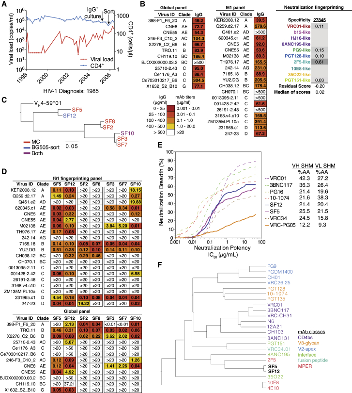
Human subjects Donor 27845 is an adult male who was diagnosed with HIV-1 in 1985. He was a study participant in Seattle Vaccine Unit Observational Protocols “Immune Determinants Favoring Non-Progression in HIV-1 Infection” and “Evaluation of HIV-Specific Immunological and Virological Responses of HIV-1 Multiply-Exposed Seronegative Individuals” (P.I. MJ McElrath) at the Fred Hutchinson Cancer Research Center and was followed from 1998 - 2006. Apart from an NIH interventional study during which Donor 27845 started and stopped anti-retroviral therapy (ART) from 1998-2001, the subject has been off ART. Samples for this study were obtained from 2005 and 2006. During the time of follow-up, viral loads ranged from 35 – 23,300 copies/ml (median: 1,640 copies/ml) and CD4 + T cell counts ranged from 291 to 1,000 cells/mm 3 (median: 590 cells/mm 3 ). Studies and procedures were approved by the Fred Hutchinson Cancer Research Center Internal Review Board (FWA00001920). Samples for analysis were obtained under protocol MNU-0628 approved by the Rockefeller University Institutional Review Board.
Humanized mice
NOD-Rag1 null IL2rg null (NRG) mice were purchased from The Jackson Laboratory, and were subsequently bred and maintained in the Dezentrales Tierhaltungsnetzwerk Weyertal at University of Cologne. NRG mice were fed ssniff complete feed 1124 during breeding and ssniff complete feed 1534 during maintenance, and kept under a 12 hr light/dark cycle with specific-pathogen-free (SPF) conditions. To determine the pharmacokinetics of antibody SF12, 6-week old non-humanized NRG mice (n = 3 per antibody, male and female mice in both groups) were injected intravenously via the tail-vein with 250 μg of antibody (SF12 or 3BNC117). Mice were bled on days 1, 3, 6, 9 and 14 after injection from the facial vein into Z-Gel Serum tubes (Sarstedt). Serum levels were determined using a previously described total IgG ELISA ( Klein et al., 2012 ). Humanized mice for treatment experiments were generated using a previously described protocol with slight modifications ( Klein et al., 2012 , Traggiai et al., 2004 ). In brief, 1-5 days old NRG mice were sublethally irradiated, and 3-6 hours later injected intrahepatically with CD34 + hematopoietic stem cells. CD34+ cells were enriched by magnetic bead-based positive selection (Miltenyi) from PBMCs obtained from human cord blood and by placental perfusion under a protocol approved by the ethics committee of the Medical Faculty of the University of Cologne (protocol #16-110). All cord blood and tissue donors provided written informed consent. Humanization screening was performed at 12 weeks post injection by flow cytometry as previously described ( Klein et al., 2012 ). For treatment experiments, humanized mice were infected with HIV-1 YU2 ( Zhang et al., 2002 ) (produced in 293T cells) intraperitoneally ( Horwitz et al., 2013 , Klein et al., 2012 ). To determine viral loads, plasma viral RNA was measured using a quantitative PCR (qPCR) assay ( Horwitz et al., 2013 , Horwitz et al., 2017 ) based on pol using primers HIV-1 Pol region F 5′-TAATGGCAGCAATTTCACCA-3′ and HIV-1 Pol region R 5′-GAATGCCAAATTCCTGCTTGA-3′, and probe 5′-/56-FAM/CCCACCAAC/ZEN/ARGCRGCCTTAACTG/3IABkFQ/-3′. The limit of accuracy of the assay (based on the standard curve used) was 384 copies/ml. Plasma viral loads were determined twice before experiment start and only mice with viral loads over 4,000 copies/ml were included in experiments. Male and female mice (18-67 weeks old) were used for treatment experiments. Treatment groups were matched primarily based on viral load and stem cell donor (previously identified as key determinants of viral load kinetics), with equal or comparable distributions of age and sex across groups. For antibody treatments, 1 mg of each antibody was administered subcutaneously as a loading dose followed by twice-weekly injections of 0.5 mg of each antibody in PBS subcutaneously for a total of 3 weeks (monotherapy) or 5 weeks (tri-mix). All mouse experiments were authorized by the State Agency for Nature, Environment and Consumer Protection (LANUV) of North Rhine-Westphalia.
Cell lines
HEK293T cells were obtained from the American Type Culture Collection (ATCC) and maintained in Dulbecco’s modified Eagle Medium (DMEM, GIBCO) with 10% fetal bovine serum (FBS, Sigma Aldrich), 1x Penicillin/Streptomycin (GIBCO), 1 mM Sodium Pyruvate (GIBCO), 2 mM L-Glutamine (Thermo Fisher Scientific) at 37°C/5% CO 2 . The HeLa-derived TZM-bl reporter cell line was sourced from the NIH AIDS Reagent Program and maintained in DMEM containing 10% fetal bovine serum, 1 mM Sodium Pyruvate, 2 mM L-Glutamine (Thermo Fisher Scientific), 50 μg/ml Gentamicin (Sigma-Aldrich), and 25 mM HEPES (Biochrom) at 37°C/5% CO 2 . HEK293EBNA1-6E (293-6E) cells were obtained from the National Research Council Canada (NRC) and maintained in Freestyle 293 Expression Medium (Thermo Fisher Scientific) containing 0.2% Penicillin/Streptomycin at 37°C /5% CO 2 with shaking at 90-120 rpm. The sex of these cell lines is unknown.
CHO Flp-In™ cells
(Invitrogen) were a kind gift from the lab of John Moore (Cornell University) and maintained in Ham’s F-12 Medium supplemented with 10% heat-inactivated FBS (Sigma-Aldrich), 200 U/ml penicillin/streptomycin, 2 mM L-glutamine, 20 mM HEPES, 0.1 mM non-essential amino acids, 1 mM sodium pyruvate (GIBCO), and further supplemented with 100 μg/ml Zeocin (Invitrogen).
Methods Details IgG isolation for polyclonal IgG neutralization testing
IgG from subject 27845 was purified from heat-inactivated (1h 56°C) plasma (late 2005 time point) using Protein G Sepharose 4 Fast Flow (GE Healthcare), buffer exchanged into phosphate buffered saline (PBS) using an Amicon Ultra 30 kDa (Millipore), and sterile-filtered.
Neutralization fingerprinting analysis
Neutralization fingerprinting ( Figure 1 B) of the polyclonal antibody response of subject 27845 was done using a panel of 20 diverse HIV-1 strains ( Doria-Rose et al., 2017 ). In brief, the neutralization fingerprint of a serum/polyclonal IgG (the potency-defined pattern of neutralization of a set of diverse viral strains) is represented as a combination of the neutralization fingerprints of a reference set of bNAbs, grouped in ten epitope-specific clusters. Using this method, the prevalence of each of the ten antibody groups can be estimated for the given serum/polyclonal IgG, with prevalence scores ranging between 0 (low) and 1 (high). Additionally, two measures (Residual score and Median of scores) are computed as a way to estimate prediction confidence for the prevalence scores ( Georgiev et al., 2013 , Raju et al., 2019 ). For neutralization fingerprinting of monoclonal antibodies ( Figure 1 F), a set of 80 viruses for which data was available for all antibodies was used. The tree was constructed with a distance metric based on the similarity of the neutralization patterns of the different antibodies. First, the correlations between the neutralization fingerprints (the antibody-specific pattern of neutralization of a set of diverse HIV-1 strains) were computed for each pair of antibodies. The antibody-antibody correlation matrix was then used as input to a hierarchical clustering algorithm to generate a neutralization fingerprinting-based antibody tree. Generally, antibodies that cluster closely together in the tree may indicate similar patterns of neutralization sensitivity/resistance for the given set of strains. B cell microculture Sorting and culturing of memory B cells was performed according to a previously published protocol ( Doria-Rose et al., 2015 , Huang et al., 2013 ). In brief, peripheral blood mononuclear cells (PBMCs) were stained with LIVE/DEAD Fixable Aqua (Invitrogen), CD19-PE-Cy7, CD16-Pacific Blue, CD3-APC-Cy7, IgM-PE-Cy5 and IgD-FITC. Gating was done on IgM - and IgD - negative B cells, and these were bulk sorted using a FACS Aria II cytometer. Bulk B cells were then diluted and plated at 2 B cells per well in 384-well plates. B cells were cultured for two weeks in the presence of IL-2 (Roche), IL-21 (Life Technologies) and CD40L-expressing NIH 3T3 cells as described ( Doria-Rose et al., 2015 , Huang et al., 2013 ). To assess culture success, a total IgG ELISA was performed on supernatants after two weeks of culture to determine the number of wells with positive IgG production. Cell supernatants were then screened in a microneutralization TZM.bl assay against viruses BaL.26 and BG505.T332N. Wells with neutralization > 50% against one or both strains were amplified using various sets of previously described primer sets for heavy chain and light chain ( Doria-Rose et al., 2015 , Scheid et al., 2011 ). Clone specific primers in the leader region were designed when necessary to obtain fully native sequences of the entire framework 1 region (FWR1). Positive bands were sanger sequenced using reverse amplification primers. In the case that multiple B cells were present (double peaks were obtained), bands were subcloned using the TOPO-TA kit (Invitrogen), colony PCR was performed, and bands were again sequenced by Sanger sequencing. Antibody sequences were analyzed using both IgBLAST and the international ImMunoGeneTics information system (IMGT) ( Lefranc et al., 2009 , Ye et al., 2013 ). Obtained heavy and light chain genes were cloned into human Igγ1-, Igκ or Igλ-expression vectors using sequence and ligation independent cloning (SLIC) ( Jeong et al., 2012 , Tiller et al., 2008 , von Boehmer et al., 2016 ). The correct heavy and light chain pairing of the SF5/SF12 antibody family was confirmed by single B cell BG505 bait sorting data. Single B cell bait-sorting BG505 SOSIP.664-Avi for B cell sorting was produced in CHO cells and purified using a PGT145 immunoaffinity column as described ( Pugach et al., 2015 , Sok et al., 2014 ). Biotinylation of BG505 was done using BirA-ligase (Avidity) according to the manufacturer’s instructions. An aliquot of BG505 SOSIP.664-Avi-biotin was freshly coupled to Streptavidin-PE (Invitrogen) using 2.5 μg and 1 μl (0.2 mg/ml) of Streptavidin-PE in a total volume of 10 ul PBS. For the sort, 20 million PBMCs were freshly thawed and stained with the following fluorophore-coupled anti-human antibodies: IgG-APC, IgM-BV605, CD19-BV421, CD20-BV421, CD3-PerCP-Cy5.5, CD14 PerCP-Cy5.5, CD335 PerCP-Cy5.5, CD606 PerCP-Cy5.5 and 1:20 Streptavidin-PE coupled BG505.SOSIP.664 mix described above. Staining was performed for 30 mins at 4°C. Sorting was done on a FACS Aria II. The gating first included singlets, followed by exclusion of unwanted cells (CD3 - , CD14 - , CD335 - , CD606 - ), selection for B cells (CD19 + , CD20 + ) and finally sorting of single IgG + PE + cells into 96-well plates containing lysis buffer. B cell antibody genes were amplified and Sanger sequenced. Antibody sequences were analyzed using both IgBLAST and the international ImMunoGeneTics information system (IMGT) ( Lefranc et al., 2009 , Ye et al., 2013 ). Sequences of interest were cloned into human Igγ1-, Igκ or Igλ-expression vectors by SLIC as described above. Phylogenetic analysis of SF family heavy chain sequences The IgV H 4 ∗ 59 ∗ 01 Homo sapiens allele sequence was obtained from the international ImMunoGeneTics information system (IMGT) ( Lefranc et al., 2009 ). Antibody heavy chain nucleotide sequences of the SF family were aligned with the IgV H 4 ∗ 59 ∗ 01 sequence in Geneious R8 (v8.1.9) using ClustalW. The maximum-likelihood tree was generated using the RAxML plugin (v 7.2.8) with a GTR Gamma model using the ‘Rapid Bootstrapping and search for best-scoring ML tree’ function with 100 bootstrap replicates. The best-scoring ML tree was then formatted using FigTree (v1.4.3). Antibody production for ELISA, neutralization assays and in vivo experiments 293-6E cells were maintained in Freestyle 293 Expression Medium (Thermo Fisher Scientific) containing 0.2% Penicillin-Streptomycin (Thermo Fisher Scientific). Paired heavy and light chain expression constructs were transfected into 293-6E cells (NRC) using branched polyethylenimine (PEI) 25 kDA (Sigma). After 7 days of culture, cells were spun down at 4200 g for 40 mins at 4°C and supernatants were filtered through 0.22 μM aPES (Thermo Nalgene Rapid-Flow). Antibodies were then purified from filtered supernatants using Protein G Sepharose 4 Fast Flow (GE Healthcare) according to standard protocols. Antibodies were buffer exchanged and concentrated into PBS using Amicon Ultra centrifugal filter (Millipore) with either a 30 or 50 kDA molecular weight cutoff (MWCO).
Enzyme-linked immunosorbent assay
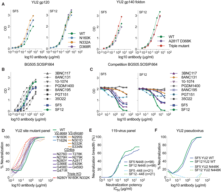
(ELISA) of (mutant) YU2 gp120/gp140 proteins Wild-type and mutant His-tagged YU2 gp120/gp140 proteins were expressed by transient transfection of 293-6E cells and purified using Ni-NTA according to manufacturer’s instructions. Corning Costar 96-Well Assay high-binding plates were coated for 1h at 37°C with 2 μg/ml of the respective protein (YU2 gp120 WT, YU2 gp120 D368R gp120 , YU2 gp120 N332A gp120 , YU2 gp120 N160K gp120 and YU2 gp140 WT, YU2 gp140 A281T gp120 /D368K gp120 , YU2 gp140 N160K gp120 A281T gp120 /D368K gp120 and N332K gp120 (triple mutant)) using a volume of 50 μl/well. Plates were washed 6x using PBS-Tween20 (0.05%), and subsequently blocked using 3% BSA in PBS for 1h at 37°C (200 μl/well). After washing, serially-diluted antibodies were added (starting at 4 or 10 ug/ml, 1:3 dilution series) at 50 μl/well in 1% PBS/BSA and incubated for 1h at room temperature or 37°C. After another wash step, anti-human IgG (Southern Biotech or Jackson Immunoresearch) was added at 1:5000 (50 μl/well) in 1% PBS/BSA for 30 mins at 37°C. Development was done using 100 μl/well ABTS 1-Step Solution (Thermo Fisher Scientific), and absorbance was measured at 405 nm on a FluoStar Omega or 415 nm on a Tecan Sunrise. BG505 SOSIP.664-His ELISAs Corning Costar 96-Well Assay high-binding plates were coated overnight at room temperature or for 1h at 37°C with 2 μg/ml anti-His-tag antibody (Abcam) in PBS (50 μl/well). Plates were washed 6x using PBS-Tween20 (0.05%), and subsequently blocked using 3% BSA in PBS or 2% milk powder in PBS for 1h at 37°C (200 μl/well). After washing, purified BG505 SOSIP.664-His ( de Taeye et al., 2015 , Sanders et al., 2013 ) was added at 2 μg/ml in 1% BSA in PBS (50 μl/well), and incubated for 1h at 37°C, followed by another washing step. Next, serially-diluted antibodies were added (starting at 4 or 10 ug/ml, 1:3 dilution series) at 50 μl/well in 1% PBS/BSA, and incubated for 1h at room temperature or 37°C. After washing, anti-human IgG (Southern Biotech) was added at 1:5000 in 1% BSA in PBS (50 μl/well) for 30 mins at 37°C. Post washing, development was done using 100 μl/well ABTS 1-Step Solution (Thermo Fisher Scientific), and absorbance was measured at 405 nm on a FluoStar Omega or 415 nm on a Tecan Sunrise.
Competition ELISAs Antibodies
SF5 and SF12 were biotinylated using the FluoReporter Mini-Biotin-XX Protein Labeling Kit (Thermo Fisher Scientific). Corning Costar 96-Well Assay high-binding plates were coated overnight at room temperature or for 1h at 37°C with 2 μg/ml anti-His-tag antibody (Abcam) in PBS (50 μl/well). Plates were washed 6x using PBS-Tween20 (0.05%), and subsequently blocked using 3% BSA in PBS for 1h at 37°C (200 μl/well). After washing, BG505 SOSIP.664 was added at 2 μg/ml in 1% BSA in PBS (50 μl/well), and incubated for 1h at 37°C, followed by another washing step. Next, serially-diluted competitor antibodies (starting at 32 μg/ml, 1:3 dilution series) were added at 50 μl/well in 1% PBS/BSA and incubated for 1h at room temperature. Plates were washed and biotinylated SF5 or SF12 were added at 0.5 μg/ml (50 μl/well in 1% PBS/BSA) and incubated for 1h room temperature. After another wash step, Streptavidin-HRP (1:1000) was added at 50 μl/well in 1% PBS/BSA for 30 mins at room temperature. Development was done using 100 μl/well ABTS 1-Step Solution (Thermo Fisher Scientific), and absorbance was measured at 405 nm on a FluoStar Omega or 415 nm on a Tecan Sunrise.
Generation of mutant HIV YU2 and HIV BG505 pseudoviruses
Point mutations were introduced into the HIV YU2 and HIV BG505T332N gp160 expression plasmids using the QuikChange site-directed mutagenesis kit (Agilent Technologies) or the Q5 Site-directed mutagenesis kit (NEB) according to manufacturer’s specifications. Pseudoviruses were produced by co-transfection with pSG3ΔEnv into HEK293T according to an established protocol ( Sarzotti-Kelsoe et al., 2014 ). In vitro neutralization assays Neutralization activities of polyclonal IgG and monoclonal antibodies were determined using a luciferase-based TZM.bl assay ( Li et al., 2005 , Sarzotti-Kelsoe et al., 2014 , Seaman et al., 2010 ), which measures the reduction of Tat-induced luciferase expression in TZM-bl reporter cells during a single round of infection. Samples were assayed at least in in duplicate. Polyclonal IgG neutralization assays were done using a starting concentration of 500 μg/ml and monoclonal antibodies were assayed at starting concentrations of 10, 25 or 50 μg/ml. IC 50 s and IC 80 s were derived 5-parameter curve fitting. Neutralization was also assessed against murine leukemia virus (MuLV) to detect unspecific activity ( Sarzotti-Kelsoe et al., 2014 ). Neutralization data for Figures 1 E and 2 E were analyzed and graphed using Antibody Database (v 2.0) ( West et al., 2013 ).
Autoreactivity and polyreactivity assays
Autoreactivity of antibodies
SF5 and SF12 and reference antibodies 4E10 and 2F5 was determined using the commercially-available HEp-2 based assay NOVA Lite kit (Inova Diagnostics) at an IgG concentration of 25 μg/ml. Slides were photographed on a Leica DMI 6000 B with an exposure of 800 ms, Gain of 10 and Intensity of 100%. Measurements were done in duplicate. Representative images are shown in Figure S1 . Polyreactivity assays were conducted using ELISA detection of non-specific binding to baculovirus extracts as described ( Hötzel et al., 2012 ). Briefly, a solution of 1% baculovirus particles in 100mM sodium bicarbonate buffer pH 9.6 was absorbed onto the wells of a 384-well ELISA plate (Nunc Maxisorp) using a Tecan Freedom Evo liquid handling robot, and the plate was incubated overnight at 4°C. The plate was then blocked with 0.5% BSA in PBS for 1 hour at room temperature. Purified IgGs (diluted to 1 μg/mL in PBS, 0.5% BSA) were added to the blocked assay plate and incubated for 3 hours at room temperature. Bound IgG was detected as the luminescence signal at 425 nm using an HRP-conjugated anti-human IgG (H&L) secondary antibody (Genscript) and SuperSignal ELISA Femto Maximum Sensitivity Substrate (Thermo Fisher Scientific). Single genome sequencing of plasma HIV-1 Env genes Amplification of genes encoding HIV-1 Env gp160 from single viral genomes was carried out as described previously with slight modifications ( Keele et al., 2008 , Salazar-Gonzalez et al., 2008 ). In brief, complementary DNA (cDNA) was synthesized using primer YB383 5′-TTTTTTTTTTTTTTTTTTTTTTTTRAAGCAC-3′ and Superscript III (Invitrogen) according to manufacturer’s instructions. cDNA was then serially diluted and HIV-1 Env was amplified in two rounds of nested PCR using Platinum Taq Green Hot Start (Thermo Fisher Scientific) and primers specifically adapted for HIV YU2NL4-3 (1 st round primers were YB383 and YB50 5′- GGCTTAGGCATCTCCTATGGCAGGAAGAA-3′; 2 nd round primers YB49 5′-TAGAAAGAGCAGAAGACAGTGGCAATGA-3′, YB52 5′-GGTGTGTAGTTCTGCCAATCAGGGAAGWAGCCTTGTG-3′). Positive Bands from amplifications with less than 30% efficiency were PCR-purified using the Nucleospin Gel and PCR-Clean Up kit (Macherey Nagel) and Sanger sequenced using a set of 8 primers. Env sequences were assembled using the Geneious 8.1.9 (Biomatters) de-novo assembly tool. Assembled sequences were cross-checked against sequencing traces again to validate assemblies, and sequences with full coverage of gp160 Env were used in downstream analyses. Protein expression and purification for structural studies Fabs from SF12, SF5 and 10-1074 IgGs were produced as described ( Scharf et al., 2015 ). Briefly, Fabs were expressed by transiently transfecting HEK293-6E cells with vectors encoding the appropriate light chain and C-terminal 6x-His tagged heavy chain genes. Secreted Fabs were purified from cell supernatants using Ni 2+ -NTA affinity chromatography (GE Healthcare), followed by size exclusion chromatography (SEC) with a Superdex200 16/60 column (GE Healthcare). Purified Fabs were concentrated and maintained at 4°C in storage buffer (20 mM Tris pH 8.0, 150 mM NaCl, 0.02% sodium azide). A gene encoding soluble B41 SOSIP.664 gp140 trimer, including SF501C gp120, T605C gp41 , and I559P gp41 substitutions, an enhanced gp120–gp41 cleavage site (REKR to RRRRRR), and a stop codon after residue 664 gp41 (Env numbering according to HXB2 nomenclature), was stably expressed in Chinese hamster ovary cells as described ( Chung et al., 2014 , Pugach et al., 2015 ). Secreted Env trimers expressed in the absence of glycosylation inhibitors were isolated from cell supernatants using 2G12 immunoaffinity chromatography by covalently coupling 2G12 IgG monomer to an activated-NHS Sepharaose column (GE Healthcare) as described ( Scharf et al., 2015 ). Trimers were eluted using 3M MgCl 2 and immediately dialyzed into storage buffer before SEC purification with a Superdex200 16/60 column (GE Healthcare) against the same buffer. Peak fractions pertaining to SOSIP trimers were pooled and repurified using the same column and buffer conditions. Individual fractions were stored separately at 4°C. Crystal structure of SF12 Fab Purified Fab was concentrated to 10-15 mg/mL by centrifugation with a 30-kDa concentrator (Amicon). Initial matrix crystallization trials were performed at room temperature using the sitting drop vapor diffusion method by mixing equal volumes of protein sample and reservoir using a TTP LabTech Mosquito robot and commercially-available screens (Hampton Research and QIAGEN). Initial hits were optimized and crystals were obtained in 0.1 M HEPES pH 7.0, 1.6 M Sodium Formate at 20°C. Crystals were cryo-protected stepwise to 3.0 M Sodium Formate before being cryopreserved in liquid nitrogen. X-ray diffraction data were collected for SF12 Fab at the Stanford Synchroton Radiation Lightsource (SSRL) beamline 12-2 on a Pilatus 6M pixel detector (Dectris). Data from a single crystal were indexed and integrated in XDS ( Kabsch, 2010 ) and merged with AIMLESS in the CCP4 software suite ( Winn et al., 2011 ). Structures were determined by molecular replacement in PHASER ( McCoy et al., 2007 ) using a single search with coordinates of the F105 Fab (PDB 1U6A ), which had ∼85% sequence identity to SF12 after removal of CDR loops. Models were refined using B-factor refinement in CNS ( Brunger, 2007 ) and Phenix ( Adams et al., 2010 ), followed by several cycles of manual building with B factor sharpening in Coot ( Emsley et al., 2010 ).
Cryo-EM sample preparation
Complexes of B41-SF12-101074 were assembled by incubating purified SF12 Fab with B41 SOSIP.664 trimer at a 1.2:1 Fab:gp120-protomer molar ratio. Following overnight incubation at RT, 10-1074 Fab was incubated with the complex at a 1.2:1 Fab:gp120-protomer molar ratio for 5 h. SF12–B41–10-1074 complexes were diluted to 0.5-0.8 mg/ml in TBS and 3μL was added to Quantifoil R2/2 300 mesh copper grids (Electron Microscopy Services) that had been freshly glow-discharged using a PELCO easiGlow (Ted Pella). Samples were immediately vitrified in 100% liquid ethane using a Mark IV Virtoblot (Thermo Fisher Scientific) by blotting for 2.5-4 s with Whatman No. 1 filter paper at 20°C and 100% relative humidity.
Cryo-EM data collection and processing
Single-particle cryo-EM data were collected on a Titan Krios transmission electron microscope (Thermo Fisher Scientific) operating at 300 kV, using the EPU automated image acquisition software (Thermo Fisher Scientific). Movies were collected on a Gatan K2 Summit direct electon detector (DED) operating in counting mode at a nominal magnification of 130,000x (1.09 Å/pixel) using a defocus range of −1.2 μm to −3.0 μm. Movies were collected over an 8 s exposure with an exposure rate of ∼4.8 e - /pixel/s, resulting in a total dose of ∼40 e - /Å 2 . Movies were motion corrected and doseweighted using the MotionCor2 frame alignment program in RELION-3 ( Zivanov et al., 2018 ). Non-doseweighted summed images were used for CTF determination using Gctf ( Zhang, 2016 ), and reference-free particle picking from 18 micrographs was achieved using Laplacian-of-Gaussian filtering in RELION-3 ( Zivanov et al., 2018 ). An initial stack of 1,907 particles was 2D classified and the best classes representing top-down and side views of Env-trimers was used for subsequent automated template-based picking in RELION-3. 676,161 particles were extracted from 2,209 dose-weighted micrographs, binned 4x4 (4.36 Å/pixel), and subjected to reference-free 2D classification in RELION-3 and a 240 Å circular mask. A total of 371,665 particles corresponding to class averages that displayed secondary-structural elements and represented views different views of Fab bound Env-trimer were extracted and re-centered prior to heterogenous ab inito model generation using cryoSPARC v2.2 ( Punjani et al., 2017 ). The generated volume was low-passed filtered to 40 Å and used as an initial model for 3D auto-refinement in RELION-3. Due to the observed low occupancy of 10-1074 Fab, particles were re-extracted unbinned (1.09 Å/pixel) and 3D classified (C1 symmetry, k = 8) with a soft mask generated from the initial model (5-pixel extension, 10-pixel soft cosine edge). Classification resulted in two distinct classes comprising three or two SF12 Fabs bound per trimer and one 10-1074 Fab bound per trimer. Particles from each class were then separately refined, followed by 3D auto-refinement using a soft mask in which Fab constant domains were masked out. Class 1 (301,920 particles) refined to a final estimaled resolution of ∼3.28 Å (SF12 3 —B41–10-1074 1 ; C1 symmetry) and class 2 (55,136 particles) refined to a final estimated resolution of ∼4.36 Å (SF12 2 —B41–10-1074 1 ; C1 symmetry) according to gold-standard FSC ( Bell et al., 2016 ). Modeling and refinement of cryo-EM structures For the final reconstruction of class 1 (SF12 3 —B41–10-1074 1 ; C1 symmetry), initial coordinates were generated by docking reference models into the cryo-EM density using UCSF Chimera v1.13 ( Goddard et al., 2007 ) (gp120-gp41, PDB 6CH9 ; 10-1074 Fab, PDB 4FQQ ; SF12 Fab, this work). Initial models were then refined into the EM maps using one round of rigid body, morphing, and simulated annealing followed by subsequent rounds of B-factor refinement in Phenix ( Adams et al., 2010 ). Models were manually built following iterative rounds of real-space and B-factor refinement in Coot ( Emsley et al., 2010 ) and Phenix ( Adams et al., 2010 ) with secondary structure restraints. Modeling of glycans was achieved by interpreting cryo-EM density at PNGS in Coot using a map with a −75 Å 2 B-factor sharpening value, contoured at 6σ due to the lower resolution of glycans at the periphery of the structure. Validation of model coordinates was performed using MolProbity ( Chen et al., 2010 ) and Privateer ( Agirre et al., 2015 ).
Structural and bioinformatic analyses
Superpositions and figures were rendered using PyMOL (Version 1.5.0.4 Schrodinger, LLC), and protein electrostatic calculations were done using APBS and PDB2PQR webservers ( Unni et al., 2011 ). Buried surface areas (BSAs) were determined with PDBePISA using a 1.4Å probe ( Krissinel and Henrick, 2007 ). Potential hydrogen bonds were assigned using a distance of < 3.6Å and an A-D-H angle of > 90°, while the maximum distance allowed for a van der Waals interaction was 4.0Å. Putative H-bonds, van der Waals assignments and total BSA should be considered tentative, owing to the relatively low structure resolutions. Computational analysis of neutralization panel data ( Table S7 ) was done as previously described ( West et al., 2013 ). For determining the difference in orientation of the antibody variable domains of the SF12-Env and VRC-PG05-Env complexes, those structures were aligned on gp120, and then the transformation relating the V H -V L domains was calculated by using TM-align ( Zhang and Skolnick, 2005 ). The corresponding screw transformation was calculated as described ( Siciliano and Khatib, 2008 ) and visualized using Antibody Database ( West et al., 2013 ).
Data and Software Availability
The accession numbers for the nucleotide sequences of SF-family members are GenBank: MK722158–MK722171 . The accession numbers for the cryo-EM reconstructions of the SF12–B41–10-1074 complexes comprising three or two SF12 Fabs are Electron Microscopy Data Bank (EMDB): EMD-20100 and EMD-20101, respectively. The accession numbers for coordinates for atomic models of the cryo-EM SF12–B41–10-1074 complex (class 1: three SF12 Fabs and one 10-1074 Fab) and the unliganded SF12 Fab crystal structure are Protein Data Bank (PDB): PDB 6OKP and PDB 6OKQ , respectively.
Experimental Model and Subject Details
Human subjects Donor 27845 is an adult male who was diagnosed with HIV-1 in 1985. He was a study participant in Seattle Vaccine Unit Observational Protocols “Immune Determinants Favoring Non-Progression in HIV-1 Infection” and “Evaluation of HIV-Specific Immunological and Virological Responses of HIV-1 Multiply-Exposed Seronegative Individuals” (P.I. MJ McElrath) at the Fred Hutchinson Cancer Research Center and was followed from 1998 - 2006. Apart from an NIH interventional study during which Donor 27845 started and stopped anti-retroviral therapy (ART) from 1998-2001, the subject has been off ART. Samples for this study were obtained from 2005 and 2006. During the time of follow-up, viral loads ranged from 35 – 23,300 copies/ml (median: 1,640 copies/ml) and CD4 + T cell counts ranged from 291 to 1,000 cells/mm 3 (median: 590 cells/mm 3 ). Studies and procedures were approved by the Fred Hutchinson Cancer Research Center Internal Review Board (FWA00001920). Samples for analysis were obtained under protocol MNU-0628 approved by the Rockefeller University Institutional Review Board.
Humanized mice
NOD-Rag1 null IL2rg null (NRG) mice were purchased from The Jackson Laboratory, and were subsequently bred and maintained in the Dezentrales Tierhaltungsnetzwerk Weyertal at University of Cologne. NRG mice were fed ssniff complete feed 1124 during breeding and ssniff complete feed 1534 during maintenance, and kept under a 12 hr light/dark cycle with specific-pathogen-free (SPF) conditions. To determine the pharmacokinetics of antibody SF12, 6-week old non-humanized NRG mice (n = 3 per antibody, male and female mice in both groups) were injected intravenously via the tail-vein with 250 μg of antibody (SF12 or 3BNC117). Mice were bled on days 1, 3, 6, 9 and 14 after injection from the facial vein into Z-Gel Serum tubes (Sarstedt). Serum levels were determined using a previously described total IgG ELISA ( Klein et al., 2012 ). Humanized mice for treatment experiments were generated using a previously described protocol with slight modifications ( Klein et al., 2012 , Traggiai et al., 2004 ). In brief, 1-5 days old NRG mice were sublethally irradiated, and 3-6 hours later injected intrahepatically with CD34 + hematopoietic stem cells. CD34+ cells were enriched by magnetic bead-based positive selection (Miltenyi) from PBMCs obtained from human cord blood and by placental perfusion under a protocol approved by the ethics committee of the Medical Faculty of the University of Cologne (protocol #16-110). All cord blood and tissue donors provided written informed consent. Humanization screening was performed at 12 weeks post injection by flow cytometry as previously described ( Klein et al., 2012 ). For treatment experiments, humanized mice were infected with HIV-1 YU2 ( Zhang et al., 2002 ) (produced in 293T cells) intraperitoneally ( Horwitz et al., 2013 , Klein et al., 2012 ). To determine viral loads, plasma viral RNA was measured using a quantitative PCR (qPCR) assay ( Horwitz et al., 2013 , Horwitz et al., 2017 ) based on pol using primers HIV-1 Pol region F 5′-TAATGGCAGCAATTTCACCA-3′ and HIV-1 Pol region R 5′-GAATGCCAAATTCCTGCTTGA-3′, and probe 5′-/56-FAM/CCCACCAAC/ZEN/ARGCRGCCTTAACTG/3IABkFQ/-3′. The limit of accuracy of the assay (based on the standard curve used) was 384 copies/ml. Plasma viral loads were determined twice before experiment start and only mice with viral loads over 4,000 copies/ml were included in experiments. Male and female mice (18-67 weeks old) were used for treatment experiments. Treatment groups were matched primarily based on viral load and stem cell donor (previously identified as key determinants of viral load kinetics), with equal or comparable distributions of age and sex across groups. For antibody treatments, 1 mg of each antibody was administered subcutaneously as a loading dose followed by twice-weekly injections of 0.5 mg of each antibody in PBS subcutaneously for a total of 3 weeks (monotherapy) or 5 weeks (tri-mix). All mouse experiments were authorized by the State Agency for Nature, Environment and Consumer Protection (LANUV) of North Rhine-Westphalia.
Cell lines
HEK293T cells were obtained from the American Type Culture Collection (ATCC) and maintained in Dulbecco’s modified Eagle Medium (DMEM, GIBCO) with 10% fetal bovine serum (FBS, Sigma Aldrich), 1x Penicillin/Streptomycin (GIBCO), 1 mM Sodium Pyruvate (GIBCO), 2 mM L-Glutamine (Thermo Fisher Scientific) at 37°C/5% CO 2 . The HeLa-derived TZM-bl reporter cell line was sourced from the NIH AIDS Reagent Program and maintained in DMEM containing 10% fetal bovine serum, 1 mM Sodium Pyruvate, 2 mM L-Glutamine (Thermo Fisher Scientific), 50 μg/ml Gentamicin (Sigma-Aldrich), and 25 mM HEPES (Biochrom) at 37°C/5% CO 2 . HEK293EBNA1-6E (293-6E) cells were obtained from the National Research Council Canada (NRC) and maintained in Freestyle 293 Expression Medium (Thermo Fisher Scientific) containing 0.2% Penicillin/Streptomycin at 37°C /5% CO 2 with shaking at 90-120 rpm. The sex of these cell lines is unknown.
CHO Flp-In™ cells
(Invitrogen) were a kind gift from the lab of John Moore (Cornell University) and maintained in Ham’s F-12 Medium supplemented with 10% heat-inactivated FBS (Sigma-Aldrich), 200 U/ml penicillin/streptomycin, 2 mM L-glutamine, 20 mM HEPES, 0.1 mM non-essential amino acids, 1 mM sodium pyruvate (GIBCO), and further supplemented with 100 μg/ml Zeocin (Invitrogen).
Methods Details IgG isolation for polyclonal IgG neutralization testing
IgG from subject 27845 was purified from heat-inactivated (1h 56°C) plasma (late 2005 time point) using Protein G Sepharose 4 Fast Flow (GE Healthcare), buffer exchanged into phosphate buffered saline (PBS) using an Amicon Ultra 30 kDa (Millipore), and sterile-filtered.
Neutralization fingerprinting analysis
Neutralization fingerprinting ( Figure 1 B) of the polyclonal antibody response of subject 27845 was done using a panel of 20 diverse HIV-1 strains ( Doria-Rose et al., 2017 ). In brief, the neutralization fingerprint of a serum/polyclonal IgG (the potency-defined pattern of neutralization of a set of diverse viral strains) is represented as a combination of the neutralization fingerprints of a reference set of bNAbs, grouped in ten epitope-specific clusters. Using this method, the prevalence of each of the ten antibody groups can be estimated for the given serum/polyclonal IgG, with prevalence scores ranging between 0 (low) and 1 (high). Additionally, two measures (Residual score and Median of scores) are computed as a way to estimate prediction confidence for the prevalence scores ( Georgiev et al., 2013 , Raju et al., 2019 ). For neutralization fingerprinting of monoclonal antibodies ( Figure 1 F), a set of 80 viruses for which data was available for all antibodies was used. The tree was constructed with a distance metric based on the similarity of the neutralization patterns of the different antibodies. First, the correlations between the neutralization fingerprints (the antibody-specific pattern of neutralization of a set of diverse HIV-1 strains) were computed for each pair of antibodies. The antibody-antibody correlation matrix was then used as input to a hierarchical clustering algorithm to generate a neutralization fingerprinting-based antibody tree. Generally, antibodies that cluster closely together in the tree may indicate similar patterns of neutralization sensitivity/resistance for the given set of strains. B cell microculture Sorting and culturing of memory B cells was performed according to a previously published protocol ( Doria-Rose et al., 2015 , Huang et al., 2013 ). In brief, peripheral blood mononuclear cells (PBMCs) were stained with LIVE/DEAD Fixable Aqua (Invitrogen), CD19-PE-Cy7, CD16-Pacific Blue, CD3-APC-Cy7, IgM-PE-Cy5 and IgD-FITC. Gating was done on IgM - and IgD - negative B cells, and these were bulk sorted using a FACS Aria II cytometer. Bulk B cells were then diluted and plated at 2 B cells per well in 384-well plates. B cells were cultured for two weeks in the presence of IL-2 (Roche), IL-21 (Life Technologies) and CD40L-expressing NIH 3T3 cells as described ( Doria-Rose et al., 2015 , Huang et al., 2013 ). To assess culture success, a total IgG ELISA was performed on supernatants after two weeks of culture to determine the number of wells with positive IgG production. Cell supernatants were then screened in a microneutralization TZM.bl assay against viruses BaL.26 and BG505.T332N. Wells with neutralization > 50% against one or both strains were amplified using various sets of previously described primer sets for heavy chain and light chain ( Doria-Rose et al., 2015 , Scheid et al., 2011 ). Clone specific primers in the leader region were designed when necessary to obtain fully native sequences of the entire framework 1 region (FWR1). Positive bands were sanger sequenced using reverse amplification primers. In the case that multiple B cells were present (double peaks were obtained), bands were subcloned using the TOPO-TA kit (Invitrogen), colony PCR was performed, and bands were again sequenced by Sanger sequencing. Antibody sequences were analyzed using both IgBLAST and the international ImMunoGeneTics information system (IMGT) ( Lefranc et al., 2009 , Ye et al., 2013 ). Obtained heavy and light chain genes were cloned into human Igγ1-, Igκ or Igλ-expression vectors using sequence and ligation independent cloning (SLIC) ( Jeong et al., 2012 , Tiller et al., 2008 , von Boehmer et al., 2016 ). The correct heavy and light chain pairing of the SF5/SF12 antibody family was confirmed by single B cell BG505 bait sorting data. Single B cell bait-sorting BG505 SOSIP.664-Avi for B cell sorting was produced in CHO cells and purified using a PGT145 immunoaffinity column as described ( Pugach et al., 2015 , Sok et al., 2014 ). Biotinylation of BG505 was done using BirA-ligase (Avidity) according to the manufacturer’s instructions. An aliquot of BG505 SOSIP.664-Avi-biotin was freshly coupled to Streptavidin-PE (Invitrogen) using 2.5 μg and 1 μl (0.2 mg/ml) of Streptavidin-PE in a total volume of 10 ul PBS. For the sort, 20 million PBMCs were freshly thawed and stained with the following fluorophore-coupled anti-human antibodies: IgG-APC, IgM-BV605, CD19-BV421, CD20-BV421, CD3-PerCP-Cy5.5, CD14 PerCP-Cy5.5, CD335 PerCP-Cy5.5, CD606 PerCP-Cy5.5 and 1:20 Streptavidin-PE coupled BG505.SOSIP.664 mix described above. Staining was performed for 30 mins at 4°C. Sorting was done on a FACS Aria II. The gating first included singlets, followed by exclusion of unwanted cells (CD3 - , CD14 - , CD335 - , CD606 - ), selection for B cells (CD19 + , CD20 + ) and finally sorting of single IgG + PE + cells into 96-well plates containing lysis buffer. B cell antibody genes were amplified and Sanger sequenced. Antibody sequences were analyzed using both IgBLAST and the international ImMunoGeneTics information system (IMGT) ( Lefranc et al., 2009 , Ye et al., 2013 ). Sequences of interest were cloned into human Igγ1-, Igκ or Igλ-expression vectors by SLIC as described above. Phylogenetic analysis of SF family heavy chain sequences The IgV H 4 ∗ 59 ∗ 01 Homo sapiens allele sequence was obtained from the international ImMunoGeneTics information system (IMGT) ( Lefranc et al., 2009 ). Antibody heavy chain nucleotide sequences of the SF family were aligned with the IgV H 4 ∗ 59 ∗ 01 sequence in Geneious R8 (v8.1.9) using ClustalW. The maximum-likelihood tree was generated using the RAxML plugin (v 7.2.8) with a GTR Gamma model using the ‘Rapid Bootstrapping and search for best-scoring ML tree’ function with 100 bootstrap replicates. The best-scoring ML tree was then formatted using FigTree (v1.4.3). Antibody production for ELISA, neutralization assays and in vivo experiments 293-6E cells were maintained in Freestyle 293 Expression Medium (Thermo Fisher Scientific) containing 0.2% Penicillin-Streptomycin (Thermo Fisher Scientific). Paired heavy and light chain expression constructs were transfected into 293-6E cells (NRC) using branched polyethylenimine (PEI) 25 kDA (Sigma). After 7 days of culture, cells were spun down at 4200 g for 40 mins at 4°C and supernatants were filtered through 0.22 μM aPES (Thermo Nalgene Rapid-Flow). Antibodies were then purified from filtered supernatants using Protein G Sepharose 4 Fast Flow (GE Healthcare) according to standard protocols. Antibodies were buffer exchanged and concentrated into PBS using Amicon Ultra centrifugal filter (Millipore) with either a 30 or 50 kDA molecular weight cutoff (MWCO).
Enzyme-linked immunosorbent assay
(ELISA) of (mutant) YU2 gp120/gp140 proteins Wild-type and mutant His-tagged YU2 gp120/gp140 proteins were expressed by transient transfection of 293-6E cells and purified using Ni-NTA according to manufacturer’s instructions. Corning Costar 96-Well Assay high-binding plates were coated for 1h at 37°C with 2 μg/ml of the respective protein (YU2 gp120 WT, YU2 gp120 D368R gp120 , YU2 gp120 N332A gp120 , YU2 gp120 N160K gp120 and YU2 gp140 WT, YU2 gp140 A281T gp120 /D368K gp120 , YU2 gp140 N160K gp120 A281T gp120 /D368K gp120 and N332K gp120 (triple mutant)) using a volume of 50 μl/well. Plates were washed 6x using PBS-Tween20 (0.05%), and subsequently blocked using 3% BSA in PBS for 1h at 37°C (200 μl/well). After washing, serially-diluted antibodies were added (starting at 4 or 10 ug/ml, 1:3 dilution series) at 50 μl/well in 1% PBS/BSA and incubated for 1h at room temperature or 37°C. After another wash step, anti-human IgG (Southern Biotech or Jackson Immunoresearch) was added at 1:5000 (50 μl/well) in 1% PBS/BSA for 30 mins at 37°C. Development was done using 100 μl/well ABTS 1-Step Solution (Thermo Fisher Scientific), and absorbance was measured at 405 nm on a FluoStar Omega or 415 nm on a Tecan Sunrise. BG505 SOSIP.664-His ELISAs Corning Costar 96-Well Assay high-binding plates were coated overnight at room temperature or for 1h at 37°C with 2 μg/ml anti-His-tag antibody (Abcam) in PBS (50 μl/well). Plates were washed 6x using PBS-Tween20 (0.05%), and subsequently blocked using 3% BSA in PBS or 2% milk powder in PBS for 1h at 37°C (200 μl/well). After washing, purified BG505 SOSIP.664-His ( de Taeye et al., 2015 , Sanders et al., 2013 ) was added at 2 μg/ml in 1% BSA in PBS (50 μl/well), and incubated for 1h at 37°C, followed by another washing step. Next, serially-diluted antibodies were added (starting at 4 or 10 ug/ml, 1:3 dilution series) at 50 μl/well in 1% PBS/BSA, and incubated for 1h at room temperature or 37°C. After washing, anti-human IgG (Southern Biotech) was added at 1:5000 in 1% BSA in PBS (50 μl/well) for 30 mins at 37°C. Post washing, development was done using 100 μl/well ABTS 1-Step Solution (Thermo Fisher Scientific), and absorbance was measured at 405 nm on a FluoStar Omega or 415 nm on a Tecan Sunrise.
Competition ELISAs Antibodies
SF5 and SF12 were biotinylated using the FluoReporter Mini-Biotin-XX Protein Labeling Kit (Thermo Fisher Scientific). Corning Costar 96-Well Assay high-binding plates were coated overnight at room temperature or for 1h at 37°C with 2 μg/ml anti-His-tag antibody (Abcam) in PBS (50 μl/well). Plates were washed 6x using PBS-Tween20 (0.05%), and subsequently blocked using 3% BSA in PBS for 1h at 37°C (200 μl/well). After washing, BG505 SOSIP.664 was added at 2 μg/ml in 1% BSA in PBS (50 μl/well), and incubated for 1h at 37°C, followed by another washing step. Next, serially-diluted competitor antibodies (starting at 32 μg/ml, 1:3 dilution series) were added at 50 μl/well in 1% PBS/BSA and incubated for 1h at room temperature. Plates were washed and biotinylated SF5 or SF12 were added at 0.5 μg/ml (50 μl/well in 1% PBS/BSA) and incubated for 1h room temperature. After another wash step, Streptavidin-HRP (1:1000) was added at 50 μl/well in 1% PBS/BSA for 30 mins at room temperature. Development was done using 100 μl/well ABTS 1-Step Solution (Thermo Fisher Scientific), and absorbance was measured at 405 nm on a FluoStar Omega or 415 nm on a Tecan Sunrise.
Generation of mutant HIV YU2 and HIV BG505 pseudoviruses
Point mutations were introduced into the HIV YU2 and HIV BG505T332N gp160 expression plasmids using the QuikChange site-directed mutagenesis kit (Agilent Technologies) or the Q5 Site-directed mutagenesis kit (NEB) according to manufacturer’s specifications. Pseudoviruses were produced by co-transfection with pSG3ΔEnv into HEK293T according to an established protocol ( Sarzotti-Kelsoe et al., 2014 ). In vitro neutralization assays Neutralization activities of polyclonal IgG and monoclonal antibodies were determined using a luciferase-based TZM.bl assay ( Li et al., 2005 , Sarzotti-Kelsoe et al., 2014 , Seaman et al., 2010 ), which measures the reduction of Tat-induced luciferase expression in TZM-bl reporter cells during a single round of infection. Samples were assayed at least in in duplicate. Polyclonal IgG neutralization assays were done using a starting concentration of 500 μg/ml and monoclonal antibodies were assayed at starting concentrations of 10, 25 or 50 μg/ml. IC 50 s and IC 80 s were derived 5-parameter curve fitting. Neutralization was also assessed against murine leukemia virus (MuLV) to detect unspecific activity ( Sarzotti-Kelsoe et al., 2014 ). Neutralization data for Figures 1 E and 2 E were analyzed and graphed using Antibody Database (v 2.0) ( West et al., 2013 ).
Autoreactivity and polyreactivity assays
Autoreactivity of antibodies
SF5 and SF12 and reference antibodies 4E10 and 2F5 was determined using the commercially-available HEp-2 based assay NOVA Lite kit (Inova Diagnostics) at an IgG concentration of 25 μg/ml. Slides were photographed on a Leica DMI 6000 B with an exposure of 800 ms, Gain of 10 and Intensity of 100%. Measurements were done in duplicate. Representative images are shown in Figure S1 . Polyreactivity assays were conducted using ELISA detection of non-specific binding to baculovirus extracts as described ( Hötzel et al., 2012 ). Briefly, a solution of 1% baculovirus particles in 100mM sodium bicarbonate buffer pH 9.6 was absorbed onto the wells of a 384-well ELISA plate (Nunc Maxisorp) using a Tecan Freedom Evo liquid handling robot, and the plate was incubated overnight at 4°C. The plate was then blocked with 0.5% BSA in PBS for 1 hour at room temperature. Purified IgGs (diluted to 1 μg/mL in PBS, 0.5% BSA) were added to the blocked assay plate and incubated for 3 hours at room temperature. Bound IgG was detected as the luminescence signal at 425 nm using an HRP-conjugated anti-human IgG (H&L) secondary antibody (Genscript) and SuperSignal ELISA Femto Maximum Sensitivity Substrate (Thermo Fisher Scientific). Single genome sequencing of plasma HIV-1 Env genes Amplification of genes encoding HIV-1 Env gp160 from single viral genomes was carried out as described previously with slight modifications ( Keele et al., 2008 , Salazar-Gonzalez et al., 2008 ). In brief, complementary DNA (cDNA) was synthesized using primer YB383 5′-TTTTTTTTTTTTTTTTTTTTTTTTRAAGCAC-3′ and Superscript III (Invitrogen) according to manufacturer’s instructions. cDNA was then serially diluted and HIV-1 Env was amplified in two rounds of nested PCR using Platinum Taq Green Hot Start (Thermo Fisher Scientific) and primers specifically adapted for HIV YU2NL4-3 (1 st round primers were YB383 and YB50 5′- GGCTTAGGCATCTCCTATGGCAGGAAGAA-3′; 2 nd round primers YB49 5′-TAGAAAGAGCAGAAGACAGTGGCAATGA-3′, YB52 5′-GGTGTGTAGTTCTGCCAATCAGGGAAGWAGCCTTGTG-3′). Positive Bands from amplifications with less than 30% efficiency were PCR-purified using the Nucleospin Gel and PCR-Clean Up kit (Macherey Nagel) and Sanger sequenced using a set of 8 primers. Env sequences were assembled using the Geneious 8.1.9 (Biomatters) de-novo assembly tool. Assembled sequences were cross-checked against sequencing traces again to validate assemblies, and sequences with full coverage of gp160 Env were used in downstream analyses. Protein expression and purification for structural studies Fabs from SF12, SF5 and 10-1074 IgGs were produced as described ( Scharf et al., 2015 ). Briefly, Fabs were expressed by transiently transfecting HEK293-6E cells with vectors encoding the appropriate light chain and C-terminal 6x-His tagged heavy chain genes. Secreted Fabs were purified from cell supernatants using Ni 2+ -NTA affinity chromatography (GE Healthcare), followed by size exclusion chromatography (SEC) with a Superdex200 16/60 column (GE Healthcare). Purified Fabs were concentrated and maintained at 4°C in storage buffer (20 mM Tris pH 8.0, 150 mM NaCl, 0.02% sodium azide). A gene encoding soluble B41 SOSIP.664 gp140 trimer, including SF501C gp120, T605C gp41 , and I559P gp41 substitutions, an enhanced gp120–gp41 cleavage site (REKR to RRRRRR), and a stop codon after residue 664 gp41 (Env numbering according to HXB2 nomenclature), was stably expressed in Chinese hamster ovary cells as described ( Chung et al., 2014 , Pugach et al., 2015 ). Secreted Env trimers expressed in the absence of glycosylation inhibitors were isolated from cell supernatants using 2G12 immunoaffinity chromatography by covalently coupling 2G12 IgG monomer to an activated-NHS Sepharaose column (GE Healthcare) as described ( Scharf et al., 2015 ). Trimers were eluted using 3M MgCl 2 and immediately dialyzed into storage buffer before SEC purification with a Superdex200 16/60 column (GE Healthcare) against the same buffer. Peak fractions pertaining to SOSIP trimers were pooled and repurified using the same column and buffer conditions. Individual fractions were stored separately at 4°C. Crystal structure of SF12 Fab Purified Fab was concentrated to 10-15 mg/mL by centrifugation with a 30-kDa concentrator (Amicon). Initial matrix crystallization trials were performed at room temperature using the sitting drop vapor diffusion method by mixing equal volumes of protein sample and reservoir using a TTP LabTech Mosquito robot and commercially-available screens (Hampton Research and QIAGEN). Initial hits were optimized and crystals were obtained in 0.1 M HEPES pH 7.0, 1.6 M Sodium Formate at 20°C. Crystals were cryo-protected stepwise to 3.0 M Sodium Formate before being cryopreserved in liquid nitrogen. X-ray diffraction data were collected for SF12 Fab at the Stanford Synchroton Radiation Lightsource (SSRL) beamline 12-2 on a Pilatus 6M pixel detector (Dectris). Data from a single crystal were indexed and integrated in XDS ( Kabsch, 2010 ) and merged with AIMLESS in the CCP4 software suite ( Winn et al., 2011 ). Structures were determined by molecular replacement in PHASER ( McCoy et al., 2007 ) using a single search with coordinates of the F105 Fab (PDB 1U6A ), which had ∼85% sequence identity to SF12 after removal of CDR loops. Models were refined using B-factor refinement in CNS ( Brunger, 2007 ) and Phenix ( Adams et al., 2010 ), followed by several cycles of manual building with B factor sharpening in Coot ( Emsley et al., 2010 ).
Cryo-EM sample preparation
Complexes of B41-SF12-101074 were assembled by incubating purified SF12 Fab with B41 SOSIP.664 trimer at a 1.2:1 Fab:gp120-protomer molar ratio. Following overnight incubation at RT, 10-1074 Fab was incubated with the complex at a 1.2:1 Fab:gp120-protomer molar ratio for 5 h. SF12–B41–10-1074 complexes were diluted to 0.5-0.8 mg/ml in TBS and 3μL was added to Quantifoil R2/2 300 mesh copper grids (Electron Microscopy Services) that had been freshly glow-discharged using a PELCO easiGlow (Ted Pella). Samples were immediately vitrified in 100% liquid ethane using a Mark IV Virtoblot (Thermo Fisher Scientific) by blotting for 2.5-4 s with Whatman No. 1 filter paper at 20°C and 100% relative humidity.
Cryo-EM data collection and processing
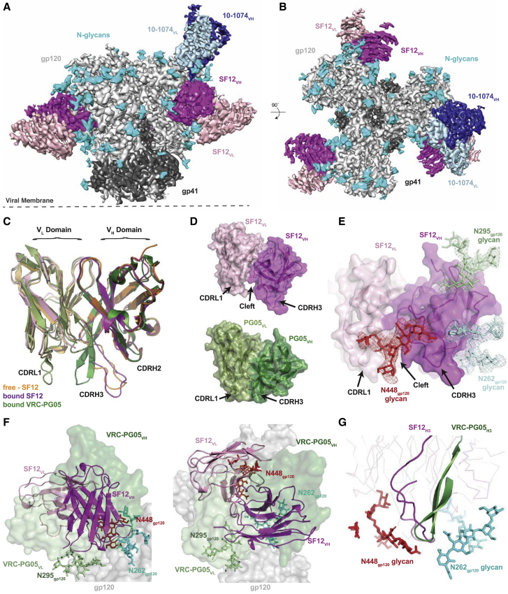
Single-particle cryo-EM data were collected on a Titan Krios transmission electron microscope (Thermo Fisher Scientific) operating at 300 kV, using the EPU automated image acquisition software (Thermo Fisher Scientific). Movies were collected on a Gatan K2 Summit direct electon detector (DED) operating in counting mode at a nominal magnification of 130,000x (1.09 Å/pixel) using a defocus range of −1.2 μm to −3.0 μm. Movies were collected over an 8 s exposure with an exposure rate of ∼4.8 e - /pixel/s, resulting in a total dose of ∼40 e - /Å 2 . Movies were motion corrected and doseweighted using the MotionCor2 frame alignment program in RELION-3 ( Zivanov et al., 2018 ). Non-doseweighted summed images were used for CTF determination using Gctf ( Zhang, 2016 ), and reference-free particle picking from 18 micrographs was achieved using Laplacian-of-Gaussian filtering in RELION-3 ( Zivanov et al., 2018 ). An initial stack of 1,907 particles was 2D classified and the best classes representing top-down and side views of Env-trimers was used for subsequent automated template-based picking in RELION-3. 676,161 particles were extracted from 2,209 dose-weighted micrographs, binned 4x4 (4.36 Å/pixel), and subjected to reference-free 2D classification in RELION-3 and a 240 Å circular mask. A total of 371,665 particles corresponding to class averages that displayed secondary-structural elements and represented views different views of Fab bound Env-trimer were extracted and re-centered prior to heterogenous ab inito model generation using cryoSPARC v2.2 ( Punjani et al., 2017 ). The generated volume was low-passed filtered to 40 Å and used as an initial model for 3D auto-refinement in RELION-3. Due to the observed low occupancy of 10-1074 Fab, particles were re-extracted unbinned (1.09 Å/pixel) and 3D classified (C1 symmetry, k = 8) with a soft mask generated from the initial model (5-pixel extension, 10-pixel soft cosine edge). Classification resulted in two distinct classes comprising three or two SF12 Fabs bound per trimer and one 10-1074 Fab bound per trimer. Particles from each class were then separately refined, followed by 3D auto-refinement using a soft mask in which Fab constant domains were masked out. Class 1 (301,920 particles) refined to a final estimaled resolution of ∼3.28 Å (SF12 3 —B41–10-1074 1 ; C1 symmetry) and class 2 (55,136 particles) refined to a final estimated resolution of ∼4.36 Å (SF12 2 —B41–10-1074 1 ; C1 symmetry) according to gold-standard FSC ( Bell et al., 2016 ). Modeling and refinement of cryo-EM structures For the final reconstruction of class 1 (SF12 3 —B41–10-1074 1 ; C1 symmetry), initial coordinates were generated by docking reference models into the cryo-EM density using UCSF Chimera v1.13 ( Goddard et al., 2007 ) (gp120-gp41, PDB 6CH9 ; 10-1074 Fab, PDB 4FQQ ; SF12 Fab, this work). Initial models were then refined into the EM maps using one round of rigid body, morphing, and simulated annealing followed by subsequent rounds of B-factor refinement in Phenix ( Adams et al., 2010 ). Models were manually built following iterative rounds of real-space and B-factor refinement in Coot ( Emsley et al., 2010 ) and Phenix ( Adams et al., 2010 ) with secondary structure restraints. Modeling of glycans was achieved by interpreting cryo-EM density at PNGS in Coot using a map with a −75 Å 2 B-factor sharpening value, contoured at 6σ due to the lower resolution of glycans at the periphery of the structure. Validation of model coordinates was performed using MolProbity ( Chen et al., 2010 ) and Privateer ( Agirre et al., 2015 ).
Structural and bioinformatic analyses
Superpositions and figures were rendered using PyMOL (Version 1.5.0.4 Schrodinger, LLC), and protein electrostatic calculations were done using APBS and PDB2PQR webservers ( Unni et al., 2011 ). Buried surface areas (BSAs) were determined with PDBePISA using a 1.4Å probe ( Krissinel and Henrick, 2007 ). Potential hydrogen bonds were assigned using a distance of < 3.6Å and an A-D-H angle of > 90°, while the maximum distance allowed for a van der Waals interaction was 4.0Å. Putative H-bonds, van der Waals assignments and total BSA should be considered tentative, owing to the relatively low structure resolutions. Computational analysis of neutralization panel data ( Table S7 ) was done as previously described ( West et al., 2013 ). For determining the difference in orientation of the antibody variable domains of the SF12-Env and VRC-PG05-Env complexes, those structures were aligned on gp120, and then the transformation relating the V H -V L domains was calculated by using TM-align ( Zhang and Skolnick, 2005 ). The corresponding screw transformation was calculated as described ( Siciliano and Khatib, 2008 ) and visualized using Antibody Database ( West et al., 2013 ).
Supplemental Information Document S1. Figures S1–S5 and Tables S1–S7 Document S2. Article plus Supplemental Information
📊 Figures
Figureu00a01
Isolation of Antibody Family from Donor 27845 by B Cell Culture and BG505 Sorting (A) Viral load and CD4 + Tu00a0cell counts of HIV-1-infected subject 27845 over time. Arrows indicate time points of B...
Figureu00a02
Antibodies SF5 and SF12 Bind a Distinct Epitope on the gp120 Portion of Env (A) ELISA of SF5 and SF12 against a gp120 monomer and a gp140 foldon trimer derived from HIV-1 strain YU2. Wild-type protein...
Figureu00a03
Structural Overview of the SF12-B41-10-1074 complex (A and B) Side-view (A) and top-view (B) of the final 3.3u00a0u00c5 single-particle cryo-EM reconstruction of the SF12-B41-10-1074 complex colored b...
Figureu00a04
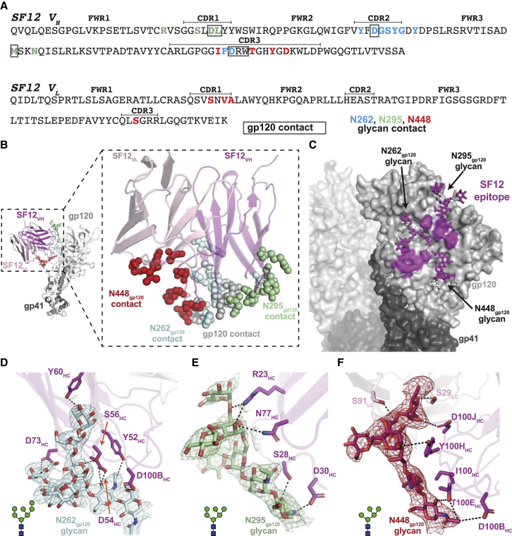
Details of SF12 Epitope and Glycan Recognition (A) Sequence of SF12 variable domains with antibody regions annotated using IMGT sequence analysis (CDR loops are bracketed). SF12 residues that contact ...
Figureu00a05
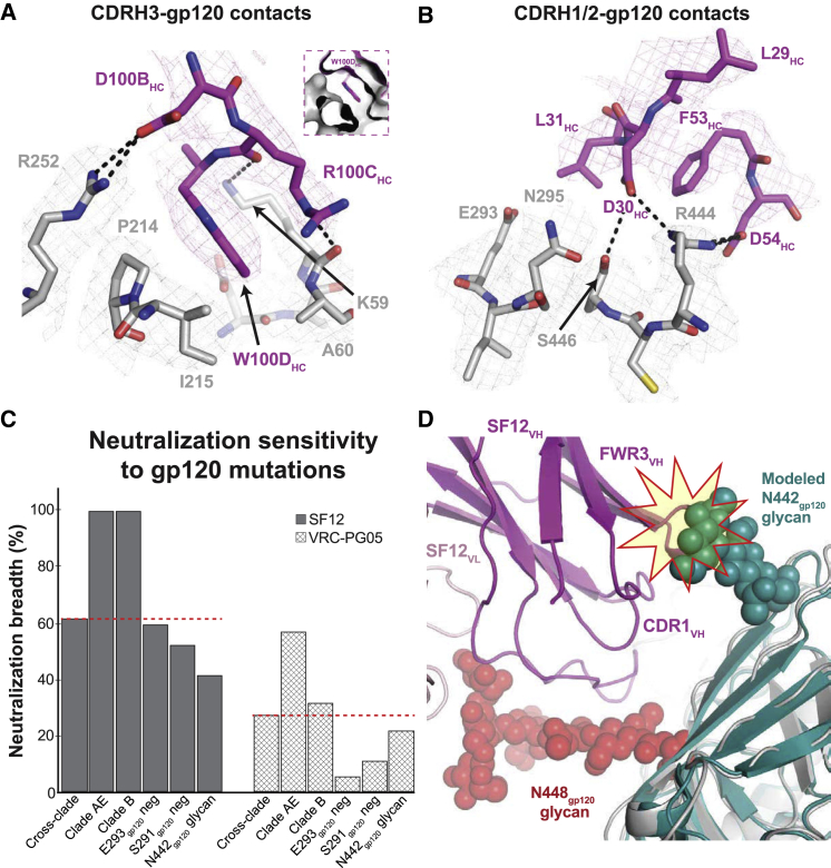
SF12 Engages Two Distinct Regions of gp120 Peptide Epitope (A) Stick representation of SF12 CDRH3 (magenta) and gp120 (gray) contacts at the SF12-Env interface. Trp100D HC inserts into a hydrophobic p...
Figureu00a06
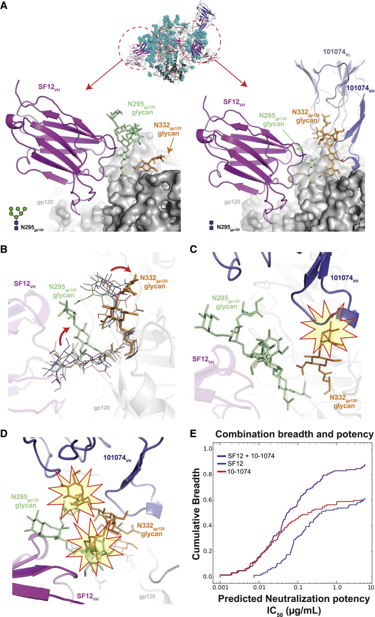
SF12-B41-10-1074 Structural Asymmetry Is Explained by N295 gp120 Glycan Heterogeneity (A) Comparison of cryo-EM density for N295 gp120 (green) and N332 gp120 (orange) glycans across protomers within t...
Figureu00a07
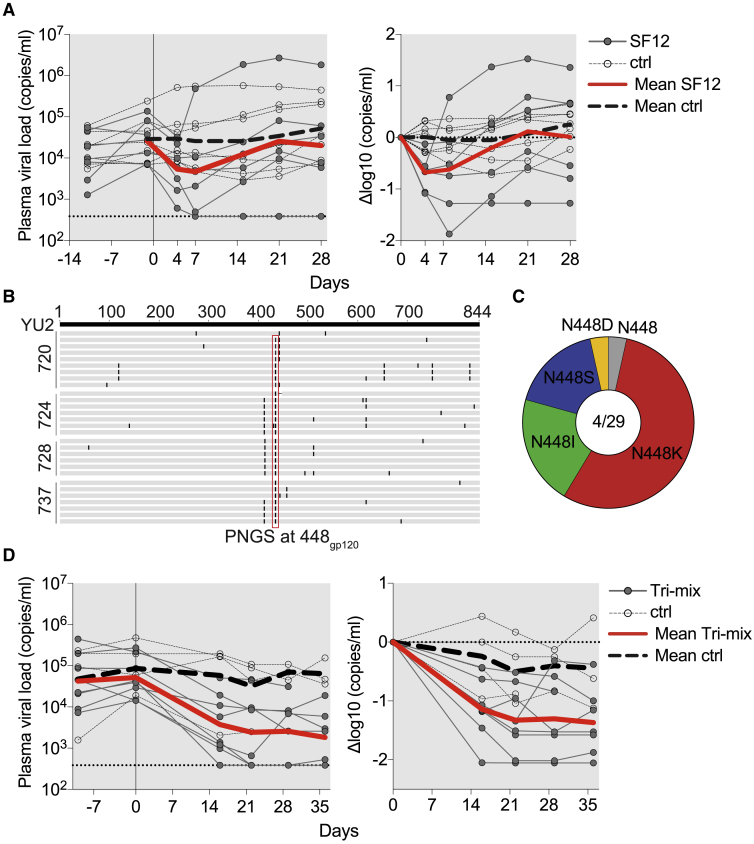
Inu00a0Vivo Evaluation of SF12 IgG in HIV YU2 -Infected Humanized Mice (A) SF12 monotherapy of humanized mice infected with HIV YU2 . The left graph shows absolute viremia (y axis) in mice treated wit...
Figure images are served from the NIH/NLM PubMed Central Open Access Subset or Europe PMC; copyright remains with the publishers and authors.
💬 Discussion
0 commentsNo comments yet. Be the first to start a discussion!
Leave a Comment