Abstract
Fluorogenic molecules are important tools for advanced biochemical and biological experiments. The extant collection of fluorogenic probes is incomplete, however, leaving regions of the electromagnetic spectrum unutilized. Here, we synthesize green-excited fluorescent and fluorogenic analogues of the classic fluorescein and rhodamine 110 fluorophores by replacement of the xanthene oxygen with a quaternary carbon. These anthracenyl "carbofluorescein" and "carborhodamine 110" fluorophores exhibit excellent fluorescent properties and can be masked with enzyme- and photolabile groups to prepare high-contrast fluorogenic molecules useful for live cell imaging experiments and super-resolution microscopy. Our divergent approach to these red-shifted dye scaffolds will enable the preparation of numerous novel fluorogenic probes with high biological utility.
🔬 Techniques
🔭 Microscopes
✨ Fluorophores
🧪 Sample Preparation
🔬 Cell Lines
🏭 Microscope Brands
🏛️ Research Organizations (ROR)
Affiliated research institutions:
📋 Methods
Chemical Synthesis
Detailed experimentals and characterization can be found in the Supporting Information . All reactions were performed under a nitrogen atmosphere in round-bottomed flasks or septum-capped crimp-top vials containing Teflon-coated magnetic stir bars. Reactions were monitored by thin layer chromatography (TLC) on precoated TLC glass plates or by LC–MS using a C18 column (ESI, positive ion mode; UV detection at 254 nm). Flash chromatography was performed on an automated purification system using prepacked silica gel columns. High-resolution mass spectrometry was performed by the Mass Spectrometry Center in the Department of Medicinal Chemistry at the University of Washington. NMR spectra were recorded on a 400 MHz spectrometer at the Janelia Farm Research Campus. 1 H and 13 C chemical shifts (δ) were referenced to TMS or residual solvent peaks, and 19 F chemical shifts (δ) were referenced to CFCl 3 .
Optical Spectroscopy and Microscopy
Spectroscopy was performed using 1-cm path length quartz cuvettes. All measurements were taken at ambient temperature (22 ± 2 °C). Absorption spectra were recorded on a Cary Model 100 spectrometer (Varian), and fluorescence spectra were recorded on a Cary Eclipse fluorometer (Varian). Absolute quantum yields (Φ) were measured using a Quantaurus-QY spectrometer (Hamamatsu). Confocal microscopy was performed on live HeLa cells using a Zeiss LSM 510 META confocal microscope. PALM imaging was performed on a custom-built instrument based on an Olympus IX81 inverted wide field microscope. Photoactivatable 32 and mEos2 fluorophores were uncaged with a 405 nm laser and excited with a 561 nm laser. AlexaFluor 647-stained samples were incubated in a redox buffer consisting of 100 mM MEA, 50 μg/mL glucose oxidase, 40 μg/mL catalase, 10% w/v glucose, pH 8.5 and excited with a 642 nm laser. Full experimental details including PALM image analysis and plotting parameters are given in the Supporting Information .
Show full methods section
Chemical Synthesis
Detailed experimentals and characterization can be found in the Supporting Information . All reactions were performed under a nitrogen atmosphere in round-bottomed flasks or septum-capped crimp-top vials containing Teflon-coated magnetic stir bars. Reactions were monitored by thin layer chromatography (TLC) on precoated TLC glass plates or by LC–MS using a C18 column (ESI, positive ion mode; UV detection at 254 nm). Flash chromatography was performed on an automated purification system using prepacked silica gel columns. High-resolution mass spectrometry was performed by the Mass Spectrometry Center in the Department of Medicinal Chemistry at the University of Washington. NMR spectra were recorded on a 400 MHz spectrometer at the Janelia Farm Research Campus. 1 H and 13 C chemical shifts (δ) were referenced to TMS or residual solvent peaks, and 19 F chemical shifts (δ) were referenced to CFCl 3 .
Optical Spectroscopy and Microscopy
Spectroscopy was performed using 1-cm path length quartz cuvettes. All measurements were taken at ambient temperature (22 ± 2 °C). Absorption spectra were recorded on a Cary Model 100 spectrometer (Varian), and fluorescence spectra were recorded on a Cary Eclipse fluorometer (Varian). Absolute quantum yields (Φ) were measured using a Quantaurus-QY spectrometer (Hamamatsu). Confocal microscopy was performed on live HeLa cells using a Zeiss LSM 510 META confocal microscope. PALM imaging was performed on a custom-built instrument based on an Olympus IX81 inverted wide field microscope. Photoactivatable 32 and mEos2 fluorophores were uncaged with a 405 nm laser and excited with a 561 nm laser. AlexaFluor 647-stained samples were incubated in a redox buffer consisting of 100 mM MEA, 50 μg/mL glucose oxidase, 40 μg/mL catalase, 10% w/v glucose, pH 8.5 and excited with a 642 nm laser. Full experimental details including PALM image analysis and plotting parameters are given in the Supporting Information .
Supplementary Material cb4000822_si_001.pdf cb4000822_si_002.pdf
📊 Figures
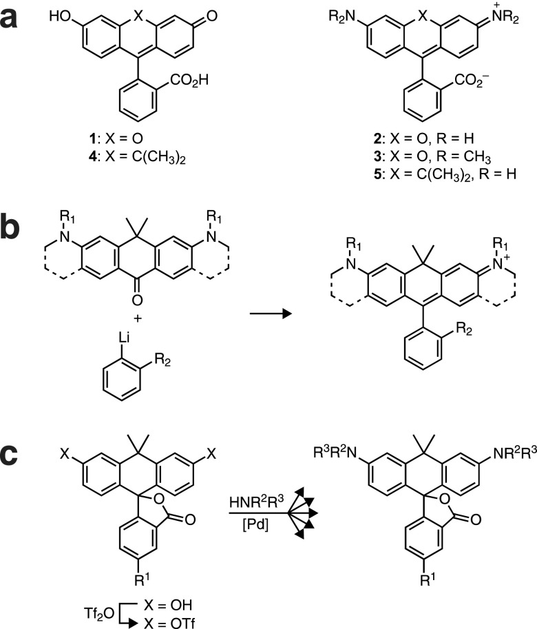
Figure 1
(a) Chemical structures of xanthene dyes and carbon-containingnisologues. (b) Existing synthetic strategy to carborhodamine dyes.n(c) Divergent synthesis of carborhodamines through carbofluoresceinnin...
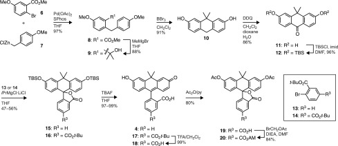
Scheme 1
Synthesis of Carbofluoresceins
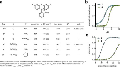
Figure 2
Properties of fluorescentndyes. (a) Spectral properties of dyes.n(b) Normalized absorbance at u03bb max versus pH for fluoresceinn( 1 ) and carbofluorescein ( 4 ). Error barsnshow standard error (SE; ...
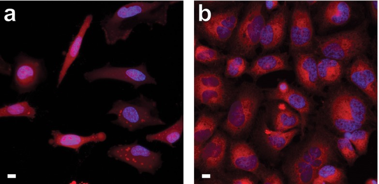
Figure 3
Confocal microscopy ofnlive, unwashed HeLa cells incubated withnesterase substrates 20 or 26 and counterstainednwith Hoechst 33342; scale bars = 10 u03bcm. (a) Compound 20 , 1 h incubation. (b) Compou...
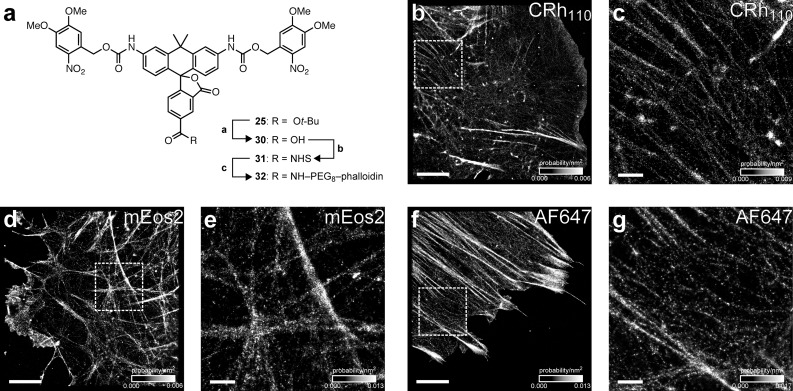
Figure 4
PALM imagingnof F-actin in fixed mouse embryonic fibroblasts. (a)nSynthesis of caged carborhodamine 110u2013phalloidin conjugate 32 . Reagents and conditions: (a) TFA/CH 2 Cl 2 , rt, 98%. (b) TSTU, DI...
Figure images are served from the NIH/NLM PubMed Central Open Access Subset or Europe PMC; copyright remains with the publishers and authors.
💬 Discussion
0 commentsNo comments yet. Be the first to start a discussion!
Leave a Comment