Abstract
Abstract Overexpression is a notorious concern in conventional and especially in super-resolution fluorescence light microscopy studies because it may cause numerous artifacts including ectopic sub-cellular localizations, erroneous formation of protein complexes and others. Nonetheless, current live cell super-resolution microscopy studies generally rely on the overexpression of a host protein fused to a fluorescent protein. Here, we establish CRISPR/Cas9-mediated generation of heterozygous and homozygous human knockin cell lines expressing fluorescently tagged proteins from their respective native genomic loci at close to endogenous levels. We tagged three different proteins, exhibiting various localizations and expression levels, with the reversibly switchable fluorescent protein rsEGFP2. We demonstrate the benefit of endogenous expression levels compared to overexpression and show that typical overexpression-induced artefacts were avoided in genome-edited cells. Fluorescence activated cell sorting analysis revealed a narrow distribution of fusion protein expression levels in genome-edited cells, compared to a pronounced variability in transiently transfected cells. Using low light intensity RESOLFT (reversible saturable optical fluorescence transitions) nanoscopy we show sub-diffraction resolution imaging of living human knockin cells. Our strategy to generate human cell lines expressing fluorescent fusion proteins at endogenous levels for RESOLFT nanoscopy can be extended to other fluorescent tags and super-resolution approaches.
🔬 Techniques
🔭 Microscopes
💻 Software
✨ Fluorophores
🧪 Sample Preparation
🔬 Cell Lines
🏭 Microscope Brands
🧪 Reagent Suppliers
📷 Detectors
💻 Software Details
💾 Data Repositories
🏛️ Research Organizations (ROR)
Affiliated research institutions:
📋 Methods
Cell culture
U2OS cells (American Type Culture Collection, Manassas, VA, USA) were cultured in Dulbecco's modified Eagle's medium (DMEM) (Invitrogen, Carlsbad, CA, USA) supplemented with 10% fetal bovine serum (PAA, Pasching, Austria), 100 units/mL penicillin, 100 μg/mL streptomycin (all Biochrom, Berlin, Germany), and 1 mM sodium pyruvate (Sigma, St. Louis, MO, USA) under constant conditions at 37°C and 5% CO 2 .
Nuclease plasmids
Design of the guide RNAs was carried out using the CRISPR Design Tool ( http://crispr.mit.edu 22 ) to minimize potential off-target effects. Oligonucleotide pairs ( Supplementary Table 1 ) were cloned into the vector pX330 17 as previously described in detail in Ref. 23 . The final bicistronic vector encoded the gRNA and the Cas9 nuclease.
Donor plasmids
DNA sequences for left homology arm
(LHA) and right homology arm (RHA) were amplified from genomic DNA using the primer pairs listed in Supplementary Table 2 . The length of the amplified homology arms was between 590 bp and 924 bp. In order to generate the Zyxin-rsEGFP2 donor plasmid, we introduced silent mutations within the Cas9 nuclease binding region of the left homology arm. To this end, a gBlock gene fragment containing the sequence was synthesized (Integrated DNA Technologies, Coralville, IA, USA). The coding sequence of rsEGFP2 was PCR amplified using the primers listed in Supplementary Table 2 . For the VIM-rsEGFP2 donor plasmid, PCR products were purified, digested with KpnI/NotI (LHA), NotI/NcoI (rsEGFP2), NcoI/SalI (RHA) and cloned into a pUC57 plasmid (Fisher Scientific, Schwerte, Germany) that was digested with KpnI/SalI by a standard four fragments ligation. For HMGA1-rsEGFP2 and ZYX-rsEGFP2, the three PCR products (and the gBlock in case of ZYX-rsEGFP2 donor) were purified and cloned into a pUC57 plasmid that was digested with EcoRV using a one-step isothermal assembly reaction 24 .
Show full methods section
Cell culture
U2OS cells (American Type Culture Collection, Manassas, VA, USA) were cultured in Dulbecco's modified Eagle's medium (DMEM) (Invitrogen, Carlsbad, CA, USA) supplemented with 10% fetal bovine serum (PAA, Pasching, Austria), 100 units/mL penicillin, 100 μg/mL streptomycin (all Biochrom, Berlin, Germany), and 1 mM sodium pyruvate (Sigma, St. Louis, MO, USA) under constant conditions at 37°C and 5% CO 2 .
Nuclease plasmids
Design of the guide RNAs was carried out using the CRISPR Design Tool ( http://crispr.mit.edu 22 ) to minimize potential off-target effects. Oligonucleotide pairs ( Supplementary Table 1 ) were cloned into the vector pX330 17 as previously described in detail in Ref. 23 . The final bicistronic vector encoded the gRNA and the Cas9 nuclease.
Donor plasmids
DNA sequences for left homology arm
(LHA) and right homology arm (RHA) were amplified from genomic DNA using the primer pairs listed in Supplementary Table 2 . The length of the amplified homology arms was between 590 bp and 924 bp. In order to generate the Zyxin-rsEGFP2 donor plasmid, we introduced silent mutations within the Cas9 nuclease binding region of the left homology arm. To this end, a gBlock gene fragment containing the sequence was synthesized (Integrated DNA Technologies, Coralville, IA, USA). The coding sequence of rsEGFP2 was PCR amplified using the primers listed in Supplementary Table 2 . For the VIM-rsEGFP2 donor plasmid, PCR products were purified, digested with KpnI/NotI (LHA), NotI/NcoI (rsEGFP2), NcoI/SalI (RHA) and cloned into a pUC57 plasmid (Fisher Scientific, Schwerte, Germany) that was digested with KpnI/SalI by a standard four fragments ligation. For HMGA1-rsEGFP2 and ZYX-rsEGFP2, the three PCR products (and the gBlock in case of ZYX-rsEGFP2 donor) were purified and cloned into a pUC57 plasmid that was digested with EcoRV using a one-step isothermal assembly reaction 24 .
Transfection and clone isolation
U2OS cells were transfected with the bicistronic nuclease plasmids and the corresponding donor plasmids using FuGENE HD transfection reagent (Promega, Mannheim, Germany). To this end, 2 × 10 5 cells per well were seeded in a 6-well plate with supplemented DMEM. The following day, transfection was carried out using a reagent to DNA ratio of 3.5 to 1 and a total DNA amount of 3 μg. Subsequently, the cells were further incubated at 37°C, 5% CO 2 . After seven days, cells were inspected by fluorescence microscopy. Wells containing cells exhibiting the expected sub-cellular localization of the rsEGFP2 fusion protein were subjected to single cell sorting into 96-well plates using a FACSAria II (BD Biosciences, Heidelberg, Germany). Within about two to three weeks after single-cell sorting, cells were split and transferred into 12-well plates containing glass cover slips. Cells expressing the fusion protein and showing the expected sub-cellular localization were identified using an epifluorescence microsope (DM6000B, Leica Microsystems, Wetzlar, Germany) equipped with an oil immersion objective (1.4 NA; 100×; Planapo; Leica) and a GFP filter cube (excitation filter: BP 470/40; emission filter: BP 525/50 nm) and were expanded for further experiments. Out-out PCR and junction PCR analysis for detection of targeted integration U2OS clonal lines that showed a specific fluorescence signal for the respective structure were further analyzed using PCR. Genomic DNA was isolated using the DNeasy Blood & Tissue Kit (QIAGEN, Hilden, Germany) from a confluent well of a 12-well plate. 100 ng genomic DNA was used as a template for an out-out PCR (primers F and R, see Fig. 1b ) or a junction PCR (primers F and GR, see Fig. 1b ) analysis with the primers listed in Supplementary Table 3 . PCR products were analyzed on a 1.5% agarose gel. Out-out PCR products of selected clonal lines were cloned into a pCR-Blunt II-TOPO vector (Invitrogen). The inserts were sequenced.
Overexpression plasmids
Cloning of overexpression constructs was carried out using the primers listed in Table Supplementary Table 4 . rsEGFP2 DNA sequences were amplified by PCR. VIM DNA was amplified from the plasmid pmKate2-vimentin (Evrogen, Moscow, Russia). HMGA1 DNA was amplified from pDONR223-HMGA1 (Human ORFeome, Internal ID: 4996). ZYX DNA was amplified from pDONR223-Zyxin (Human ORFeome, Internal ID: 4546). The respective PCR products were purified and used for one-step isothermal assembly 24 with EcoRV-digested pFLAG-CMV-5.1 (Sigma Aldrich). In this plasmid, the fusion protein expression was driven by a CMV promoter. The peptide linkers between the rsEGFP2 and the respective host protein were identical in plasmid based expression and native expression. For transfection using FuGENE HD transfection reagent (Promega), 2 × 10 5 cells per well were seeded in a 6-well plate with supplemented DMEM. The following day, transfection was carried out using a reagent to DNA ratio of 3.5 to 1 and a total DNA amount of 3 μg. Images were recorded 1 to 3 days after transfection.
Western blotting
For lysate preparation, cells (one confluent well of a 6-well plate) were washed two times in cold phosphate-buffered saline (PBS). Cells were scraped from the growth surface and resuspended in cold radioimmunoprecipitation assay (RIPA) buffer containing EDTA and complete protease inhibitor cocktail (Roche, Basel, Switzerland). After 20 min incubation on ice, the suspension was centrifuged at 13,000 rpm at 4°C for 30 min. The supernatant was removed and the protein concentration was measured using the Bradford dye-binding method (Bio-Rad, CA, USA). Samples were separated by 10% or 15% SDS-PAGE and transferred to a nitrocellulose membrane (GE Healthcare, Freiburg, Germany) in transfer buffer (25 mM Tris, 190 mM glycine, 20% methanol) over night. The membrane was rinsed in Tris-buffered saline (TBS) with 0.1% Tween 20 (TBST) and incubated in 5% blocking buffer (5 g skim milk per 100 ml TBST) at room temperature for 1 h. Primary antibodies were diluted in blocking buffer and incubated with the membrane at room temperature for 1 h. The following primary antibodies were used: anti-HMGA1 (EPR7839; 1:5000; Abcam, Cambridge, UK), anti-Vimentin (V9; 1:1000; Santa Cruz Biotechnology, Heidelberg, Germany), anti-Zyxin (ZOL301, 1:1000, Abcam), anti-Actin (AC74; 1:3000, Sigma-Aldrich), anti-GFP (JL-8; 1:3000, Clontech, Saint-Germain-en-Laye, France). After washing with TBST the membranes were incubated at room temperature for 1 h with HRP-conjugated anti-rabbit or anti-mouse secondary antibodies (Dianova, Hamburg, Germany) diluted 1:5000 in blocking buffer. After washing with TBST the membrane was incubated with Pierce ECL western blotting substrate (Fisher Scientific) and exposed to a CCD camera. Membranes were stripped using mild stripping buffer (15 g Glycine, 0.001% SDS, 0.01% Tween 20, pH 2.2) followed by the described protocol for reprobing with a different antibody.
Immunostaining and confocal imaging
U2OS cells were grown on glass coverslips in 6-well plates overnight. Vimentin staining was carried out on cold methanol (−20°C) fixed cells. For Zyxin or HMG-I staining, cells were fixed in 4% formaldehyde. After cell fixation, coverslips were incubated in 2% blocking buffer (2 g BSA per 100 ml PBS). Primary antibodies were diluted in blocking buffer and incubated with the coverslips at room temperature for 1 h. The following primary antibodies were used: anti-HMG-I (EPR7839; 1:400; Abcam), anti-Vimentin (V9; 1:100; Santa Cruz Biotechnology), anti-Zyxin (ZOL301, 1:400, Abcam). After washing in blocking buffer, KK114-coupled secondary antibodies 25 were diluted 1:50 and added for incubation at room temperature for 1 h. After three PBS washing steps, cells were mounted in Mowiol for imaging. Cells were visualized with a confocal microscope (TCS SP5, Leica) equipped with an oil objective (HCX PL APO CS 63× oil immersion objective) and a 633 nm HeNe continuous wave laser. Each image was averaged at least twice.
RESOLFT microscope
The home-built RESOLFT microscope utilized three separate beam paths for generating co-aligned focal spots: two at a wavelength of 491 nm for excitation and OFF-switching, and one at 405 nm for ON-switching. The two focal spots at 491 nm comprised: (i) a normally focused pulsed beam for reading out the fluorescence signal; (ii) a ‘doughnut-shaped’ focal intensity distribution with a central minimum (‘zero’) for OFF-switching at the focal periphery in the xy-plane, obtained by passing a continuous wave beam through a vortex phase mask (463 nm mask, vortex plate VPP-A, RPC Photonics, Rochester, NY). The two focal intensity spots were generated by two different lasers diodes: one for OFF-switching (50 mW, continuous wave, Calypso 50, Cobolt, Stockholm, Sweden) and the second (10 mW, 80–100 ps pulse width PicoQuant, Berlin, Germany) for fluorescence readout. The third focal spot, again with a regularly focused profile, was generated by a laser diode at 405 nm wavelength (30 mW, BCL-030-405-S, CrystaLaser, Reno, NV, USA) and used for the ON-switching of the fluorescent protein. An oil-immersion objective lens (HCX PC APO, 100×, 1.4 NA, oil; Leica Microsystems, Wetzlar, Germany) was used to image the different cell lines. A piezo actuator (ENV40/20, Piezosystem Jena, Jena, Germany) was used to move the objective lens along the optical axis in a range of 120 μm. A separate piezo stage (NV40, Piezosystem Jena) was implemented to translate the sample with nanometer precision in the xy-plane. The fluorescence signal was filtered by a bandpass filter (532/70 nm) and detected by an epitaxial silicon single photon avalanche diode SPAD (MPD, Bolzano, Italy); fluorescence photons were counted only when the 491 nm pulse read-out beam was switched on. The individual laser beam paths were triggered either by an acousto-optic modulator (MTS 130A3, Pegasus Optik GmbH, Wallenhorst, Germany) or by an acousto-optic tunable filter (AOTF.nC/TN, Pegasus Optik GmbH). The pulse sequence and duration were defined by a pulse generator (Model 9514, QUANTUM COMPOSERS, Bozeman, MT, USA) and triggered by a time-correlated single photon counting module (Becker & Hickl, Berlin, Germany) pixel by pixel.
RESOLFT imaging
Each image was recorded by applying a specific pulse scheme, pixel by pixel. For details on all shown images, see Supplementary Table 5 . All intensity values are referring to the light intensities in the focal plane. Image acquisition was performed with the software ImSpector.
📊 Figures
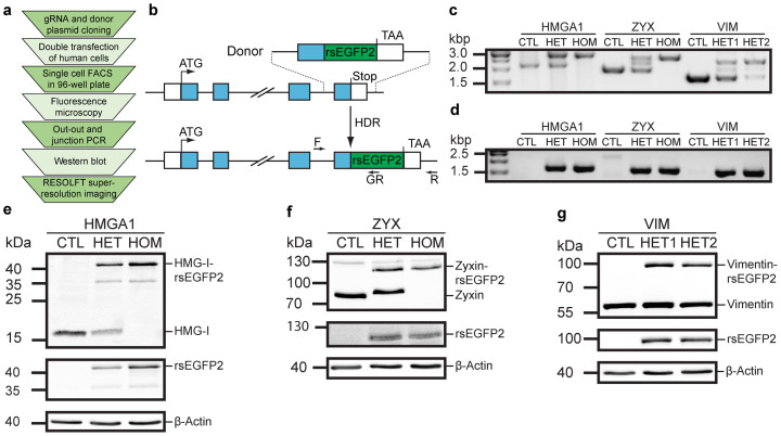
Figure 1
CRISPR/Cas9-mediated knockin of rsEGFP2 at three genomic loci in human U2OS cells.
(a) Workflow for the generation of monoclonal human knockin cell lines for RESOLFT super-resolution microscopy. (b) Schematic representation of the integration strategy for generating C-terminally tag...
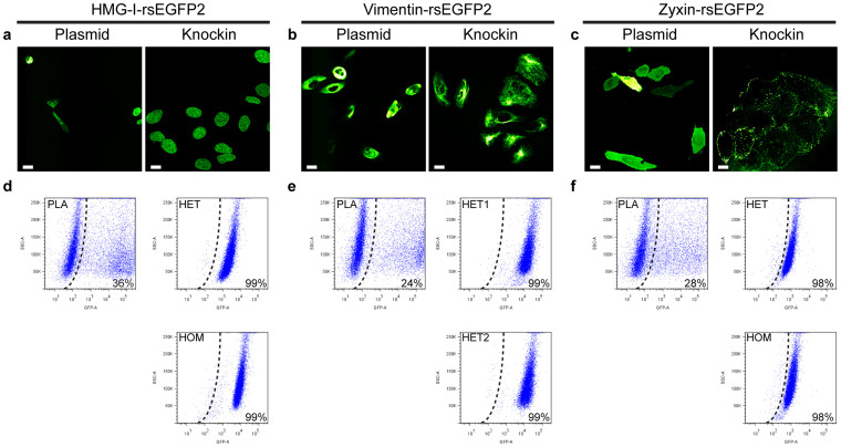
Figure 2
Expression level variability in knockin cells and upon transient expression.
(au2013c) Representative confocal images of U2OS cells expressing HMG-I-rsEGFP2 (a), Vimentin-rsEGFP2 (b) and Zyxin-rsEGFP2 (c) from a transiently transfected plasmid or from the respective native loc...
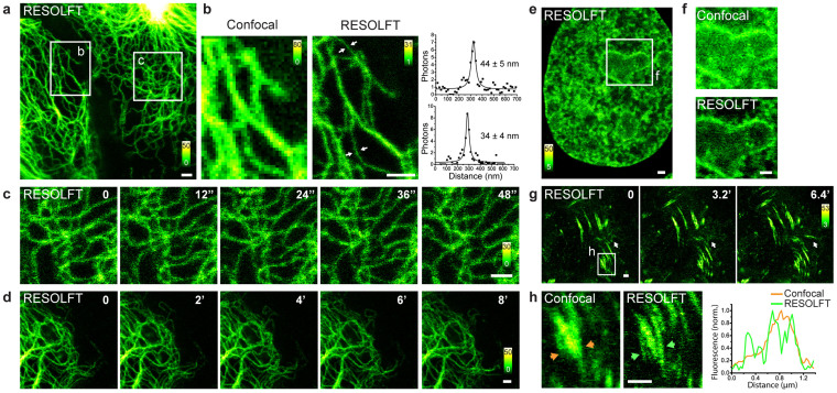
Figure 3
Live cell RESOLFT nanoscopy of CRISPR/Cas9 genome-edited rsEGFP2 knockin cells.
(au2013d) Heterozygous U2OS cells expressing Vimentin-rsEGFP2 from the endogenous locus. (a) Overview of two adjacent cells. (b) Comparison of confocal and RESOLFT images. The images have been recorde...
Figure images are served from the NIH/NLM PubMed Central Open Access Subset or Europe PMC; copyright remains with the publishers and authors.
💬 Discussion
0 commentsNo comments yet. Be the first to start a discussion!
Leave a Comment