Abstract
AbstractTick-borne encephalitis virus (TBEV) causes serious, potentially fatal neurological infections that affect humans in endemic regions of Europe and Asia. Neurons are the primary target for TBEV infection in the central nervous system. However, knowledge about this viral infection and virus-induced neuronal injury is fragmental. Here, we directly examined the pathology that occurs after TBEV infection in human primary neurons. We exploited the advantages of advanced high-pressure freezing and freeze-substitution techniques to achieve optimal preservation of infected cell architecture. Electron tomographic (ET) reconstructions elucidated high-resolution 3D images of the proliferating endoplasmic reticulum and individual tubule-like structures of different diameters in the endoplasmic reticulum cisternae of single cells. ET revealed direct connections between the tubule-like structures and viral particles in the endoplasmic reticulum. Furthermore, ET showed connections between cellular microtubules and vacuoles that harbored the TBEV virions in neuronal extensions. This study was the first to characterize the 3D topographical organization of membranous whorls and autophagic vacuoles in TBEV-infected human neurons. The functional importance of autophagy during TBEV replication was studied in human neuroblastoma cells; stimulation of autophagy resulted in significantly increased dose-dependent TBEV production, whereas the inhibition of autophagy showed a profound, dose-dependent decrease of the yield of infectious virus.
🔬 Techniques
✨ Fluorophores
🧪 Sample Preparation
🔬 Cell Lines
🏭 Microscope Brands
🧪 Reagent Suppliers
📷 Detectors
💻 Software Details
🏛️ Research Organizations (ROR)
Affiliated research institutions:
📋 Methods
Virus and cells
The virus used in this study was the TBEV strain, Neudoerfl, kindly provided by Professor F. X. Heinz from the Medical University in Vienna. The strain was originally isolated from the tick Ixodes ricinus in Austria in 1971. The virus represents a prototype strain of the European subtype of TBEV and was characterized extensively, including its genome sequence and the 3D structure of its envelope protein E 42 . The virus underwent four passages in the brains of suckling mice before use in this study. Human neurons (HNs) were isolated from the human brain and characterized by immunofluorescence with antibodies specific to neurofilament, microtubule associated protein 2 (MAP2), and β-tubulin III (purchased at passage zero from ScienCell Research Laboratories, Carlsbad, CA). HNs were propagated in Neuronal Medium (ScienCell) with 1% neuronal growth supplement and 1% penicillin/streptomycin (ScienCell) at 37 °C and 5% CO 2 . In all experiments, cells were used at passage zero. Human neuroblastoma cells UKF-NB-4 14 were cultured at 37 °C and 5% CO 2 in Iscove’s modified Dulbecco’s medium (IMDM) supplemented with 10% fetal bovine serum (Sigma) and 1% mixture of penicillin and streptomycin (Sigma). Porcine kidney stable (PS) cells 43 were cultured in L-15 medium supplemented with 3% newborn calf serum and 1% penicillin/streptomycin (Sigma-Aldrich) at 37 °C.
Viral growth in HNs
HNs were seeded onto gelatin (0.2%) coated wells of 96-well plates at 20,000–25,000 cells/cm 2 . After establishing the culture, cells were inoculated with the virus diluted in culture medium to 5 multiplicity of infection units (MOI).
Show full methods section
Virus and cells
The virus used in this study was the TBEV strain, Neudoerfl, kindly provided by Professor F. X. Heinz from the Medical University in Vienna. The strain was originally isolated from the tick Ixodes ricinus in Austria in 1971. The virus represents a prototype strain of the European subtype of TBEV and was characterized extensively, including its genome sequence and the 3D structure of its envelope protein E 42 . The virus underwent four passages in the brains of suckling mice before use in this study. Human neurons (HNs) were isolated from the human brain and characterized by immunofluorescence with antibodies specific to neurofilament, microtubule associated protein 2 (MAP2), and β-tubulin III (purchased at passage zero from ScienCell Research Laboratories, Carlsbad, CA). HNs were propagated in Neuronal Medium (ScienCell) with 1% neuronal growth supplement and 1% penicillin/streptomycin (ScienCell) at 37 °C and 5% CO 2 . In all experiments, cells were used at passage zero. Human neuroblastoma cells UKF-NB-4 14 were cultured at 37 °C and 5% CO 2 in Iscove’s modified Dulbecco’s medium (IMDM) supplemented with 10% fetal bovine serum (Sigma) and 1% mixture of penicillin and streptomycin (Sigma). Porcine kidney stable (PS) cells 43 were cultured in L-15 medium supplemented with 3% newborn calf serum and 1% penicillin/streptomycin (Sigma-Aldrich) at 37 °C.
Viral growth in HNs
HNs were seeded onto gelatin (0.2%) coated wells of 96-well plates at 20,000–25,000 cells/cm 2 . After establishing the culture, cells were inoculated with the virus diluted in culture medium to 5 multiplicity of infection units (MOI).
Virus-mediated cytopathic effect
(CPE) was investigated with light microscopy. At 0, 3, 5, 7, and 12 days p.i., supernatant medium from appropriate wells was collected and frozen at −70 °C. Viral titers were determined by plaque assay.
Plaque assay
PS cells were used to determine virus titers according to a protocol described previously, with minor modifications 44 . Tenfold dilutions of the virus samples were placed in 24-well tissue culture plates, and suspended PS cells were added (5×10 4 cells per well). After incubating for 4 h, the suspension was overlaid with carboxymethylcellulose (1.5% in L-15 medium). After incubating for 5 days at 37 °C, the plates were washed with PBS, and the cells were stained with naphthalene black (Sigma Aldrich). Virus titer was expressed in units of pfu/ml.
Immunofluorescence staining TBEV-infected and control
HNs were grown on slides. Then, cells were fixed in 4% formaldehyde for 1 h at room temperature, rinsed three times in 0.1 M phosphate buffer (PB) with 0.02 M glycine, permeabilized with 0.1% Triton X-100 with 1% normal goat serum in 0.1 M PB for 30 min, and blocked with 5% normal goat serum. Cells were labeled with flavivirus-specific mouse mAb (1:250; Millipore) for 1.5 h at 37 °C. Flavivirus-specific mAb and rabbit anti-PDIA3 antibody (1:250, Sigma-Aldrich) were used for double labeling. After washing with ten–fold diluted blocking solution, the cells were labeled with goat anti-mouse secondary antibody conjugated with FITC (1:500, Sigma-Aldrich) or goat anti-rabbit secondary antibody conjugated with Atto 550 NHS (1:500, Sigma-Aldrich) for 1.5 h at 37 °C. The cells were counterstained with DAPI (1 μg/ml, Sigma-Aldrich) for 10 min at 37 °C, mounted in 2.5% 1,4-diazabicyclo(2.2.2)octane (Sigma-Aldrich), and examined with an Olympus BX-51 fluorescence microscope equipped with an Olympus DP-70 CCD camera. Confocal microscopy was performed with an Olympus FV-1000; serial Z-series images were acquired in blue, red, and green channels.
Transmission electron microscopy and electron tomography
TBEV-infected and control HNs were grown on sapphire discs. At either 3 or 12 days p.i., cells were high-pressure frozen in the presence of 20% BSA diluted in Neuronal Medium with a Leica EM PACT2 high-pressure freezer. Freeze substitution was carried out in 2% osmium tetroxide diluted in 100% acetone, as described previously 19 , with a Leica EM AFS2 at −90 °C for 16 h. The samples were than warmed at a rate of 5 °C/h, incubated at −20 °C for 14 h, and finally warmed again at the same rate to a final temperature of 4 °C. The samples were rinsed three times in anhydrous acetone at room temperature and infiltrated stepwise in acetone mixed with SPI-pon resin (SPI) (acetone:SPI ratios of 2:1, 1:1, 1:2, for 1 h at each step). The samples in pure resin were polymerized at 60 °C for 48 h. Sections were prepared with a Leica Ultracut UCT microtome and collected on 300 mesh cooper grids. Staining was performed with alcoholic uranic acetate for 30 min, and then, lead citrate for 20 min. Images were obtained with a JEOL 2100F or JEOL 1010 transmission electron microscope. For electron tomography, protein A-conjugated 10 nm gold nanoparticles (Aurion) were added to both sides of each section as fiducial markers. Tilt series images were collected in the range of ±60° to 65°, with 0.6° to 1° increments. Images were acquired with a 200 kV JEOL 2100F transmission electron microscope equipped with a high-tilt stage and a Gatan camera (Orius SC 1000) and controlled with SerialEM automated acquisition software 45 . Images were aligned based on the fiducial markers. Electron tomograms were reconstructed with the IMOD software package. Manual masking of the area of interest was employed to generate 3D surface models 46 . Autophagy stimulation and inhibition, and microtubule disruption Stocks of rapamycin (Sigma-Aldrich) as an autophagy stimulator, spautin-1 (Sigma-Aldrich), an autophagy inhibitor, and nocodazole (Sigma-Aldrich) as a microtubule disruptor, were prepared in DMSO. Monolayers of human neuroblastoma cells in 96-well plates were pretreated with different concentrations of either drug (or DMSO as a negative control) for 30 min at 37 °C and infected with TBEV at a multiplicity of infection of 0.1 pfu per cell. Infection was always performed in triplicate. Supernatants were harvested at 24, 48, and 72 hours p.i. and titers were determined by plaque assay as described above.
Statistical analysis
Statistical analyses were performed using version 5.04 of the GraphPad Prism5 (GraphPad Software, Inc., USA). Data were transformed by use of the X’ = log(X) formula and analyzed using one-way ANOVA (Dunnett’s Multiple Comparison Test). p-values < 0.05 were considered significant.
Supplementary Material Supplementary Information Supplementary Movie S1 Supplementary Movie S2 Supplementary Movie S3 Supplementary Movie S4 Supplementary Movie S5 Supplementary Movie S6 Supplementary Movie S7 Supplementary Movie S8 Supplementary Movie S9
📊 Figures
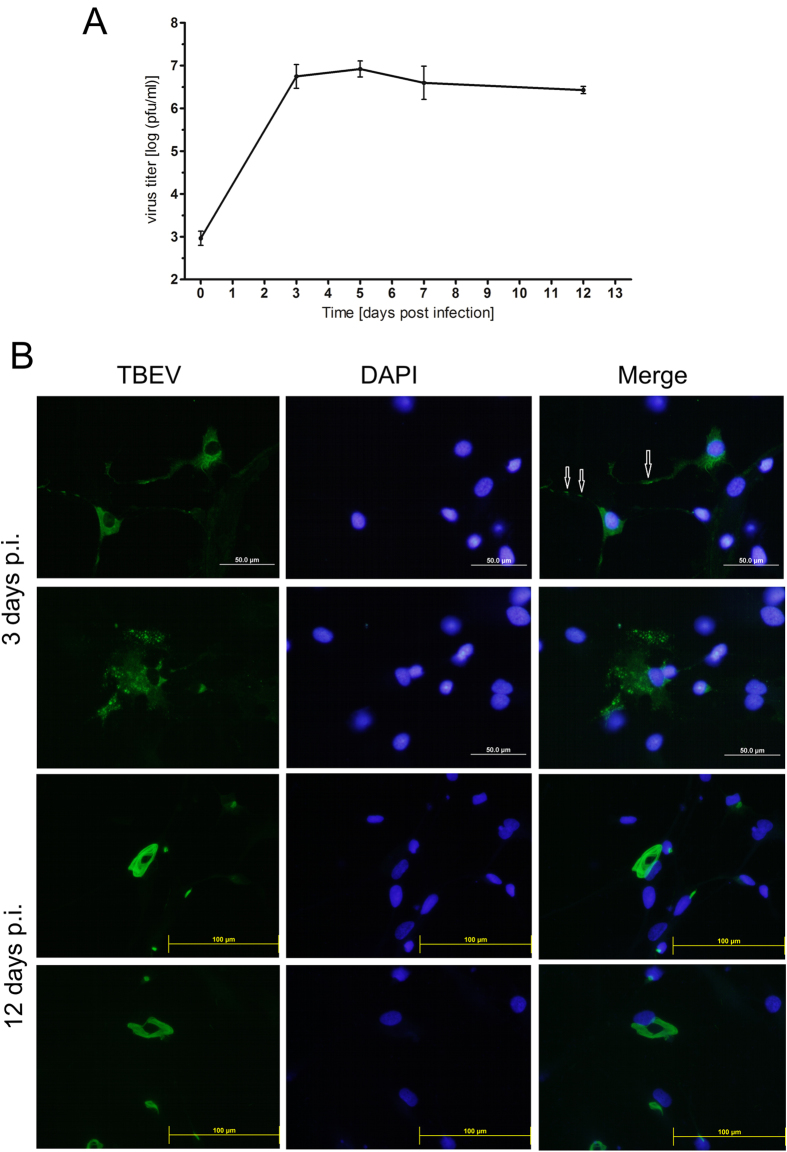
Figure 1
TBEV can infect human neurons.
( A ) TBEV titers in culture supernatants from HNs collected at 0, 3, 5, 7, and 12 days post-infection (p.i.) were determined in plaque assays with porcine kidney stable cells. Viral titers are expres...
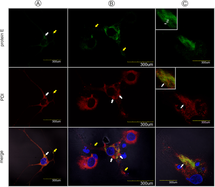
Figure 2
Confocal images of human neurons infected with TBEV.
Neurons were fixed at ( A , B ) 3 days p.i. and ( C ) 12 days p.i. and double immunolabeled with antibodies against TBEV protein E (green) ( top panels ), PDIA3 (red) ( middle panels ), and ( merge pa...
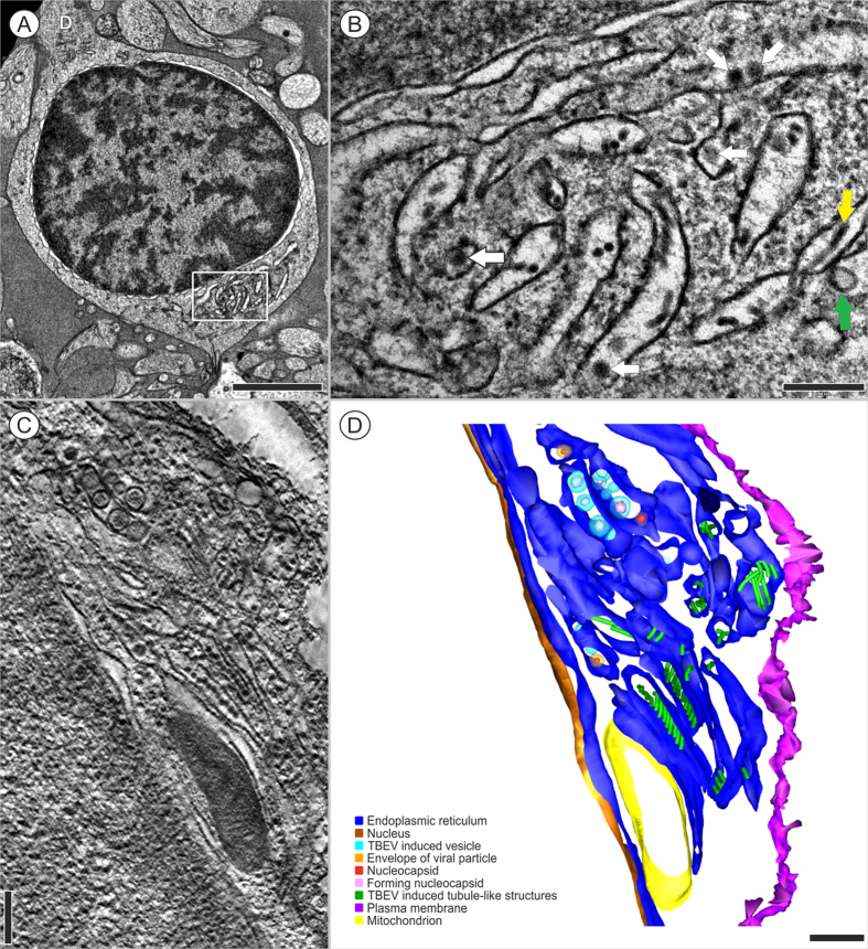
Figure 3
HNs infected with TBEV for 3 days.
( A ) A neuron with two cytoplasmic extensions on opposite sides of the cell body. D- Dendrite. ( B ) Detail of the boxed region in ( A ) shows the RER, which contains viral particles (44.75u2009nm, n...
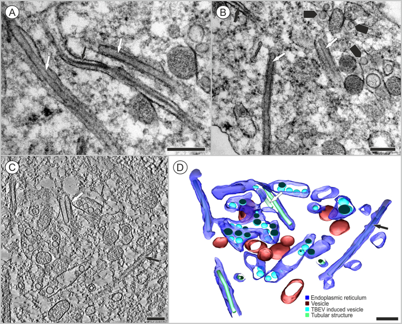
Figure 4
Tubule-like structures of different diameters were localized inside a single neuron infected with TBEV.
Transmission electron microscope images were acquired at ( A ) 3 days p.i. or ( B ) 12 days p.i. ( A ) Inside the ER, tubules of 43.8u2009u00b1u20094.3u2009nm (Nu2009=u20097) in diameter (white arrows...
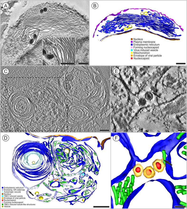
Figure 5
Proliferation of the endoplasmic reticulum observed in HNs infected with TBEV.
Transmission electron microscope images were acquired at ( A , B ) 3 days p.i. and ( C u2013 E ) 12 days p.i. ( A ) TBEV particles and TBEV-induced vesicles are located inside the proliferated and reo...
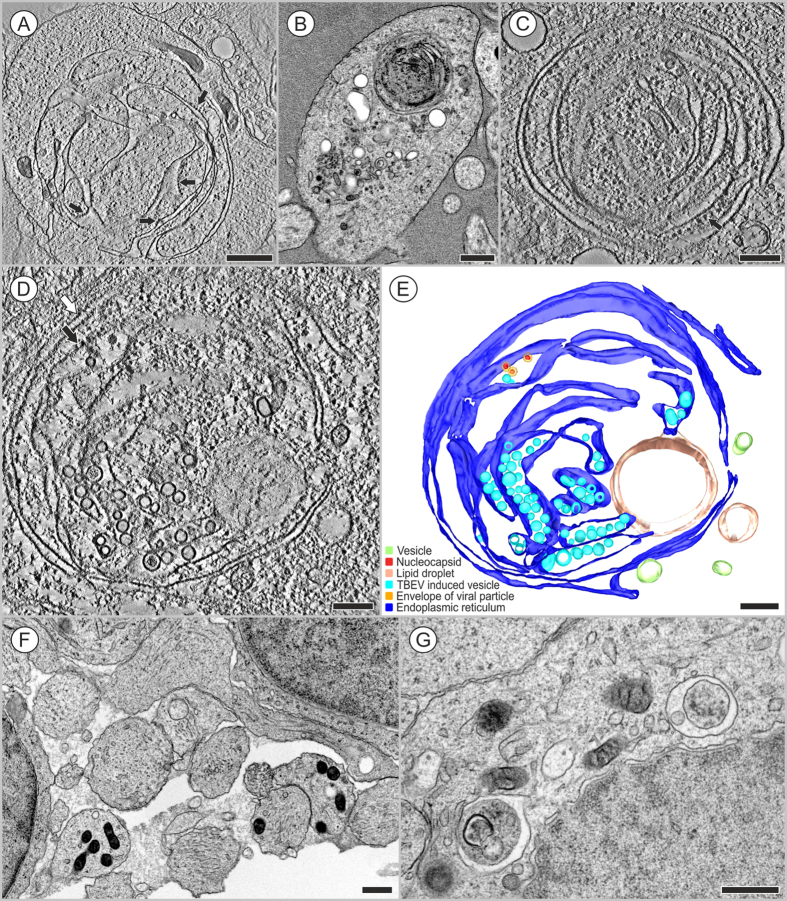
Figure 6
Formation of autophagic vacuoles in HNs infected with TBEV.
Transmission electron microscope images were acquired at ( A u2013 C ) 3 days p.i., ( D , E ) 12 days p.i., and in ( F , G ) mock-infected cells. ( A , B ) The RER (ribosomes are indicated with black ...
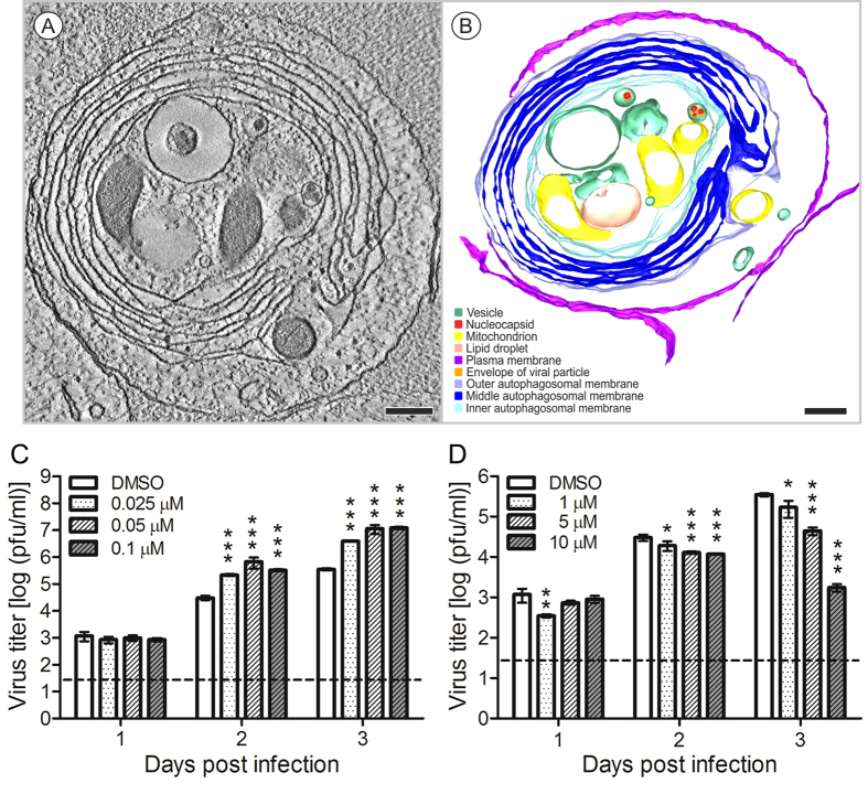
Figure 7
An autophagosome in a neural extension observed 3 days after TBEV infection.
The cytoplasm contains mitochondria and TBEV particles in vacuoles, encircled by several double membranous structures, apparently of RER origin (but devoid of ribosomes). ( A ) A slice of a single axi...
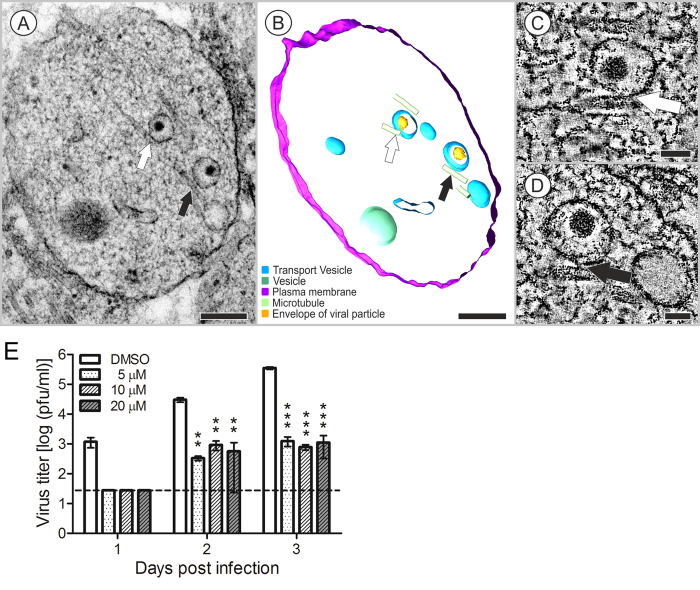
Figure 8
Two vacuoles that accommodated TBEV particles in a neuronal extension at 12 days after infection.
( A u2013 D ) Arrows indicate connections between vacuoles and microtubules. ( A ) The projection image and ( B ) the 3D model. ( C , D ) Images show slices of a double-axis tomogram acquired with a u...
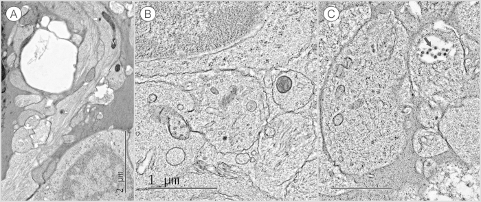
Figure 9
Mock infected HNs examined at 3 days p.i.
( A , B ) HN extensions contained a few probable secretion granules with electron-dense cores, with diameters of ( A ) about 70u2009nm and ( B ) 90u2009nm, that were in direct contact with the cytopla...
Figure images are served from the NIH/NLM PubMed Central Open Access Subset or Europe PMC; copyright remains with the publishers and authors.
💬 Discussion
0 commentsNo comments yet. Be the first to start a discussion!
Leave a Comment