Abstract
Fusion of HIV-1 with the membrane of its target cell, an obligate first step in virus infectivity, is mediated by binding of the viral envelope (Env) spike protein to its receptors, CD4 and CCR5/CXCR4, on the cell surface. The process of viral fusion appears to be fast compared with viral egress and has not been visualized by EM. To capture fusion events, the process must be curtailed by trapping Env-receptor binding at an intermediate stage. We have used fusion inhibitors to trap HIV-1 virions attached to target cells by Envs in an extended pre-hairpin intermediate state. Electron tomography revealed HIV-1 virions bound to TZM-bl cells by 2-4 narrow spokes, with slightly more spokes present when evaluated with mutant virions that lacked the Env cytoplasmic tail. These results represent the first direct visualization of the hypothesized pre-hairpin intermediate of HIV-1 Env and improve our understanding of Env-mediated HIV-1 fusion and infection of host cells.
🔬 Techniques
✨ Fluorophores
🧪 Sample Preparation
🔬 Cell Lines
🏭 Microscope Brands
🧪 Reagent Suppliers
📷 Detectors
💻 Software Details
💾 Data Repositories
🏛️ Research Organizations (ROR)
Affiliated research institutions:
📋 Methods
Experimental design
A previous study used ET to visualize HIV-1 and SIV virions in contact with target cells after promoting a temperature-arrested state ( Mkrtchyan et al., 2005 ) in which viruses can remain attached to cells prior to fusion ( Sougrat et al., 2007 ). For that study, target cells were incubated with virus at 4°C to allow binding but not fusion, warmed to 37°C, and then fixed after incubations ranging from 15 min to 3 hr ( Sougrat et al., 2007 ). At all time points after warming, viruses were found attached to target cells by a cluster of 5–7 ‘rods,’ each ~100 Å long and ~100 Å wide. The fact that the attachment structure was not found when the viruses and target cells were incubated in the presence of C34, a gp41 N-trimer–targeting C-peptide inhibitor related to T20 ( Sougrat et al., 2007 ), suggests that the rod structure that was trapped during the temperature-arrested state did not involve the pre-hairpin intermediate. We hypothesized that addition of an HIV-1 fusion inhibitor that binds to the exposed gp41 N-trimer after host cell receptor and coreceptor binding would slow or stop virus-host cell membrane fusion such that we could visualize pre-hairpin intermediate structures by ET ( Figure 1b ). We characterized three fusion inhibitors of different sizes and potencies that target the exposed gp41 N-trimer region of the pre-hairpin intermediate for attempts to visualize the pre-hairpin intermediate: T1249-Fc, a C-peptide–based inhibitor that we linked to human Fc (MW = 65 kDa), D5 IgG (MW = 150 kDa) ( Miller et al., 2005 ), and CPT31, a high-affinity D-peptide inhibitor linked to cholesterol (MW = 9 kDa) ( Redman et al., 2018 ; Welch et al., 2010 ; Figure 1a ; Figure 1—figure supplement 1a ). We measured their neutralization potencies using in vitro HIV-1 pseudovirus neutralization assays ( Montefiori, 2009 ) against the SC4226618 and 6535 viral strains. We found potencies ranging from 50% inhibitory concentration (IC 50 ) values of ~0.13 ng/mL for CPT31 to ≥40 µg/mL for D5 IgG ( Figure 1—figure supplement 1a ). T1249-Fc exhibited intermediate potencies (IC 50 s = 0.99 µg/mL; 17 µg/mL) ( Figure 1—figure supplement 1a ), higher than IC 50 s measured for T1249 peptide alone, consistent with limited steric accessibility resulting in decreased potencies for larger fusion inhibitors ( Hamburger et al., 2005 ). Incubating with a fusion inhibitor at 37°C obviated the need for a 4°C incubation of virus and target cells, which we reasoned was desirable since low temperatures alter membrane fluidity ( Avery et al., 1995 ; Simons and Vaz, 2004 ; Quinn, 1988 ), which could affect one or more steps in membrane fusion. Since target cells for HIV-1 are several microns in height, much thicker than the 0.5–1 µm limit for cryo-ET ( Beck and Baumeister, 2016 ), we used stained, plastic-embedded samples that could be cut into 300–400 nm sections using a microtome, and then examined the samples in 3-D using ET. Although ET of stained, plastic-embedded sections results in lower resolution than cryo-ET, the minimal effects of radiation damage in plastic sections ( Glaeser, 2016 ) was an advantage for locating rare attached virions. Thus, more cells could be assayed in plastic sections than in samples prepared by cryo-ET methods (e.g. by examining thin leading edges of cells or using focused-ion-beam milling [ Villa et al., 2013 ] to prepare a sufficiently thin sample), therefore allowing for statistically significant observations of virion attachment events. We prepared samples by light fixation followed by high-pressure freezing/freeze substitution fixation (HPF-FSF) instead of the traditional chemical fixation protocol used previously ( Sougrat et al., 2007 ) because HPF vitrifies cells at ~10,000°/s, stopping all cellular movement within ms and allowing optimal preservation of ultrastructural features ( Kellenberger, 1991 ; McIntosh et al., 2005 ; Sartori et al., 1993 ; Dahl and Staehelin, 1989 ). By contrast, chemical fixation immobilizes elements in the cell at different rates, and movement and rearrangement of transmembrane proteins may continue even in the presence of aldehyde fixatives ( Brock et al., 1999 ; Stanly et al., 2016 ; Tanaka et al., 2010 ). Following HPF-FSF, samples were plastic embedded and stained with uranyl acetate and lead citrate as described in our previous ET studies of HIV-1 in infected tissues ( Kieffer et al., 2017b ; Ladinsky et al., 2019 ; Ladinsky et al., 2014 ). Since biosafety requirements for the current study necessitated the use of HIV-1 pseudoviruses instead of infectious HIV-1, we verified that the ultrastructure of HIV-1 pseudoviruses, including approximate numbers and dimensions of Env trimer spikes and the presence of collapsed (in mature virions) versus C-shaped (in immature virions) cores ( Carlson et al., 2008 ; Benjamin et al., 2005 ; Ganser, 1999 ; Wright et al., 2007 ), was preserved during the fixation, embedding, and staining procedures ( Figure 2—figure supplement 1a ) consistent with our previous publications involving ET of infectious HIV-1 in tissue samples ( Kieffer et al., 2017b ; Ladinsky et al., 2019 ; Ladinsky et al., 2014 ). These results are also consistent with previous direct comparisons of tomograms of stained and plastic-embedded versus unstained and cryopreserved SIV virions ( Sougrat et al., 2007 ). We conducted ET experiments by first incubating TZM-bl cells, a HeLa cell line that stably expresses high levels of human CD4 and coreceptors CCR5 and CXCR4 ( Platt et al., 1998 ), with 130 µg/mL of inhibitor (either T1249-Fc, D5, or CPT31) and ~5000 TCID 50 /mL of HIV-1 pseudovirus at 37 °C for 2, 4, or 48 hr, followed by HPF, FSF, plastic embedding, sectioning, and visualization by ET. In order to verify that results were not dependent upon a particular viral strain, we used pseudoviruses derived from two primary isolate HIV-1 strains: SC4226618 (Tier 2) and 6535 (Tier 1B) ( Li et al., 2005 ), chosen for their sensitivity to the fusion inhibitors and because we had both wild-type and Env cytoplasmic tail-deleted forms of the 6535 pseudovirus ( Figure 1—figure supplement 1a ). TZM-bl cells are contaminated with ecotropic murine leukemia virus ( Takeuchi et al., 2008 ), which does not affect their use for HIV-1 in vitro neutralization assays ( Platt et al., 2009 ). In our surveys of TZM-bl cells incubated in the presence or absence of fusion inhibitors, we occasionally observed budding MLV virions ( Figure 2—figure supplement 1b ). As MLV serves as a control for non-specific inhibition in TZM-bl–based HIV-1 in vitro neutralization assays ( Montefiori, 2005 ), the fusion inhibitors used in our experiments are known to have no effect on MLV fusion, thus contaminating MLV virions were not captured during fusion in our experiments. To identify attached virions by EM ( Figure 2 ; Figure 2—figure supplement 1 ), the peripheries of TZM-bl cells were scanned to locate roughly spherical objects with diameters ~ 100 nm that were near a cell surface. Regions of interest were then examined at higher magnification and at tilts of 0°, 35° and −35° to verify that the objects were spherical, as expected for an HIV-1 virion ( Figure 2—figure supplement 1c ). Potential virions were then observed through a defocus series to detect core structures found inside authentic virions: that is a bullet-shaped core in mature HIV-1 and a C-shaped core in immature HIV-1 ( Benjamin et al., 2005 ; Wright et al., 2007 ). Once verified as a virion, tilt series for 3-D reconstructions were collected. Control experiments in which pseudovirus and TZM-bl cells were incubated without inhibitor, with an irrelevant IgG, or with a low concentration of inhibitor, were prepared and analyzed in the same way ( Figure 2—figure supplement 1d ). Figure 2.
Show full methods section
Experimental design
A previous study used ET to visualize HIV-1 and SIV virions in contact with target cells after promoting a temperature-arrested state ( Mkrtchyan et al., 2005 ) in which viruses can remain attached to cells prior to fusion ( Sougrat et al., 2007 ). For that study, target cells were incubated with virus at 4°C to allow binding but not fusion, warmed to 37°C, and then fixed after incubations ranging from 15 min to 3 hr ( Sougrat et al., 2007 ). At all time points after warming, viruses were found attached to target cells by a cluster of 5–7 ‘rods,’ each ~100 Å long and ~100 Å wide. The fact that the attachment structure was not found when the viruses and target cells were incubated in the presence of C34, a gp41 N-trimer–targeting C-peptide inhibitor related to T20 ( Sougrat et al., 2007 ), suggests that the rod structure that was trapped during the temperature-arrested state did not involve the pre-hairpin intermediate. We hypothesized that addition of an HIV-1 fusion inhibitor that binds to the exposed gp41 N-trimer after host cell receptor and coreceptor binding would slow or stop virus-host cell membrane fusion such that we could visualize pre-hairpin intermediate structures by ET ( Figure 1b ). We characterized three fusion inhibitors of different sizes and potencies that target the exposed gp41 N-trimer region of the pre-hairpin intermediate for attempts to visualize the pre-hairpin intermediate: T1249-Fc, a C-peptide–based inhibitor that we linked to human Fc (MW = 65 kDa), D5 IgG (MW = 150 kDa) ( Miller et al., 2005 ), and CPT31, a high-affinity D-peptide inhibitor linked to cholesterol (MW = 9 kDa) ( Redman et al., 2018 ; Welch et al., 2010 ; Figure 1a ; Figure 1—figure supplement 1a ). We measured their neutralization potencies using in vitro HIV-1 pseudovirus neutralization assays ( Montefiori, 2009 ) against the SC4226618 and 6535 viral strains. We found potencies ranging from 50% inhibitory concentration (IC 50 ) values of ~0.13 ng/mL for CPT31 to ≥40 µg/mL for D5 IgG ( Figure 1—figure supplement 1a ). T1249-Fc exhibited intermediate potencies (IC 50 s = 0.99 µg/mL; 17 µg/mL) ( Figure 1—figure supplement 1a ), higher than IC 50 s measured for T1249 peptide alone, consistent with limited steric accessibility resulting in decreased potencies for larger fusion inhibitors ( Hamburger et al., 2005 ). Incubating with a fusion inhibitor at 37°C obviated the need for a 4°C incubation of virus and target cells, which we reasoned was desirable since low temperatures alter membrane fluidity ( Avery et al., 1995 ; Simons and Vaz, 2004 ; Quinn, 1988 ), which could affect one or more steps in membrane fusion. Since target cells for HIV-1 are several microns in height, much thicker than the 0.5–1 µm limit for cryo-ET ( Beck and Baumeister, 2016 ), we used stained, plastic-embedded samples that could be cut into 300–400 nm sections using a microtome, and then examined the samples in 3-D using ET. Although ET of stained, plastic-embedded sections results in lower resolution than cryo-ET, the minimal effects of radiation damage in plastic sections ( Glaeser, 2016 ) was an advantage for locating rare attached virions. Thus, more cells could be assayed in plastic sections than in samples prepared by cryo-ET methods (e.g. by examining thin leading edges of cells or using focused-ion-beam milling [ Villa et al., 2013 ] to prepare a sufficiently thin sample), therefore allowing for statistically significant observations of virion attachment events. We prepared samples by light fixation followed by high-pressure freezing/freeze substitution fixation (HPF-FSF) instead of the traditional chemical fixation protocol used previously ( Sougrat et al., 2007 ) because HPF vitrifies cells at ~10,000°/s, stopping all cellular movement within ms and allowing optimal preservation of ultrastructural features ( Kellenberger, 1991 ; McIntosh et al., 2005 ; Sartori et al., 1993 ; Dahl and Staehelin, 1989 ). By contrast, chemical fixation immobilizes elements in the cell at different rates, and movement and rearrangement of transmembrane proteins may continue even in the presence of aldehyde fixatives ( Brock et al., 1999 ; Stanly et al., 2016 ; Tanaka et al., 2010 ). Following HPF-FSF, samples were plastic embedded and stained with uranyl acetate and lead citrate as described in our previous ET studies of HIV-1 in infected tissues ( Kieffer et al., 2017b ; Ladinsky et al., 2019 ; Ladinsky et al., 2014 ). Since biosafety requirements for the current study necessitated the use of HIV-1 pseudoviruses instead of infectious HIV-1, we verified that the ultrastructure of HIV-1 pseudoviruses, including approximate numbers and dimensions of Env trimer spikes and the presence of collapsed (in mature virions) versus C-shaped (in immature virions) cores ( Carlson et al., 2008 ; Benjamin et al., 2005 ; Ganser, 1999 ; Wright et al., 2007 ), was preserved during the fixation, embedding, and staining procedures ( Figure 2—figure supplement 1a ) consistent with our previous publications involving ET of infectious HIV-1 in tissue samples ( Kieffer et al., 2017b ; Ladinsky et al., 2019 ; Ladinsky et al., 2014 ). These results are also consistent with previous direct comparisons of tomograms of stained and plastic-embedded versus unstained and cryopreserved SIV virions ( Sougrat et al., 2007 ). We conducted ET experiments by first incubating TZM-bl cells, a HeLa cell line that stably expresses high levels of human CD4 and coreceptors CCR5 and CXCR4 ( Platt et al., 1998 ), with 130 µg/mL of inhibitor (either T1249-Fc, D5, or CPT31) and ~5000 TCID 50 /mL of HIV-1 pseudovirus at 37 °C for 2, 4, or 48 hr, followed by HPF, FSF, plastic embedding, sectioning, and visualization by ET. In order to verify that results were not dependent upon a particular viral strain, we used pseudoviruses derived from two primary isolate HIV-1 strains: SC4226618 (Tier 2) and 6535 (Tier 1B) ( Li et al., 2005 ), chosen for their sensitivity to the fusion inhibitors and because we had both wild-type and Env cytoplasmic tail-deleted forms of the 6535 pseudovirus ( Figure 1—figure supplement 1a ). TZM-bl cells are contaminated with ecotropic murine leukemia virus ( Takeuchi et al., 2008 ), which does not affect their use for HIV-1 in vitro neutralization assays ( Platt et al., 2009 ). In our surveys of TZM-bl cells incubated in the presence or absence of fusion inhibitors, we occasionally observed budding MLV virions ( Figure 2—figure supplement 1b ). As MLV serves as a control for non-specific inhibition in TZM-bl–based HIV-1 in vitro neutralization assays ( Montefiori, 2005 ), the fusion inhibitors used in our experiments are known to have no effect on MLV fusion, thus contaminating MLV virions were not captured during fusion in our experiments. To identify attached virions by EM ( Figure 2 ; Figure 2—figure supplement 1 ), the peripheries of TZM-bl cells were scanned to locate roughly spherical objects with diameters ~ 100 nm that were near a cell surface. Regions of interest were then examined at higher magnification and at tilts of 0°, 35° and −35° to verify that the objects were spherical, as expected for an HIV-1 virion ( Figure 2—figure supplement 1c ). Potential virions were then observed through a defocus series to detect core structures found inside authentic virions: that is a bullet-shaped core in mature HIV-1 and a C-shaped core in immature HIV-1 ( Benjamin et al., 2005 ; Wright et al., 2007 ). Once verified as a virion, tilt series for 3-D reconstructions were collected. Control experiments in which pseudovirus and TZM-bl cells were incubated without inhibitor, with an irrelevant IgG, or with a low concentration of inhibitor, were prepared and analyzed in the same way ( Figure 2—figure supplement 1d ). Figure 2.
Identification of attached
HIV-1 virions. ( a ) Montaged projection overview of a field of cultured TZM-bl cells from a 400 nm section. Note extensive blebbing and surface projections that are typical of the cell type. Inset: Projection detail of a HIV-1 virion adjacent to TZM-bl cell surface. ( b ) Slice (5.6 nm) from a tomographic reconstruction of the virion shown in the inset of panel a (from a dataset collected with the T1249-Fc inhibitor). The bullet-shaped core identifies the particle as mature HIV-1 (see also Figure 3—figure supplement 1 ). Two pre-hairpin intermediate ‘spokes’ (red arrowheads) attach the virion to the cell surface. ( c ) 3-D isosurface rendering of the spokes shown in panel b. ( d ) Examples of extra densities observed in some data sets collected using the D5 IgG inhibitor. These appear as ‘hook-like’ structures projecting from the sides of spokes, adjacent to the virion surface, which are visible in two sequential tomographic slices (small and large black arrows). Extra densities may represent portions of D5 IgGs attached to the prehairpin intermediate. Similar densities were not seen in experiments with the T1249-Fc or CPT31 inhibitors. Figure 2—figure supplement 1. Confirmation of pseudovirions in tomograms and experimental controls. ( a ) Identification of free pseudovirions in tomographic reconstructions. Virions were identified by visualizing a cone-shaped core structure in mature virions (upper left) and a C-shaped core structure in immature virions (lower left) and Env spikes on virion surfaces. Modeled contours highlight the core (blue) and Envs (black) in the mature virion (upper right) and in the immature virion (lower right). ( b ) TZM-bl cells are contaminated with ecotropic murine leukemia virus. Two examples of nascent MLV particles emerging from the cell surface (top) and a cellular projection (bottom). Left panels show overviews of the cell with the locations of MLV budding events indicated by black rectangles. Right panels show tomographic details of the MLV budding profiles. ( c ) Locating and identifying attached virions prior to electron tomography. Left: Overview of a TZM-bl cell in a 400 nm section. To find virions, the peripheries of cells are scanned at low magnification (2600x – 5600x) to identify structures that appear spherical, ~100 nm in diameter and proximal to the cell’s plasma membrane. Two candidate objects are indicated by the square. Upper middle and upper right: Tilted projection views of candidate objects. Once candidates are located, they are observed at high magnification (12,000x – 15,000x) first at 0° (view perpendicular to the electron beam) and then at +/- 35°. Virions maintain a spherical appearance at both tilted views, while non-viral objects (e.g. blebs and other cellular extensions) appear tubular or oblong in the high tilted view. Lower middle: Projection image of a candidate virion at −5 µm defocus. Candidate objects are further examined at high magnification through a defocus series in order to distinguish core structures (a cone-shaped core in mature virions or a C-shaped core in immature virions). Objects with a distinguishable core (arrowhead) were classified as virions and imaged by dual-axis tomography. Lower right: 5.5 nm slice from a tomographic reconstruction of the virion in the lower middle panel. ( d ) Control experiments showed no attached virions. No virions were found attached to cell surfaces when TZM-bl cells and pseudoviruses were incubated at 37°C with either no inhibitor, with an irrelevant Fc-containing protein (Z004, an anti-Zika virus IgG [ Robbiani et al., 2017 )], or with the T1249-Fc inhibitor at a concentration equivalent to 0.01x of its neutralization potency (i.e., its IC 50 value). Very few free virions were present in the samples with fewer still in proximity to cells (10 cells in close proximity). These regions were extracted from the resin wafer with a microsurgical scalpel and glued to plastic sectioning stubs. Semi-thick (300–400 nm) serial sections were cut with a UC-6 ultramicrotome (Leica Microsystems, Vienna) using a diamond knife (Diatome, Ltd., Switzerland). Sections were collected onto formvar-coated copper-rhodium 1 mm slot EM grids (Electron Microscopy Sciences) and stained with uranyl acetate and lead citrate. Colloidal gold particles (10 nm) were placed on both surfaces of the grid to serve as fiducial markers for subsequent tomographic image alignment. Grids were placed in a dual-axis tomography holder (Model 2040; E.A. Fischione Instruments, Export, PA) and imaged with a Tecnai TF-30ST transmission electron microscope (Thermo-Fisher Scientific) operating at 300 keV. Images were recorded with a XP1000 CCD camera (Gatan, Inc). For dual-axis tomography, grids were tilted +/- 64° and images taken at 1° intervals. The grid was then rotated 90° and a similar tilt-series was taken about the orthogonal axis. Tilt-series datasets were acquired automatically using the SerialEM software package ( Mastronarde, 2005 ). Tomograms were calculated, analyzed and modeled (including isosurface renderings) using the IMOD software package ( Kremer et al., 1996 ; Mastronarde, 2008 ; Mastronarde and Held, 2017 ) on Mac Pro and iMac Pro computers (Apple, Inc). Briefly, individual tomograms were prepared from the aligned tilt series based on models of the positions of ~50 fiducial markers in each tilted image, and then re-projected using an R-weighted back-projection algorithm ( Sandberg and Brega, 2007 ). Individual tomograms were joined to form a single reconstruction with less information loss than a single-axis tomogram due to the smaller missing wedge.
Identification and imaging of HIV-1 virions
Prior to collecting tomographic data, HIV-1 virions were identified as follows ( Figure 2 ; Figure 2—figure supplement 1 ): Thick sections were observed in the electron microscope and peripheries of cells were surveyed at medium magnification (3900x – 6500x). Objects that appeared to be spherical, were estimated to have a diameter of ~100 nm, and were proximal to a cell surface were examined at higher magnification (12,000x – 15,000x). These objects were observed at 0° tilt (perpendicular to the beam) and at +35° and −35° tilts to confirm that they were indeed spherical. Nonspherical objects, such as thin cellular projections or microspikes, would appear oblong or tubular at one or both high-tilt views ( Figure 2—figure supplement 1c ). Objects that remained spherical were further evaluated by observing through a defocus series to detect core structures that would be indicative of a HIV-1 virion (i.e. a bullet-shaped mature core or a C-shaped immature core [ Benjamin et al., 2005 ; Wright et al., 2007 ]). Detection of core structures allowed the object to be classified as a HIV-1 particle, and it was subsequently imaged for dual-axis tomography ( Figure 2—figure supplement 1c ). In most sample preparations, attached virions were found at an incidence of ~1 per every five cells in a given 400 nm section. On rare occasions, several (2-3) virions were found attached to a single cell, and each virion was recorded as a separate dataset. Spoke counts were determined from 3-D tomographic reconstructions ( Figure 3d ; Figure 3—source data 1 ). Significance evaluations of spoke count differences between 6535-ΔCT and wild-type pseudoviruses was performed using a two-sample t -test assuming unequal variances.
Additional files Transparent reporting form
📊 Figures
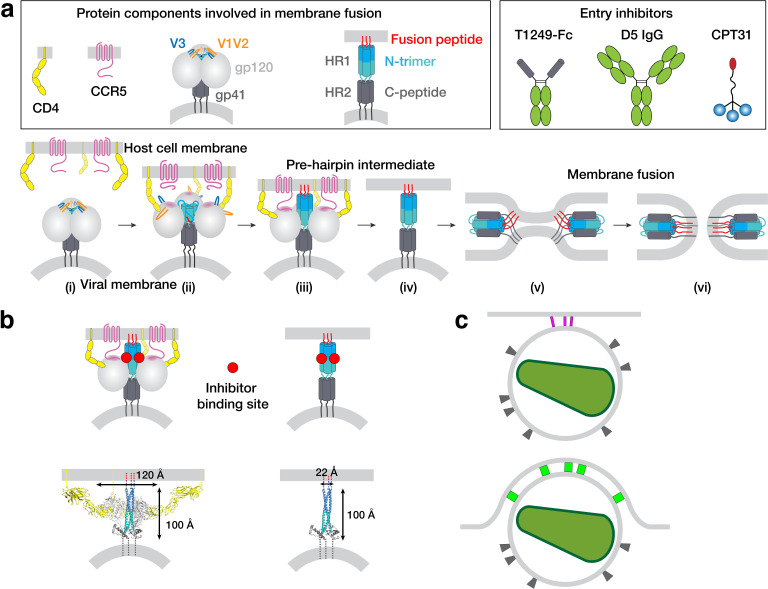
Figure 1.
HIV-1 Env-mediated fusion between viral and host cell membranes.
( a ) Top: Schematics of host receptors, HIV-1 Env trimer, pre-hairpin intermediate, and fusion inhibitors. Bottom: steps in fusion: (i) Closed, prefusion structure of HIV-1 Env trimer in which the V1...
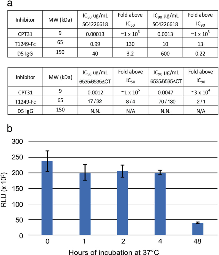
Figure 1u2014figure supplement 1.
Characterization of fusion inhibitors and viral infectivity.
( a ) Entry inhibitors are listed with their molecular weights, IC 50 and IC 90 values for neutralization potencies against SC4226618 and 6535 (and for T1249-Fc, also against 6535-u0394CT), and the fo...
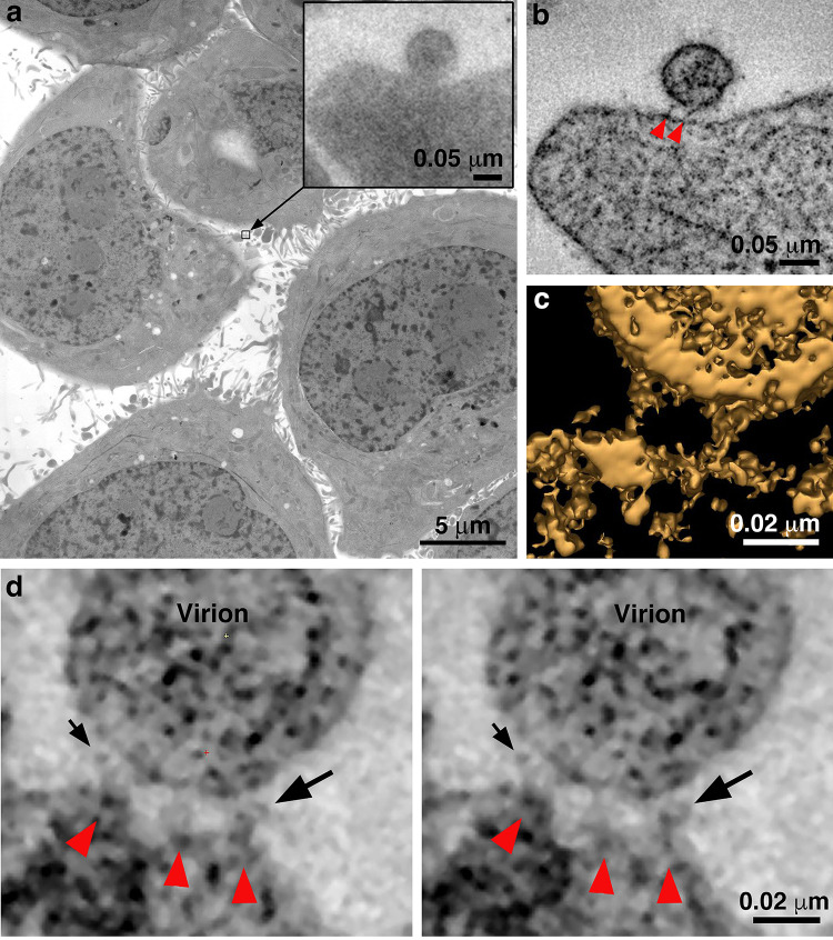
Figure 2.
Identification of attached HIV-1 virions.
( a ) Montaged projection overview of a field of cultured TZM-bl cells from a 400 nm section. Note extensive blebbing and surface projections that are typical of the cell type. Inset: Projection detai...
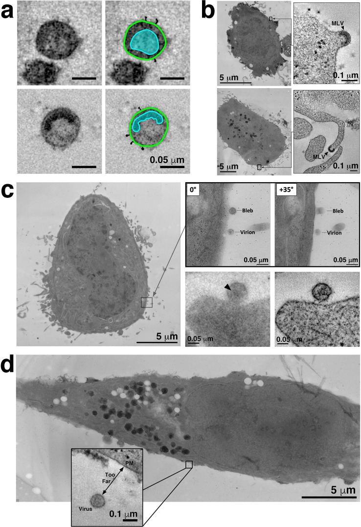
Figure 2u2014figure supplement 1.
Confirmation of pseudovirions in tomograms and experimental controls.
( a ) Identification of free pseudovirions in tomographic reconstructions.u00a0Virions were identified by visualizing a cone-shaped core structure in mature virions (upper left) and a C-shaped core st...
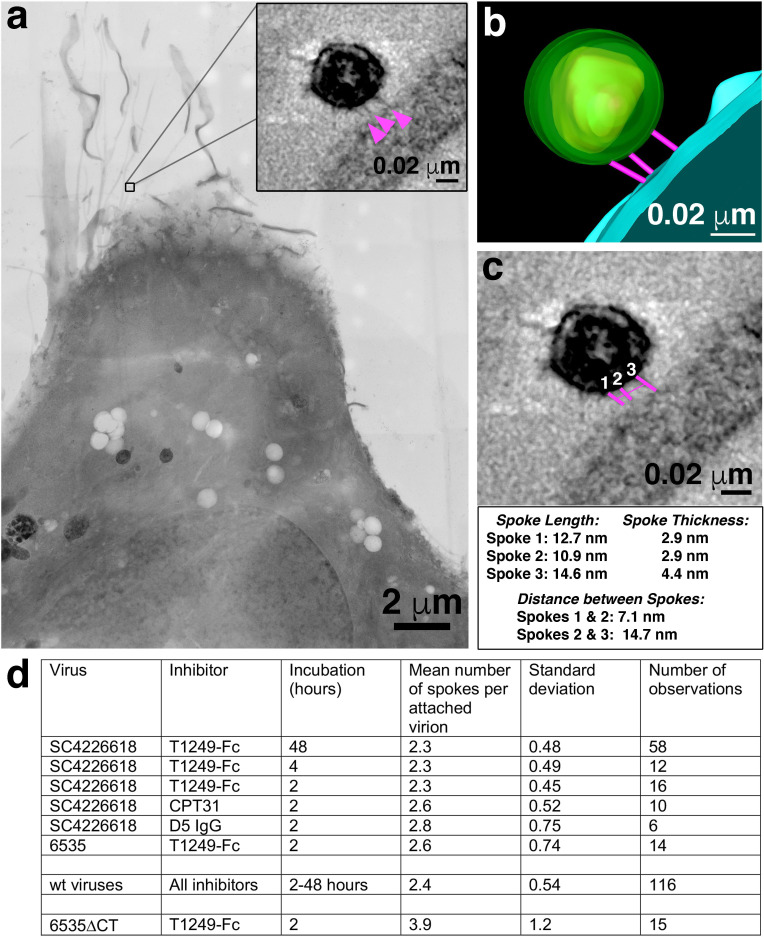
Figure 3.
Characteristics of virions attached to target cells in the presence of a fusion inhibitor.
( a ) 2-D projection image of TZM-bl cell incubated with SC4226618 pseudovirions in the presence of the T1249-Fc fusion inhibitor for 2 hr at 37u00b0C. Inset shows a tomographic slice of the attached ...
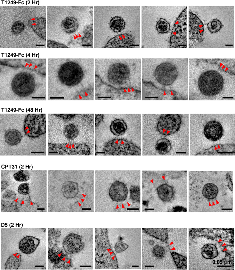
Figure 3u2014figure supplement 1.
Gallery of attachment sites formed using different fusion inhibitors and different incubation times.
In examples shown here, attachments consisted of either two or three spokes (red arrowheads) linking virions to cell surfaces. All scale barsu00a0=u00a00.05 u03bcm. See also Figure 3u2014source data 1...
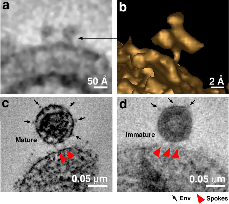
Figure 4.
Free Env trimers can be visualized on attached virions.
( a ) Example of densities observed for free HIV-1 Env trimers in a tomographic slice. ( b ) 3-D isosurface rendering of an individual free Env trimer. ( cu2013d ) Examples of attached mature (panel c...
Video 1.
Tomographic reconstruction of a mature HIV-1 pseudovirus attached to a TZM-bl cell surface by two narrow spokes.
In this experiment, the T1249-Fc inhibitor was incubated with cells and SC4226618 virus at 37 u00b0C for 2 hr. The movie presents the full volume of a 3-D reconstruction, advances at 1-pixel (0.5 nm) ...
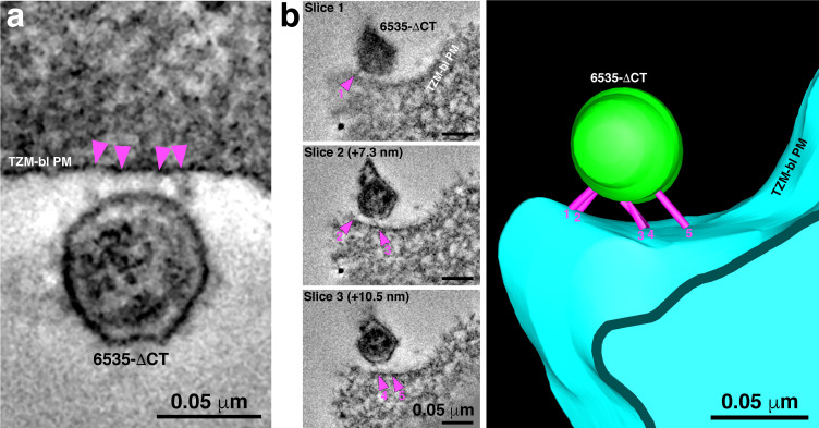
Figure 5.
6535u0394CT pseudoviruses are often attached to target cells with more than 2u20133 spokes.
( A ) An example of a 6535u0394CT virion attached to the plasma membrane of a TZM-bl cell (cell and virus were treated with the T1249-Fc fusion inhibitor for 2 hr) by four distinct spokes (arrowheads)...
Figure images are served from the NIH/NLM PubMed Central Open Access Subset or Europe PMC; copyright remains with the publishers and authors.
💬 Discussion
0 commentsNo comments yet. Be the first to start a discussion!
Leave a Comment