Abstract
The Plasmodium falciparum reticulocyte-binding protein homolog 5 (PfRH5) is the leading target for next-generation vaccines against the disease-causing blood-stage of malaria. However, little is known about how human antibodies confer functional immunity against this antigen. We isolated a panel of human monoclonal antibodies (mAbs) against PfRH5 from peripheral blood B cells from vaccinees in the first clinical trial of a PfRH5-based vaccine. We identified a subset of mAbs with neutralizing activity that bind to three distinct sites and another subset of mAbs that are non-functional, or even antagonistic to neutralizing antibodies. We also identify the epitope of a novel group of non-neutralizing antibodies that significantly reduce the speed of red blood cell invasion by the merozoite, thereby potentiating the effect of all neutralizing PfRH5 antibodies as well as synergizing with antibodies targeting other malaria invasion proteins. Our results provide a roadmap for structure-guided vaccine development to maximize antibody efficacy against blood-stage malaria.
🔬 Techniques
🔭 Microscopes
🧬 Organisms
💻 Software
✨ Fluorophores
🧪 Sample Preparation
🔬 Cell Lines
🏭 Microscope Brands
🧪 Reagent Suppliers
🔎 Objectives
💻 Software Details
💾 Data Repositories
🏷️ Research Resource Identifiers (RRIDs)
Verified research resources used in this paper:
🏛️ Research Organizations (ROR)
Affiliated research institutions:
📋 Methods
Key Resources Table REAGENT or RESOURCE SOURCE IDENTIFIER
Antibodies c2AC7 Simon J. Draper, Oxford University; Douglas et al., 2019 N/A c4BA7 Simon J. Draper, Oxford University; Douglas et al., 2019 N/A c9AD4 Simon J. Draper, Oxford University N/A QA1 Simon J. Draper, Oxford University; Douglas et al., 2014 N/A α-EBOV Simon J. Draper, Oxford University; Rijal et al., 2019 N/A Anti-Human IgG (γ-chain specific)−Alkaline Phosphatase antibody produced in goat Sigma-Aldrich Cat#A3188; RRID: AB_258057 Anti-Mouse IgG (whole molecule)−Alkaline Phosphatase antibody produced in goat Sigma-Aldrich Cat#A9316-.25ML; RRID: AB_258446 Anti-Rabbit IgG (whole molecule)–Alkaline Phosphatase antibody produced in goat Sigma-Aldrich Cat#A8025-.5ML; RRID: AB_258372 Rabbit polyclonal anti-PfRH5FL IgG This paper N/A Rabbit polyclonal anti-PfRH5ΔNL IgG This paper N/A Rabbit polyclonal PfAMA1 IgG Simon J. Draper, Oxford University; Douglas et al., 2011 N/A Rabbit polyclonal anti-PfRipr IgG This paper N/A Rabbit polyclonal anti-PfRH4 IgG Simon J. Draper, Oxford University; Douglas et al., 2011 N/A Rabbit polyclonal anti-PfMSP1 IgG Simon J. Draper, Oxford University; Douglas et al., 2011 N/A Rabbit polyclonal anti-PfCyRPA IgG This paper N/A Rat polyclonal anti-PfCyRPA IgG This paper N/A Bacterial and Virus Strains Mix & Go Competent Cells - Strain DH5α Escherichia coli Zymo Research Cat#T3007 Stellar HST08 strain Escherichia coli Competent Cells Takara Cat#636763 Biological Samples PBMC from PfRH5 Phase I trial donors Simon J. Draper, Oxford University; Payne et al., 2017 N/A Serum from PfRH5 Phase I trial donors Simon J. Draper, Oxford University; Payne et al., 2017 N/A Human O + RBC used in GIA assays In-house volunteer donations and NHS Blood and Transplant Non-clinical Issue Cat#NC15-Research Red Cells Serum from PfRH5-vaccinated rabbits GenScript N/A Serum from PfRH5ΔNL-vaccinated rabbits GenScript N/A Serum from PfAMA1-vaccinated rabbits Agro-Bio N/A Serum from PfRipr-vaccinated rabbit GenScript N/A Serum from PfRH4-vaccinated rabbits Agro-Bio N/A Serum from PfMSP1-vaccinated rabbits Agro-Bio N/A Serum from PfCyRPA-vaccinated rabbits Cambridge Research Biochemicals N/A Serum from PfCyRPA-vaccinated rat GenScript N/A Serum from liver-humanized FRGN mice This paper N/A Chemicals, Peptides, and Recombinant Proteins PfRipr This paper N/A PfCyRPA This paper N/A PfP113Nt Gavin J. Wright; Galaway et al., 2017 N/A PfRH5Nt Gavin J. Wright; Galaway et al., 2017 N/A PfRH5FL Simon J. Draper, Oxford University; Hjerrild et al., 2016 N/A PfRH5ΔNL This paper N/A PfRH5FL Y147H This paper N/A PfRH5FL H148D This paper N/A PfRH5FL S197Y This paper N/A PfRH5FL C203Y This paper N/A PfRH5FL I410M This paper N/A Basigin Matthew K. Higgins, University of Oxford; Wright et al., 2014 N/A RNasin Ribonuclease Inhibitor (Native) Promega Cat#N2111 Polyethyleneimine, linear, M.W. 25,000 Alfa Aesar Cat#43896-03 Borane dimethylamine complex (ABC) Sigma-Aldrich Cat#180238-25G Blocker Casein in PBS Thermo Fisher Scientific Cat#37528 SIGMA FAST BCIP/NBT alkaline phosphatase substrate Sigma-Aldrich Cat#B5655-25TAB SIGMA FAST p-Nitrophenyl phosphate alkaline phosphatase substrate Sigma-Aldrich Cat#N1891-50SET 62 biotinylated 20-mer PfRH5FL peptides Synthesized by Mimotopes, obtained from Simon J. Draper, Oxford University; Payne et al., 2017 N/A Clophosome-A clodronate liposomes FormuMax Scientific Cat#F70101C-A-2 AddaVax vaccine adjuvant InvivoGen Cat#vac-adx-10 Critical Commercial Assays EasySep Human B Cell Enrichment Kit StemCell Technologies Cat#19054 Sensiscript RT kit QIAGEN Cat#205213 Pierce Fab Preparation Kit Thermo Fisher Scientific Cat#44985 JCSG- plus crystallization screen Molecular Dimensions Cat#MD1-37 NeXtal JCSG Core Suite IV crystallization screen QIAGEN Cat#130924 Morpheus crystallization screen Molecular Dimensions Cat#MD1-46 Silver bullet crystallization additives Hampton Research Cat#HR2-096 ExpiFectamine 293 Transfection Kit Thermo Fisher Scientific Cat#A14525 Deposited Data R5.004 Fab fragment This paper PDB: 6RCO R5.011 Fab fragment This paper PDB: 6RCQ R5.016 Fab fragment This paper PDB: 6RCS PfRH5ΔNL:R5.004:R5.016 complex This paper PDB: 6RCU PfRH5ΔNL:R5.011:R5.016 complex This paper PDB: 6RCV Experimental Models: Cell Lines Human Expi293F Thermo Fisher Scientific Cat#A14527 Human HEK293T ATCC Cat#CRL-3216 Drosophila S2 cell line expressing PfRH5FL Simon J. Draper, Oxford University; Hjerrild et al., 2016 N/A Drosophila S2 cell line expressing PfRH5ΔNL This paper N/A Experimental Models: Organisms/Strains P. falciparum 3D7 Carole Long, NIAID N/A P. falciparum FVO Carole Long, NIAID N/A P. falciparum GB4 Carole Long, NIAID N/A P. falciparum M-Camp Carole Long, NIAID N/A P. falciparum Dd2 Carole Long, NIAID N/A P. falciparum Cp806 Carole Long, NIAID; Williams et al., 2012 N/A P. falciparum Cp845 Carole Long, NIAID; Williams et al., 2012 N/A P. falciparum NF54HT-GFP-luc Stefan H.I. Kappe; Foquet et al., 2018 N/A Liver-humanized FRGN KO mice Yecuris N/A Sprague Dawley Rat GenScript N/A New Zealand White rabbits GenScript, Agro-Bio, Cambridge Research Biochemicals N/A Oligonucleotides Primers for Vγ, Vλ, and Vκ antibody heavy and light chain sequence reverse-transcription and nested PCR Hedda Wardemann; Tiller et al., 2008 N/A Primers for amplifying and cloning human Vγ, Vλ, and Vκ antibody heavy and light chain sequences This paper N/A Recombinant DNA AbVec-hIg expression plasmids Patrick C. Wilson, University of Chicago; Wrammert et al., 2008 N/A pExpreS 2 -1 Drosophila S2 expression plasmid Simon J. Draper, Oxford University; Hjerrild et al., 2016 N/A Software and Algorithms XDS http://xds.mpimf-heidelberg.mpg.de/ RRID: SCR_015652 Phaser https://www.phenix-online.org/documentation/reference/phaser.html RRID: SCR_014219 BUSTER https://www.globalphasing.com/buster/ RRID: SCR_015653 PHENIX http://www.phenix-online.org RRID: SCR_014224 COOT https://www2.mrc-lmb.cam.ac.uk/personal/pemsley/coot/ RRID: SCR_014222 PyMOL version 2.3.0 Schrödinger ( https://pymol.org/2/ ) RRID: SCR_000305 GraphPad Prism version 5-7 GraphPad ( https://www.graphpad.com/ ) RRID: SCR_002798 IgBLAST https://www.ncbi.nlm.nih.gov/igblast/ RRID: SCR_002873 Other Octet Biosensor / Streptavidin (SA) FortéBio Cat#18-5019 Biacore Biotin CAPture Kit GE Healthcare Cat#28920233 Biacore Sensor Chip CM5 GE Healthcare Cat#BR-1000-12 Biacore Sensor Chip Protein A GE Healthcare Cat#29127558 Lead Contact and Materials Availability Further information and requests for resources should be directed to and will be fulfilled by the Lead Contact, Simon J. Draper ( simon.draper@ndm.ox.ac.uk ).
Show full methods section
Key Resources Table REAGENT or RESOURCE SOURCE IDENTIFIER
Antibodies c2AC7 Simon J. Draper, Oxford University; Douglas et al., 2019 N/A c4BA7 Simon J. Draper, Oxford University; Douglas et al., 2019 N/A c9AD4 Simon J. Draper, Oxford University N/A QA1 Simon J. Draper, Oxford University; Douglas et al., 2014 N/A α-EBOV Simon J. Draper, Oxford University; Rijal et al., 2019 N/A Anti-Human IgG (γ-chain specific)−Alkaline Phosphatase antibody produced in goat Sigma-Aldrich Cat#A3188; RRID: AB_258057 Anti-Mouse IgG (whole molecule)−Alkaline Phosphatase antibody produced in goat Sigma-Aldrich Cat#A9316-.25ML; RRID: AB_258446 Anti-Rabbit IgG (whole molecule)–Alkaline Phosphatase antibody produced in goat Sigma-Aldrich Cat#A8025-.5ML; RRID: AB_258372 Rabbit polyclonal anti-PfRH5FL IgG This paper N/A Rabbit polyclonal anti-PfRH5ΔNL IgG This paper N/A Rabbit polyclonal PfAMA1 IgG Simon J. Draper, Oxford University; Douglas et al., 2011 N/A Rabbit polyclonal anti-PfRipr IgG This paper N/A Rabbit polyclonal anti-PfRH4 IgG Simon J. Draper, Oxford University; Douglas et al., 2011 N/A Rabbit polyclonal anti-PfMSP1 IgG Simon J. Draper, Oxford University; Douglas et al., 2011 N/A Rabbit polyclonal anti-PfCyRPA IgG This paper N/A Rat polyclonal anti-PfCyRPA IgG This paper N/A Bacterial and Virus Strains Mix & Go Competent Cells - Strain DH5α Escherichia coli Zymo Research Cat#T3007 Stellar HST08 strain Escherichia coli Competent Cells Takara Cat#636763 Biological Samples PBMC from PfRH5 Phase I trial donors Simon J. Draper, Oxford University; Payne et al., 2017 N/A Serum from PfRH5 Phase I trial donors Simon J. Draper, Oxford University; Payne et al., 2017 N/A Human O + RBC used in GIA assays In-house volunteer donations and NHS Blood and Transplant Non-clinical Issue Cat#NC15-Research Red Cells Serum from PfRH5-vaccinated rabbits GenScript N/A Serum from PfRH5ΔNL-vaccinated rabbits GenScript N/A Serum from PfAMA1-vaccinated rabbits Agro-Bio N/A Serum from PfRipr-vaccinated rabbit GenScript N/A Serum from PfRH4-vaccinated rabbits Agro-Bio N/A Serum from PfMSP1-vaccinated rabbits Agro-Bio N/A Serum from PfCyRPA-vaccinated rabbits Cambridge Research Biochemicals N/A Serum from PfCyRPA-vaccinated rat GenScript N/A Serum from liver-humanized FRGN mice This paper N/A Chemicals, Peptides, and Recombinant Proteins PfRipr This paper N/A PfCyRPA This paper N/A PfP113Nt Gavin J. Wright; Galaway et al., 2017 N/A PfRH5Nt Gavin J. Wright; Galaway et al., 2017 N/A PfRH5FL Simon J. Draper, Oxford University; Hjerrild et al., 2016 N/A PfRH5ΔNL This paper N/A PfRH5FL Y147H This paper N/A PfRH5FL H148D This paper N/A PfRH5FL S197Y This paper N/A PfRH5FL C203Y This paper N/A PfRH5FL I410M This paper N/A Basigin Matthew K. Higgins, University of Oxford; Wright et al., 2014 N/A RNasin Ribonuclease Inhibitor (Native) Promega Cat#N2111 Polyethyleneimine, linear, M.W. 25,000 Alfa Aesar Cat#43896-03 Borane dimethylamine complex (ABC) Sigma-Aldrich Cat#180238-25G Blocker Casein in PBS Thermo Fisher Scientific Cat#37528 SIGMA FAST BCIP/NBT alkaline phosphatase substrate Sigma-Aldrich Cat#B5655-25TAB SIGMA FAST p-Nitrophenyl phosphate alkaline phosphatase substrate Sigma-Aldrich Cat#N1891-50SET 62 biotinylated 20-mer PfRH5FL peptides Synthesized by Mimotopes, obtained from Simon J. Draper, Oxford University; Payne et al., 2017 N/A Clophosome-A clodronate liposomes FormuMax Scientific Cat#F70101C-A-2 AddaVax vaccine adjuvant InvivoGen Cat#vac-adx-10 Critical Commercial Assays EasySep Human B Cell Enrichment Kit StemCell Technologies Cat#19054 Sensiscript RT kit QIAGEN Cat#205213 Pierce Fab Preparation Kit Thermo Fisher Scientific Cat#44985 JCSG- plus crystallization screen Molecular Dimensions Cat#MD1-37 NeXtal JCSG Core Suite IV crystallization screen QIAGEN Cat#130924 Morpheus crystallization screen Molecular Dimensions Cat#MD1-46 Silver bullet crystallization additives Hampton Research Cat#HR2-096 ExpiFectamine 293 Transfection Kit Thermo Fisher Scientific Cat#A14525 Deposited Data R5.004 Fab fragment This paper PDB: 6RCO R5.011 Fab fragment This paper PDB: 6RCQ R5.016 Fab fragment This paper PDB: 6RCS PfRH5ΔNL:R5.004:R5.016 complex This paper PDB: 6RCU PfRH5ΔNL:R5.011:R5.016 complex This paper PDB: 6RCV Experimental Models: Cell Lines Human Expi293F Thermo Fisher Scientific Cat#A14527 Human HEK293T ATCC Cat#CRL-3216 Drosophila S2 cell line expressing PfRH5FL Simon J. Draper, Oxford University; Hjerrild et al., 2016 N/A Drosophila S2 cell line expressing PfRH5ΔNL This paper N/A Experimental Models: Organisms/Strains P. falciparum 3D7 Carole Long, NIAID N/A P. falciparum FVO Carole Long, NIAID N/A P. falciparum GB4 Carole Long, NIAID N/A P. falciparum M-Camp Carole Long, NIAID N/A P. falciparum Dd2 Carole Long, NIAID N/A P. falciparum Cp806 Carole Long, NIAID; Williams et al., 2012 N/A P. falciparum Cp845 Carole Long, NIAID; Williams et al., 2012 N/A P. falciparum NF54HT-GFP-luc Stefan H.I. Kappe; Foquet et al., 2018 N/A Liver-humanized FRGN KO mice Yecuris N/A Sprague Dawley Rat GenScript N/A New Zealand White rabbits GenScript, Agro-Bio, Cambridge Research Biochemicals N/A Oligonucleotides Primers for Vγ, Vλ, and Vκ antibody heavy and light chain sequence reverse-transcription and nested PCR Hedda Wardemann; Tiller et al., 2008 N/A Primers for amplifying and cloning human Vγ, Vλ, and Vκ antibody heavy and light chain sequences This paper N/A Recombinant DNA AbVec-hIg expression plasmids Patrick C. Wilson, University of Chicago; Wrammert et al., 2008 N/A pExpreS 2 -1 Drosophila S2 expression plasmid Simon J. Draper, Oxford University; Hjerrild et al., 2016 N/A Software and Algorithms XDS http://xds.mpimf-heidelberg.mpg.de/ RRID: SCR_015652 Phaser https://www.phenix-online.org/documentation/reference/phaser.html RRID: SCR_014219 BUSTER https://www.globalphasing.com/buster/ RRID: SCR_015653 PHENIX http://www.phenix-online.org RRID: SCR_014224 COOT https://www2.mrc-lmb.cam.ac.uk/personal/pemsley/coot/ RRID: SCR_014222 PyMOL version 2.3.0 Schrödinger ( https://pymol.org/2/ ) RRID: SCR_000305 GraphPad Prism version 5-7 GraphPad ( https://www.graphpad.com/ ) RRID: SCR_002798 IgBLAST https://www.ncbi.nlm.nih.gov/igblast/ RRID: SCR_002873 Other Octet Biosensor / Streptavidin (SA) FortéBio Cat#18-5019 Biacore Biotin CAPture Kit GE Healthcare Cat#28920233 Biacore Sensor Chip CM5 GE Healthcare Cat#BR-1000-12 Biacore Sensor Chip Protein A GE Healthcare Cat#29127558 Lead Contact and Materials Availability Further information and requests for resources should be directed to and will be fulfilled by the Lead Contact, Simon J. Draper ( simon.draper@ndm.ox.ac.uk ).
Experimental Model and Subject Details Human blood sample collection
Healthy, malaria-naive males and non-pregnant females aged 18-50 were invited to participate in the VAC057 study of the PfRH5-based vaccine ( Payne et al., 2017 ). VAC057 was a first-in-human, open-label, non-randomized, dose escalation Phase Ia clinical trial evaluating the safety and immunogenicity of the viral vectored vaccines ChAd63 RH5 and MVA RH5 in a heterologous prime-boost regime with an eight week interval. The study was conducted in the UK at the Centre for Clinical Vaccinology and Tropical Medicine (CCVTM), University of Oxford, Oxford, and the NIHR Wellcome Trust Clinical Research Facility (WTCRF) in Southampton. The study received ethical approval from the Oxfordshire Research Ethics Committee A in the UK (REC reference 14/SC/0120). The study was also reviewed and approved by the UK Medicines and Healthcare products Regulatory Agency (MHRA, reference 21584/0331/001-0001). Volunteers signed written consent forms and consent was verified before each vaccination. The trial was registered on Clinicaltrials.gov ( NCT02181088 ) and was conducted according to the principles of the current revision of the Declaration of Helsinki 2008 and in full conformity with the ICH guidelines for Good Clinical Practice (GCP). The primary endpoint of the study was to assess the safety of ChAd63 RH5 and MVA RH5, with a secondary endpoint to assess immunogenicity. Human blood samples were collected into lithium heparin-treated vacutainer blood collection systems (Becton Dickinson). PBMC were isolated and used within 6 hours in fresh assays, otherwise excess cells were frozen in fetal calf serum (FCS) containing 10% dimethyl sulfoxide and stored in liquid nitrogen. Plasma samples were stored at −80°C. For serum preparation, untreated blood samples were stored at room temperature and then the clotted blood was centrifuged for 5 min at 1000 g . Serum was stored at −80°C.
Experimental animal models Sprague Dawley rat and female New Zealand
White rabbit immunizations (PfRH5FL, PfRH5ΔNL and PfRipr) were carried out by Genscript (Piscataway, NJ, USA). GenScript holds a valid and current Animal Welfare Assurance in compliance with the Public Health Service (PHS) Policy on humane Care and Use of Laboratory Animals as granted by the Office of Laboratory Animal Welfare (OLAW). Female New Zealand White rabbit immunizations (PfCyRPA) were carried out by Cambridge Research Biochemicals (Billingham, UK) in compliance with the UK Animals (Scientific Procedures) 1986 Act (ASPA). Female New Zealand White rabbit immunizations (PfRH4, PfMSP1 and PfAMA1) were carried out by Agro-Bio (La Ferté Saint Aubin, France) according to the current French version of the European Directive 2010/63/EU and following a protocol approved by their internal ethics committee. The study using liver-humanized FRGN KO mice was carried out in accordance with the recommendations of the NIH Office of Laboratory Animal Welfare standards (OLAW welfare assurance # A3640-01). The protocol was approved by the Center for Infectious Disease Research Institutional Animal Care and Use Committee (IACUC) under protocol SK-16.
Cell lines Adherent
HEK293T cells were grown in Dulbecco’s Modified Eagle Medium (DMEM) (Sigma-Aldrich) supplemented with 2 mM L-glutamine, 1 mM sodium pyruvate, 100 U/mL penicillin, 0.1 mg/mL streptomycin and 10% ultra-low IgG fetal bovine serum (FBS) (all from Thermo Fisher Scientific) in a static incubator at 37°C, 8% CO 2 . Expi293F HEK cells were cultured in suspension in Expi293 expression medium (Thermo Fisher Scientific) at 37°C, 8% CO 2 , on an orbital shaker set at 125 RPM. Drosophila S2 were cultured in suspension in EX-CELL 420 medium (Sigma-Aldrich) supplemented with 100 U/mL penicillin, 0.1 mg/mL streptomycin and 10% FBS at 25°C.
Method Details Generation of monoclonal antibodies Plasmablast isolation and sorting
Volunteers from a Phase Ia safety and immunogenicity clinical trial were bled seven days after the second immunization using MVA (modified vaccinia virus Ankara) encoding PfRH5FL ( Payne et al., 2017 ). Blood was collected from volunteers in heparinized tubes and centrifuged in Leucosep tubes (Greiner Bio one) to separate the peripheral blood mononuclear cells (PBMC). The PBMC were enriched for B cells using a human pan-B cell enrichment kit (StemCell Technologies, Inc.) and resuspended in DMEM before staining with a CD19 + , CD10 – , CD21 – , CD27 + , CD20 – , CD38 + , IgG + fluorophore-conjugated antibody panel. Plasmablasts were single-cell sorted using a MoFlo cell sorter (Dako cytomation) into 96-well PCR plates containing 10 μL of 10 mM Tris HCl buffer containing 40 U/mL of RNase inhibitor (Promega). The study received ethical approval from the Oxfordshire Research Ethics Committee A in the UK (REC reference 14/SC/0120). The volunteers signed written consent forms and consent was verified before each vaccination.
Antibody variable gene amplification
In each well of a 96-well plate containing a single antibody-secreting cell (ASC), a two-step RT-PCR was carried out with a first reverse transcription (RT) step using a Sensiscript RT kit (QIAGEN) and degenerate primers 1-17 (modified from Tiller et al. [2008 ]) (see Table S4 ). Next, a first PCR (PCR1) was performed on 1 μL of the RT reaction product using the same set of primers used before (1-17) which cover the diversity of all Vγ, Vκ and Vλ sequences using Phusion HF master mix (New England Biolabs). Following this, a second PCR (PCR2) was performed using primers 18-51 ( Table S4 ), also using Phusion HF master mix, on 1 μL of the previous product diluted 1:100 to amplify inserts which contain plasmid-homologous extensions designed for circular polymerase extension cloning (CPEC) ( Quan and Tian, 2009 ). Cloning The AbVec-hIgG1/AbVec-hIgKappa/AbVec-hIgLambda expression plasmids were a kind gift from Patrick C. Wilson (University of Chicago) ( Wrammert et al., 2008 ). These plasmids were 5′ digested using BshTI and at the 3′ using SalI (AbVec-hIgG1), XhoI (AbVec-hIgLambda) and Pfl23II (AbVec-hIgKappa) to yield linear products. CPEC assembly was done by mixing 100 ng of a 1:1 molar ratio of insert:plasmid in 20 μL containing 1x Phusion HF polymerase master mix and assembled using an 8-cycle CPEC protocol (8 cycles: 98°C 10 s, slow ramp anneal 70°C → 55°C at 0.1°C/s, 72°C 35 s). Full nicked plasmids were subsequently transformed into Zymo 5α Mix & go competent Escherichia coli (Zymo Research) according to manufacturer’s instructions, streaked on LB agar Petri dishes containing 100 μg/mL carbenicillin and grown at 37°C overnight in a static incubator. Colonies were screened by PCR for inserts of the correct size.
Screening
Exponential growth-phase adherent HEK293T cells were resuspended in DMEM (Sigma-Aldrich) supplemented with 2 mM L-glutamine, 1 mM sodium pyruvate, 100 U/mL penicillin, 0.1 mg/mL streptomycin and 10% ultra-low IgG FBS (all from Thermo Fisher Scientific) and seeded at 4 × 10 4 cells/well in 100 μL 24 h prior to transfection in Costar 96-well cell culture plates (Corning). On the day of transfection, for each well, 50 μL of 60 μg/mL linear 25 kDa PEI (Alfa Aesar) was mixed with 200 ng of cognate heavy- and light-chain coding plasmid in a volume of 50 μL and shaken at 20°C for 30 min. The DNA-PEI complexes were then added to the HEK293T cells. The next day, an additional 50 μL of supplemented DMEM (as described above) was added to each well. Supernatants were screened for PfRH5FL binding by indirect ELISA as described in the ELISA methods section.
Protein expression and purification
The recombinant PfRH5 sequence used in all experiments except for those involving BLI ( Figures 1 C, 3 A, S3 A, and S3B; Table S1 ) was based on the 3D7 clone P. falciparum reference sequence and encoded amino acids E26-Q526. The sequence also encoded a C-terminal four-amino acid purification tag (C-tag: EPEA) and four mutations to delete N-linked glycosylation sequons (T40A, T216A, T286A and T299A) and was named “PfRH5FL” (this recombinant protein is also known as “RH5.1”) ( Jin et al., 2018 ). This protein was expressed as secreted protein by a stable monoclonal Drosophila S2 cell line ( Hjerrild et al., 2016 ) and affinity purified using CaptureSelect C-tag affinity matrix ( Jin et al., 2017 ) (Thermo Fisher Scientific). A further size-exclusion chromatography (SEC) polishing step was done on a HiLoad 16/60 Superdex 200 pg column (GE Healthcare) to separate monomers from oligomers and contaminants as well as to buffer-exchange the protein into 20 mM Tris, 150 mM NaCl, pH 7.5. A detailed description of the production of PfRH5FL was described by Jin et al., 2018. The recombinant PfRH5 sequence (also named “PfRH5FL” for simplicity) used in BLI experiments also encoded amino acids E26-Q526 of the 3D7 clone P. falciparum reference sequence, with only two mutations to delete N-linked glycosylation (N38Q and N214Q). This sequence is identical to the vaccine sequence ( Payne et al., 2017 ) and was expressed with an additional C-terminal AviTag and Strep-II® tag in tandem. This protein was expressed in Expi293F HEK cells as a secreted, monobiotinylated protein as described previously by Bushell et al. (2008) . The recombinant PfRH5ΔNL sequence used was based on the 3D7 clone P. falciparum reference sequence and encoded amino acids K140-K247 and N297-Q526 with two mutations to delete N-linked glycosylation sequons (T216A and T299A) and with the addition of a C-terminal C-tag. PfRH5ΔNL was expressed as secreted protein from stably transfected polyclonal Drosophila S2 cells. Its purification is detailed in the X-ray crystallography experimental procedures section. The PfRipr expression construct used for rabbit vaccination to yield IgG used in Figures 5 E and S5 F comprised amino acids M1-N1086 with eight mutations introduced to ablate N-linked glycosylation (N103Q, N144Q, N228Q, N334Q, N480Q, N498Q, N506Q, N526Q, N646Q, N647Q, N964Q and N1021Q) followed by a C-terminal C-tag. PfRipr was purified by C-tag affinity chromatography followed by SEC as detailed above for PfRH5. The expression construct used for monomeric PfCyRPA production in Figure 3 C and rat vaccination in Figure S5 F was based on the 3D7 clone P. falciparum sequence and comprised amino acids D29-E362 with three mutations introduced to ablate N-linked glycosylation (S147A, T324A and T340A) and also included a C-terminal GGGS linker followed by a 4-amino acid C-tag (EPEA). The PfCyRPA immunogen sequence used for rabbit vaccination in Figure 5 E was identical to that described above with the C-tag replaced by a C-terminal CD4 tag comprising rat domains 3 and 4 (CD4d3+4) tag followed by a hexahistidine (His6) tag. The protein was expressed as secreted protein from Expi293F HEK cells and purified by C-tag affinity chromatography followed by SEC on a HiLoad 16/60 Superdex 200 pg column (GE Healthcare). Basigin protein comprised of immunoglobulin domains 1 and 2 of the short isoform (residues A22-H205) and was expressed from E. coli and purified by Ni 2+ -affinity and size exclusion chromatography. Further details can be found in Wright et al., 2014. PfP113Nt, encoding amino acids Y23-K219 of PfP113 (3D7) and PfRH5Nt encoding amino acids F25-K140 of PfRH5 (3D7), were expressed encoding C-terminal tags comprising CD4d3+4, a biotin acceptor peptide and a His6 tag in tandem. For further details see Galaway et al. (2017) . Recombinant monoclonal antibodies were transiently expressed in Expi293F HEK cells. Cognate heavy and light chain-coding plasmids were co-transfected at a 1:1 ratio. Supernatants were harvested by centrifuging the culture at 2500 x g for 15 min and filtering the supernatant with a 0.22 μm vacuum filter. All mAbs were purified using a 5 mL Protein G HP column (GE Healthcare) on an ÄKTA start FPLC system or an ÄKTA Pure FPLC system (both GE Healthcare). Equilibration and wash steps were performed with Dulbecco’s PBS and mAbs were eluted in 0.1 M glycine pH 2.7. The eluates were pH equilibrated to 7.4 using 1.0 M Tris HCl pH 9.0 and immediately buffer-exchanged into Dulbecco’s PBS and concentrated using an Amicon ® ultra centrifugal concentrator (Millipore) with a molecular weight cut-off of 30 kDa. IgG from serum in Figures 3 E, 3F, 3H, 5C, 5 E, S5 B, and S5F was purified on drip columns packed with Pierce Protein G agarose resin (Thermo Fisher Scientific). Pierce protein G IgG binding buffer (Thermo Fisher Scientific) was used to dilute the serum 1:1 before loading as well as for equilibration and wash steps. Bound IgG was subsequently eluted, neutralized and concentrated as above. Bispecific DVD-Ig 1611 was constructed by cloning R5.016 variable regions upstream (5′) of R5.011 variable regions, separated by ASTKGPSVFPLAP and TVAAPSVFIFPP linkers for the heavy and light chains, respectively. 1611 DVD-Ig expression and purification was conducted as described above for monospecific mAbs.
X-Ray crystallography Complex preparation
PfRH5ΔNL was purified from Drosophila S2 culture supernatant using C-tag affinity chromatography and glycosylated contaminants were removed by a subsequent lectin chromatography step with a HiTrap ConA 4B column (GE Healthcare). Disordered regions were trimmed by an overnight incubation at 20°C with endoproteinase gluC (New England Biolabs) at a final concentration of 1 μg/mL. Fab fragments were generated by papain digestion using a Pierce Fab Preparation Kit (Thermo Fisher Scientific) following the manufacturer’s recommendations. Complexes were prepared by mixing each Fab fragment with PfRH5ΔNL at a 1:1 molar ratio and were methylated with 1 M ABC (Borane dimethylamine complex) and 1 M formaldehyde (both Sigma-Aldrich) ( Walter et al., 2006 ). The methylated complexes were subjected to SEC on a HiLoad 16/60 Superdex 200 pg column (GE Healthcare) at 4°C in 20 mM Tris pH 7.4, 150 mM NaCl. The complex-containing fractions were pooled and concentrated using an Amicon ® ultra centrifugal concentrator (Millipore) with a molecular weight cut-off of 30 kDa. Crystallization, data collection and processing Crystallization was achieved using vapor diffusion in sitting drops. Crystals were obtained for the Fab fragments of mAbs R5.004, R5.011 and R5.016 alone, as well as for complexes consisting of PfRH5ΔNL:R5.004:R5.016 and PfRH5ΔNL:R5.011:R5.016. In each case, a TTP Labtech Mosquito LCP robot was employed to mix 100 nL of each protein complex at a concentration of 10 mg/mL with 100 nL of well solutions from commercially available crystal screens. Crystals of R5.004 Fab fragments were obtained in the JCSG-plus crystallization screen (Molecular Dimensions) and were optimized with a final well solution of 0.2 M lithium sulfate, 0.1 M NaAc pH 4.5, 30% PEG 8000 and 0.06% hexamminecobalt(III) chloride, 12 mM MES monohydrate, 12 mM PIPES, 4 mM HEPES chloride (Silver bullet additive D3, Hampton Research). They were cryo-protected by transfer into well solution supplemented with 25% glycerol, then cryo-cooled by plunging into liquid nitrogen. Data were collected on beamline I-03 at Diamond Light Source (Harwell, UK), leading to a complete dataset at a resolution of 1.7 Å. Crystals of R5.016 Fab fragments were obtained in the JCSG-plus crystallization screen (Molecular Dimensions), with a final well solution of 0.2 M lithium sulfate, 0.1 M NaAc pH 4.5, 50% PEG 400. Crystals were cryo-cooled directly in the well solution by plunging into liquid nitrogen. Data were collected on beamline I04-1 at Diamond Light Source (Harwell, UK), leading to a complete dataset at a resolution of 2.1 Å. Crystals of R5.011 Fab fragments were obtained in the NeXtal JCSG-IV crystallization screen (QIAGEN), with a final well solution of 0.16 M ZnAc, 0.108 M Na cacodylate pH 6.5, 14.4% PEG 8000, 20% glycerol. They were cryo-protected by transfer into well solution supplemented with 25% glycerol, then cryo-cooled by plunging into liquid nitrogen. Data were collected on beamline PROXIMA-1 at SOLEIL (Saint-Aubin, France), leading to a complete dataset at a resolution of 2.3 Å. Crystals of the PfRH5ΔNL:R5.004:R5.016 complex were obtained in the JCSG-plus crystallization screen (Molecular Dimensions), with a final well solution of 0.15 M DL malic acid, 20% PEG 3350. They were cryo-protected by transfer into well solution supplemented with 25% glycerol, then cryo-cooled by plunging into liquid nitrogen. Data were collected on beamline I04 at Diamond Light Source (Harwell, UK), leading to a complete dataset at a resolution of 4.0 Å. Crystals of the PfRH5ΔNL:R5.011:R5.016 complex were obtained in the Morpheus crystallization screen (Molecular Dimensions), with a final well solution of 10% PEG 20000, 20% PEG 550MME, 0.02 M amino acids, 0.1 M MES/imidazole pH 6.5. Crystals were cryo-cooled directly in the well solution by plunging into liquid nitrogen. Data were collected on beamline ID23 at the European Synchrotron Radiation Facility (Grenoble, France), leading to a complete dataset at a resolution of 3.6 Ε. In each case, data reduction was performed using XDS ( Kabsch, 2010 ). Molecular replacement solutions were found for each individual Fab fragment, using Phaser ( McCoy et al., 2007 ) with the most closely related Fab fragment structure in the PDB, split into their constant and variable domains, as search models (4KQ3 for R5.004, 4HK0 for R5.011 and 5K9O for R5.016). This led to a cycle of model building and refinement using COOT ( Emsley et al., 2010 ) and BUSTER ( Bricogne et al., 2011 ). For R5.004, this resulted in a complete model for the Fab fragment. For R5.011, this resulted in a complete model for the Fab fragment, with the exception of residues 143-148 in the heavy chain, which were disordered in the electron density map. For R5.016, this resulted in a complete model for the Fab fragment, with the exception of residues 103-108 in the heavy chain and 1-7, 25-29 and 55-58 in the light chain, all of which were disordered in the electron density map. The structure of the PfRH5ΔNL:R5.011:R5.016 complex was obtained to 3.6 Å resolution using Phaser with the structure of PfRH5ΔNL (PDB code: 4U0R ) and those of Fab fragments obtained above, split into their constant and variable domains, as search models. All domains were well resolved, although significant changes in conformation were observed in CDR H3 of both R5.011 and R5.016, as a result of PfRH5ΔNL binding. COOT, BUSTER and PHENIX ( Afonine et al., 2012 ) were used for refinement, including application of restraints that derived from the higher resolution structures of PfRH5ΔNL and the shared regions of the Fab fragments. This allowed the production of a final model in which all of the Fab fragment domains, and all of the CDR loops, were clearly resolved. The structure of the PfRH5ΔNL:R5.004:R5.016 complex was obtained at 4 Å resolution, using Phaser. The structure of PfRH5ΔNL bound to the variable domain of R5.016, obtained from the structure of the PfRH5ΔNL:R5.011:R5.016 complex, and the structure of the variable domain of R5.004 obtained above, were used as search models in Phaser. No significant changes were observed in the CDR loops of the Fab fragment of R5.004. Placement of the constant domains of R5.016 and R5.004 was challenging, due to anisotropy resulting from disorder within the crystal. These domains were therefore placed using real space docking after refinement in BUSTER of a structure consisting of PfRH5ΔNL and the variable domains of R5.004 and R5.016. This gave unambiguous electron density for the regions of the constant domains which lie close to the contact with the variable domains, but, after refinement, electron density is still absent for the PfRH5ΔNL-distal parts of the constant domains. Refinement in COOT, BUSTER and PHENIX allowed the production of a final model in which the Fab fragment domains, and all of the CDR loops, were clearly resolved. Humanized mouse passive transfer P. falciparum sporozoite production and mouse infection The luciferase expressing strain P. falciparum NF54HT-GFP-luc was maintained in RPMI 1640 supplemented with 25 mM HEPES, 2 mM L-glutamine, 50 μM hypoxanthine, 10% human serum and sub-cultured in 5% human group O RhD positive human RBC. Briefly, asexual cultures were inoculated at 1% parasitemia with no further sub-culturing and daily media changes to induce gametocytogenesis. Mature gametocytes were fed to 4-day old Anopheles stephensi mosquitoes to initiate infection. Mosquitoes were incubated at 27°C and 75% humidity for 14 d and given 8% dextrose + PABA to foster parasite growth. Liver humanized FRGN KO mice were purchased from Yecuris Corp. and showed human hepatocyte repopulation levels above 70% determined by human serum albumin levels. Liver humanized mice were cycled on NTBC once a month for 3 d at 8 μg/mL in the drinking water to maintain health. Animals did not receive NTBC three weeks prior to and during the infection study. For mosquito bite infection, 3-5 liver humanized FRGN KO mice were anesthetised and placed on top of a mosquito cage containing 150-250 infected mosquitoes for 20 min. P. falciparum liver-to-blood stage transition On the day of challenge, liver humanized FRGN KO mice were injected both in the retro-orbital plexus and the peritoneal cavity with 50 μL clodronate-containing liposomes (Clophosome®-A, FormuMax Scientific), 100 mg/kg cyclophosphamide (Sigma-Aldrich). Five days after challenge, the animals were bled a volume of approximately 200 μL, and 500 μL hRBC was injected in the retro-orbital plexus (70% O+ human erythrocytes in RPMI 1640 supplemented with 25 mM HEPES, 2 mM L-glutamine, 50 μM hypoxanthine and 10% human serum). The next day, mice were bled 200 μL and received an intraperitoneal injection of 700 μL hRBC. mAb transfer was done on day 6 to precede the establishment of blood-stage infection. Dosage was 15 mg total rabbit IgG per mouse formulated in PBS and delivered intravenously. The clodronate liposome and cyclophosphamide injections were repeated on days 5, 9, 11, 13. Human RBC were injected daily in a volume of 300-700 μL to keep the percentage of hRBC stable around 50%–60%. If the percentage reached more than 70%, the mice were not injected with hRBC to limit morbidity. On days 9, 11 and 13 each mouse was bled from the retro-orbital plexus to sample antibody levels.
Quantification of parasite burden
Luciferase activity was measured in the mice using the IVIS Lumina II animal imager (Perkin Elmer). The abdomen of mice was shaved to enhance detection. Mice were injected intraperitoneally with 100 μL of luciferase substrate RediJect D-Luciferin (Perkin Elmer) to quantitate specific enzymatic activity. Animals were anesthetized and imaged within 5 min of substrate injection. Signal was acquired for 5 min using a field of view of 10 cm and medium binning factor. Living Image 3.0 software was used to measure total flux (photons/second) of a region of interest, which was placed around each mouse. A comprehensive description of the mouse model was published by Foquet et al., 2018. PfCyRPA/PfP113/BSG SPR blocking assays Data were collected on a Biacore X100 (GE Healthcare). All data used were reference subtracted from a non-immobilized flow cell (Fc 2-1). Binding values were measured manually in the Biacore X100 control software. PfRH5-basigin binding Experiments were performed at 25°C in SPR running buffer (PBS + 0.005% Polysorbate-20, GE Healthcare). Approximately 670 RU of basigin was amine-coupled to a CM5 chip (GE Healthcare) on flow cell 2 (Fc 2) using standard NHS/EDC chemistry. PfRH5FL-mAb complexes were made by mixing PfRH5FL and mAb to a final concentration of 0.5 μM PfRH5FL and 1 μM mAb in SPR running buffer. Complexes were injected over Fc 1 and Fc 2 at a flow rate of 10 μL/min for 30 s and the surface was regenerated with 10 mM glycine pH 1.5 for at a flow rate of 10 μL/min for 60 s. Between experiments, one injection of 0.5 μM PfRH5FL was made to assess basigin degradation caused by regeneration. PfRH5-PfCyRPA binding Experiments were performed at 25°C in SPR running buffer (PBS + 0.005% Polysorbate-20, GE Healthcare) using a sensor chip protein A (GE Healthcare). mAb was injected at a concentration of 20 nM at a flow rate of 5 μL/min for 35 s over Fc 2 on a protein A coated chip (GE Healthcare). Next, PfRH5FL was injected over Fc 1 and Fc 2 at a concentration of 50 nM at a flow rate of 10 μL/min for 120 s before injecting PfCyRPA at a concentration of 1 μM for 120 s, also at a flow rate of 10 μL/min. The chip surface was regenerated with 10 mM glycine pH 1.5 for at a flow rate of 10 μL/min for 60 s. PfRH5-PfP113 binding Experiments were performed at 37°C in SPR running buffer (PBS + 0.005% Polysorbate-20, GE Healthcare) using a Sensor chip CAP (GE Healthcare). The whole experiment was run at a flow rate of 5 μL/min. Approximately 1500 RU of CAP reagent (GE Healthcare) was captured on Fc 1 and Fc 2. On Fc 1, approximately 500 RU of a biotinylated control CD4d3+4-tagged protein was immobilized. On Fc 2, each experiment consisted of capturing 1500 RU of CAP reagent in an 80 s injection followed by approximately 1800 RU of PfP113Nt in an 80 s injection at a concentration of 20 μg/mL. After this, PfRH5FL-mAb complex (both at 1 μM) was flowed over Fc 1 and Fc 2. Flow cell 2 was regenerated between experiments in a 110 s injection of 6 M guanidine + 250 mM NaOH, as per the manufacturer’s instructions. Bio-Layer Interferometry All BLI was carried out on an OctetRED384 (FortéBio) using streptavidin-coated biosensors (FortéBio) to immobilize PfRH5FL enzymatically monobiotinylated on a C-terminal AviTag. Assays were carried out in 96-well format in black plates (Greiner). For assaying mAb binding to PfRH5FL variants ( Figure 1 C), the experiment followed a four-step sequential assay: Baseline (PBS, 30 s); Protein immobilization (neat supernatant, 180 s); Wash (PBS, 60 s); and mAb binding (150 nM mAb, 120 s). Graphed data show the fold-change in binding of each mAb to the 3D7 PfRH5FL reference protein relative to the binding of each mAb to each mutant protein after correction for PfRH5FL immobilization level on each biosensor. The fold-change values which were inferior to 1 were plotted as their inverse to avoid skewing in their data representation compared to fold-change values greater than 1. For epitope binning studies in Figures 3 A and S3 A and Table S1 the experiment followed a six-step sequential assay: Baseline (PBS, 30 s); Protein immobilization (neat supernatant, 120 s); Wash (PBS, 60 s); mAb1 binding (300 nM mAb1, 120 s); Wash (PBS, 60 s); mAb2 binding (150 nM mAb2, 120 s). “Relative binding” shown in Table S1 shows the ratio (Signal mAb2 with mAb1 bound)/(Signal mAb2 with no mAb1) where “Signal mAb2 ” was normalized for the amount of PfRH5FL bound to the biosensor such that “Signal mAb2 ” = the raw signal in “mAb2 binding” divided by the raw signal in the “Protein immobilization” phase. The resulting “binding profile” for any given mAb corresponds to the column of “relative binding values” under that mAb in the “relative binding” table. To establish the epitope bins, binding profiles between each mAb pair was correlated using a person product-moment correlation coefficient, the values of which are shown in the “binding profile correlation” table in Table S1 . mAb pairs whose binding profile correlation was > 0.7 were grouped into the same epitope bin. ELISA Qualitative mAb binding ELISAs such as those used in Figures 1 D, 3 D, and S3 F were carried out by coating PfRH5FL, PfRH5ΔNL or PfRH5Nt on Maxisorp flat-bottom 96-well ELISA plates (Nunc) at 2 μg/mL in 50 μL at 4°C overnight. In Figure 1 D, PfRH5FL was heat-treated by incubation at 90°C for 10 min. Plates were then washed twice with PBS-Tween 20 and blocked with 200 μL of Blocker Casein (Thermo Fisher Scientific) for 1 h. Next, wells were incubated with 1000 ng/mL of mAb for approximately 45 min at 20°C then washed 4 times with PBS-Tween 20 before the addition of 50 μL of goat anti-human gamma-chain alkaline phosphatase-conjugated secondary antibody (goat anti-mouse IgG alkaline phosphatase-conjugated secondary antibody for QA1) (both Sigma-Aldrich) for 45 min at 20°C. Wells were then washed 6 times with PBS-Tween 20 and developed with 100 μL of pNPP substrate at 1 mg/mL (Sigma-Aldrich) and read at 405 nm. Peptide ELISAs in Figure S3 E were carried out with a set of sixty-two custom-synthesized biotinylated 20-mer peptides of PfRH5 overlapping by 12 amino acids (Mimotopes). The list of peptide sequences was described previously by Payne et al., 2017. Briefly, the peptides were designed to be oriented and tethered by their N-terminal biotinylated linker peptide (biotin-SGSG) with the exception the first peptide which contained the biotinylated linker at its C terminus to preserve potential binding activity to the most N-terminal residues. These peptides were coated to the bottom wells of a streptavidin-coated 96-well plate in 50 μL. The next day wells were blocked with 200 μL of Blocker Casein (Thermo Fisher Scientific) and washed five times with PBS-Tween 20. Bound mAbs were detected using an anti-human gamma-chain-specific alkaline phosphatase-conjugated secondary antibody (Sigma-Aldrich) or the mouse equivalent for QA1 in 50 μL for 30 min at a dilution of 1:1000 in PBS and washed six times with PBS-Tween 20 before being developed with 100 μL of pNPP alkaline phosphatase substrate for 20 min and read at 405 nm. To determine total PfRH5FL-specific IgG as in Figures 3 F and 3I, standardized methodology was used as described previously ( Sheehy et al., 2011 , Payne et al., 2017 ). Responses measured in AU are reported in μg/mL following generation of a conversion factor by calibration-free concentration analysis (CFCA). For the calculation of the conversion factor, see Williams et al. (2012) . Animal immunization In Figures 3 F–3H and S5 B using PfRH5-reactive IgG, two groups of six outbred New Zealand White rabbits were immunized three times three weeks apart and terminally exsanguinated two weeks following the final vaccination. Vaccines were formulated as equimolar doses (29 μg of PfRH5FL or 20 μg of PfRH5ΔNL per dose) in 50% v/v AddaVax adjuvant (InvivoGen), a squalene-based oil-in-water nano-emulsion and administered by intramuscular (i.m.) route by GenScript. In Figures 5 E and S5 F, anti-PfAMA1, anti-PfMSP1 and anti-PfRH4 antisera were generated in rabbits as previously reported ( Douglas et al., 2011 , Williams et al., 2012 ). For PfCyRPA, rabbit immunizations to generate the IgG used in Figure 5 E were carried out by Cambridge Research Biochemicals. New Zealand white female rabbits (n = 2) were immunized i.m. on day 0 with 100 μg of PfCyRPA protein formulated in AddaVax adjuvant (InvivoGen) followed by two i.m. booster immunizations on days 28 and 56. Serum was collected 1 week after the final immunization on day 63. To generate the PfCyRPA-reactive IgG used in Figure S5 F, a single Sprague Dawley rat was immunized three times on day 0, day 28, day 56 and terminally exsanguinated two weeks following the final vaccination. Vaccines were formulated as 50 μg doses in complete Freund’s adjuvant for the initial vaccination and incomplete Freund’s adjuvant for the following two, and administered i.m by GenScript. To generate the PfRipr-reactive IgG used in Figures 5 E and S5 F, a single New Zealand White rabbit was immunized four times on day 0, day 14, day 28, day 42 and terminally exsanguinated two weeks following the final vaccination. Vaccines were formulated as 20 μg doses in complete Freund’s adjuvant for the initial vaccination and incomplete Freund’s adjuvant for the following three, and administered i.m by GenScript. Affinity determination by SPR Data were collected on a Biacore X100 (GE Healthcare). Experiments were performed at 25°C in Dulbecco’s PBS + 0.005% Polysorbate-20 (GE Healthcare). In Figure 1 B and Figure S1 B, a sensor chip protein A (GE Healthcare) was used to capture 50-100 RU of purified mAb diluted in SPR running buffer at a flow rate of 5 μL/min on flow cell 2. Next, an appropriate range (typically 20 nM-0.625 nM) of six 2-fold dilutions with one replicate of PfRH5FL was injected for 90 s at 60 μL/min and dissociation was measured for 1600 s (7200 s when necessary). The PfRH5FL analyte was > 95% pure as assessed by SDS-PAGE. Specific binding of the PfRH5FL protein to mAb was obtained by reference-subtracting the response of a blank surface from that of the mAb-coated surface. The sensor surface was regenerated with a 60 s pulse of 10 mM glycine-HCl pH 1.5 (GE Healthcare). In Figure S6 E, sensor chips CM5 (GE Healthcare) were amine-coupled with approximately 90 RU of R5.004 or R5.016 antibody on flow cell 2. Five two-fold dilutions of PfRH5FL or PfRH5FL-R5.011 Fab fragment complex (made by co-incubating PfRH5FL with a 3-fold molar excess of R5.011 Fab fragment for 30 min at room temperature; all PfRH5FL was bound by R5.011 Fab fragment as assessed by SEC) were injected for 90 s at 60 μL/min at concentrations ranging from 10 nM to 0.625 nM and dissociation was measured for 800 s. The R5.004 sensor chip surface was regenerated with a 30 s pulse of 10 mM glycine-HCl pH 3.0 (GE Healthcare) whereas the R5.016 sensor chip surface was regenerated with a 30 s pulse of 10 mM glycine-HCl pH 1.5. Sensorgrams were fitted to a global Langmuir 1:1 interaction model, allowing determination of the kinetic association and dissociation rate constants using Biacore X100 evaluation software. AVEXIS blocking assay All AVEXIS assays were conducted at the Wellcome Trust Sanger Institute, Cambridge, UK ( Bushell et al., 2008 ). Biotinylated monomeric bait protein was captured on streptavidin-coated flat-bottomed 96-well microtiter plates in 50 μL volumes for 1 h at 20°C. Plates were then washed 3 times with PBS and incubated with 50 μL of human PfRH5-specific mAb for 1 h at 20°C. The plates were washed 3 more times in PBS before the addition of 50 μL of pentameric β-lactamase-tagged prey proteins for a further 1 h at 20°C. All wells were subsequently washed twice with PBS-Tween 20 and then twice with PBS before the addition of 150 μL/well of the β-lactamase substrate nitrocefin. Absorbance readings were made at 485 nm. The protein constructs were all full-length ectodomains with threonine to alanine mutations to remove N-linked glycosylation sequons (except for basigin) as previously reported ( Crosnier et al., 2011 , Galaway et al., 2017 ): PfRH5 amino acids F25-Q526; PfCyRPA amino acids D29–E362; PfP113 amino acids Y23-K942; and basigin isoform 2 amino acids M1-A23 followed by G140-L322. All bait and prey proteins were expressed as fusion proteins N-terminal of a CD4d3+4 tag, a biotin acceptor peptide and a His6 tag. Prey proteins were expressed with a C-terminal with a collagen oligomeric matrix protein (COMP) peptide to promote pentamerization and a β-lactamase enzyme for quantification purposes. Assay of GIA All mAb GIA assays in Figures 2 A–2C and 3 E were performed at the GIA Reference Center, NIAID, NIH. Test mAbs were buffer exchanged against RPMI 1640 (KD Medical) and concentrated to 6 mg/mL. The one-cycle GIA was performed at indicated concentrations of mAbs in duplicate wells and a biochemical measurement using a P. falciparum lactate dehydrogenase assay was used to quantify parasitemia which has been described previously ( Malkin et al., 2005 ). GIA assays performed with rabbit and rat polyclonal antibody and mAb combinations in Figures 3 F, 5 , and S5 were performed at the Jenner Institute, University of Oxford using identical protocols and procedures to those at the NIAID but in triplicate wells. Polyclonal IgG resulting from animal immunization were pre-incubated with human group O RhD-positive RBC to eliminate spurious GIA results caused by hemagglutination.
Live-cell microscopy
Highly synchronous 3D7 parasite cultures at 4% hematocrit were diluted to 0.16% in warmed RPMI media and 0.2 mL of this was added to each well of a Lab-Tek 8-well Chambered Coverglass (Thermo Fisher Scientific). IgGs were added at concentrations indicated for each figure and the chamber was immediately placed on into a preheated (37°C) incubator stage of a Zeiss AxioObserver Z1 fluorescence microscope supplied with humidified gas (94% N 2 , 1% O 2 , and 5% CO 2 ). Late stage schizonts that appeared ready to rupture ( Crick et al., 2013 ) were imaged with a Zeiss LCI Plan-NEOFLUAR 63x/1.3 DIC 1 mm Korr objective at 4 frames per second with an AxioCam MRm camera. The schizonts were imaged for 20 minutes and if they did not rupture, a new schizont in a new well was selected. The image files were cropped, time stamped and then converted to AVI video format in Zen microscopy software (Zeiss). The behavior of invading merozoites was manually viewed in FIJI and the invasion statistics were analyzed and graphed in Prism (Graphpad). Dot blot Briefly, 1.5 μL of anti-PfRH5 mAb, a human anti- Zaire Ebolavirus GP IgG1 mAb (α-EBOV) ( Rijal et al., 2019 ), recombinant PfRH5FL (all at 1 mg/mL) and PBS were spotted onto 0.2 μm nitrocellulose membrane and air-dried for 10 min. Afterward, the membrane was blocked in 3% BSA + 3% skimmed milk in PBS for 1 h and washed in PBS. It was then immersed in P. falciparum 3D7 culture supernatant for 1 h and washed again in PBS. Bound PfRH5FL was detected by incubating the membrane in PfRH5FL-immunized rabbit serum diluted 2000-fold in PBS followed by an alkaline phosphatase-conjugated anti-rabbit IgG mAb (clone RG-96, Sigma-Aldrich) also diluted 1:2000, separated by two wash steps in PBS. After a final series of five PBS washes, the dot blot was developed with Sigmafast BCIP/NBT alkaline phosphatase substrate at 1 mg/mL (Sigma-Aldrich).
Hydrogen-deuterium exchange mass spectrometry
HDX-MS was performed using a Waters HDX platform composed of a liquid handling robotic setup (LEAP technologies) for sample preparation and a nano-Acquity UPLC coupled to a Synapt G2-Si (Waters) mass spectrometer. Samples were prepared by 11-fold dilutions from 7 μM of apo PfRH5FL or PfRH5FL-mAb complex in deuterated or non-deuterated 20 mM HEPES, 150 mM NaCl pH 7.4 buffer. The pH of the sample was brought down to 2.3 by adding 50% vol/vol 150 mM HCl. Non-deuterated and deuterated samples were loaded by the robot. The apo PfRH5FL protein or PfRH5FL-mAb complex was digested in-line using a pepsin-immobilized column at 20°C. The peptides generated from pepsin digestion were trapped on a micro peptide trap for 2 min for the removal of salts at a flow rate of 200 μL/min and then separated using a C18 column with a linear gradient of 5%–80% acetonitrile (CH 3 CN) and water both supplemented with 0.1% formic acid for 12 min at flow rate of 40 μL/min. The liquid chromatography temperature was set at 0°C to reduce back-exchange. Sequence coverage and deuterium uptake were analyzed by using ProteinLynx Global Server (Waters) and DynamX (Waters) programmes, respectively. Peptide mapping was obtained by using nondeuterated samples in triplicates and only unique peptides present in all three data files were selected for deuterium uptake data analysis. Leucine enkephalin at a continuous flow rate of 5 μL/min was sprayed as a lock mass for mass correction. Apo PfRH5FL protein digests provided a list of 2056 peptides, after applying several selection filters and manual inspection only 127 peptides were selected for analysis. These peptides provided > 93% sequence coverage with many overlapping peptides. The samples were labeled for 20 s, 10 min and 2 h. All HDX-MS experiments were performed in duplicate.
Quantification and Statistical Analysis
Data were analyzed using GraphPad Prism versions 5.04, 7.0.5 and 8.0.1 for Windows (GraphPad Software Inc.). In Figures 2 B, 2C, and 3 F, a four-parameter sigmoidal dose-response curve was fitted to the relationship between Log10(antibody concentration) and percentage GIA for each dataset and used to interpolate EC 50 values. In Figure S2 , the nonparametric Spearman’s rank correlation coefficient (ρ) was used to assess a correlation between the variables K on / K off / K D and GIA EC 30 . In Figures 7 A, 7B, and S7 B, a nonparametric Kolmogorov-Smirnov test was used to compare the cumulative frequency distribution of two groups. In all statistical tests, reported P -values are two-tailed with p > 0.05 not considered significant.
Data and Code Availability
The crystal structures of unliganded R5.004, R5.011 and R5.016 Fab fragments have been deposited in the Protein Data Bank (PDB) under ID codes 6RCO, 6RCQ and 6RCS, respectively. The crystal structure of R5.004 and R5.016 Fab fragments bound to PfRH5 as well as that of R5.011 and R5.016 Fab fragments bound to PfRH5 can be accessed under PDB ID codes 6RCU and 6RCV, respectively.
Lead Contact and Materials Availability
Further information and requests for resources should be directed to and will be fulfilled by the Lead Contact, Simon J. Draper ( simon.draper@ndm.ox.ac.uk ).
Experimental Model and Subject Details Human blood sample collection
Healthy, malaria-naive males and non-pregnant females aged 18-50 were invited to participate in the VAC057 study of the PfRH5-based vaccine ( Payne et al., 2017 ). VAC057 was a first-in-human, open-label, non-randomized, dose escalation Phase Ia clinical trial evaluating the safety and immunogenicity of the viral vectored vaccines ChAd63 RH5 and MVA RH5 in a heterologous prime-boost regime with an eight week interval. The study was conducted in the UK at the Centre for Clinical Vaccinology and Tropical Medicine (CCVTM), University of Oxford, Oxford, and the NIHR Wellcome Trust Clinical Research Facility (WTCRF) in Southampton. The study received ethical approval from the Oxfordshire Research Ethics Committee A in the UK (REC reference 14/SC/0120). The study was also reviewed and approved by the UK Medicines and Healthcare products Regulatory Agency (MHRA, reference 21584/0331/001-0001). Volunteers signed written consent forms and consent was verified before each vaccination. The trial was registered on Clinicaltrials.gov ( NCT02181088 ) and was conducted according to the principles of the current revision of the Declaration of Helsinki 2008 and in full conformity with the ICH guidelines for Good Clinical Practice (GCP). The primary endpoint of the study was to assess the safety of ChAd63 RH5 and MVA RH5, with a secondary endpoint to assess immunogenicity. Human blood samples were collected into lithium heparin-treated vacutainer blood collection systems (Becton Dickinson). PBMC were isolated and used within 6 hours in fresh assays, otherwise excess cells were frozen in fetal calf serum (FCS) containing 10% dimethyl sulfoxide and stored in liquid nitrogen. Plasma samples were stored at −80°C. For serum preparation, untreated blood samples were stored at room temperature and then the clotted blood was centrifuged for 5 min at 1000 g . Serum was stored at −80°C.
Experimental animal models Sprague Dawley rat and female New Zealand
White rabbit immunizations (PfRH5FL, PfRH5ΔNL and PfRipr) were carried out by Genscript (Piscataway, NJ, USA). GenScript holds a valid and current Animal Welfare Assurance in compliance with the Public Health Service (PHS) Policy on humane Care and Use of Laboratory Animals as granted by the Office of Laboratory Animal Welfare (OLAW). Female New Zealand White rabbit immunizations (PfCyRPA) were carried out by Cambridge Research Biochemicals (Billingham, UK) in compliance with the UK Animals (Scientific Procedures) 1986 Act (ASPA). Female New Zealand White rabbit immunizations (PfRH4, PfMSP1 and PfAMA1) were carried out by Agro-Bio (La Ferté Saint Aubin, France) according to the current French version of the European Directive 2010/63/EU and following a protocol approved by their internal ethics committee. The study using liver-humanized FRGN KO mice was carried out in accordance with the recommendations of the NIH Office of Laboratory Animal Welfare standards (OLAW welfare assurance # A3640-01). The protocol was approved by the Center for Infectious Disease Research Institutional Animal Care and Use Committee (IACUC) under protocol SK-16.
Cell lines Adherent
HEK293T cells were grown in Dulbecco’s Modified Eagle Medium (DMEM) (Sigma-Aldrich) supplemented with 2 mM L-glutamine, 1 mM sodium pyruvate, 100 U/mL penicillin, 0.1 mg/mL streptomycin and 10% ultra-low IgG fetal bovine serum (FBS) (all from Thermo Fisher Scientific) in a static incubator at 37°C, 8% CO 2 . Expi293F HEK cells were cultured in suspension in Expi293 expression medium (Thermo Fisher Scientific) at 37°C, 8% CO 2 , on an orbital shaker set at 125 RPM. Drosophila S2 were cultured in suspension in EX-CELL 420 medium (Sigma-Aldrich) supplemented with 100 U/mL penicillin, 0.1 mg/mL streptomycin and 10% FBS at 25°C.
Experimental animal models Sprague Dawley rat and female New Zealand
White rabbit immunizations (PfRH5FL, PfRH5ΔNL and PfRipr) were carried out by Genscript (Piscataway, NJ, USA). GenScript holds a valid and current Animal Welfare Assurance in compliance with the Public Health Service (PHS) Policy on humane Care and Use of Laboratory Animals as granted by the Office of Laboratory Animal Welfare (OLAW). Female New Zealand White rabbit immunizations (PfCyRPA) were carried out by Cambridge Research Biochemicals (Billingham, UK) in compliance with the UK Animals (Scientific Procedures) 1986 Act (ASPA). Female New Zealand White rabbit immunizations (PfRH4, PfMSP1 and PfAMA1) were carried out by Agro-Bio (La Ferté Saint Aubin, France) according to the current French version of the European Directive 2010/63/EU and following a protocol approved by their internal ethics committee. The study using liver-humanized FRGN KO mice was carried out in accordance with the recommendations of the NIH Office of Laboratory Animal Welfare standards (OLAW welfare assurance # A3640-01). The protocol was approved by the Center for Infectious Disease Research Institutional Animal Care and Use Committee (IACUC) under protocol SK-16.
Method Details Generation of monoclonal antibodies Plasmablast isolation and sorting
Volunteers from a Phase Ia safety and immunogenicity clinical trial were bled seven days after the second immunization using MVA (modified vaccinia virus Ankara) encoding PfRH5FL ( Payne et al., 2017 ). Blood was collected from volunteers in heparinized tubes and centrifuged in Leucosep tubes (Greiner Bio one) to separate the peripheral blood mononuclear cells (PBMC). The PBMC were enriched for B cells using a human pan-B cell enrichment kit (StemCell Technologies, Inc.) and resuspended in DMEM before staining with a CD19 + , CD10 – , CD21 – , CD27 + , CD20 – , CD38 + , IgG + fluorophore-conjugated antibody panel. Plasmablasts were single-cell sorted using a MoFlo cell sorter (Dako cytomation) into 96-well PCR plates containing 10 μL of 10 mM Tris HCl buffer containing 40 U/mL of RNase inhibitor (Promega). The study received ethical approval from the Oxfordshire Research Ethics Committee A in the UK (REC reference 14/SC/0120). The volunteers signed written consent forms and consent was verified before each vaccination.
Antibody variable gene amplification
In each well of a 96-well plate containing a single antibody-secreting cell (ASC), a two-step RT-PCR was carried out with a first reverse transcription (RT) step using a Sensiscript RT kit (QIAGEN) and degenerate primers 1-17 (modified from Tiller et al. [2008 ]) (see Table S4 ). Next, a first PCR (PCR1) was performed on 1 μL of the RT reaction product using the same set of primers used before (1-17) which cover the diversity of all Vγ, Vκ and Vλ sequences using Phusion HF master mix (New England Biolabs). Following this, a second PCR (PCR2) was performed using primers 18-51 ( Table S4 ), also using Phusion HF master mix, on 1 μL of the previous product diluted 1:100 to amplify inserts which contain plasmid-homologous extensions designed for circular polymerase extension cloning (CPEC) ( Quan and Tian, 2009 ). Cloning The AbVec-hIgG1/AbVec-hIgKappa/AbVec-hIgLambda expression plasmids were a kind gift from Patrick C. Wilson (University of Chicago) ( Wrammert et al., 2008 ). These plasmids were 5′ digested using BshTI and at the 3′ using SalI (AbVec-hIgG1), XhoI (AbVec-hIgLambda) and Pfl23II (AbVec-hIgKappa) to yield linear products. CPEC assembly was done by mixing 100 ng of a 1:1 molar ratio of insert:plasmid in 20 μL containing 1x Phusion HF polymerase master mix and assembled using an 8-cycle CPEC protocol (8 cycles: 98°C 10 s, slow ramp anneal 70°C → 55°C at 0.1°C/s, 72°C 35 s). Full nicked plasmids were subsequently transformed into Zymo 5α Mix & go competent Escherichia coli (Zymo Research) according to manufacturer’s instructions, streaked on LB agar Petri dishes containing 100 μg/mL carbenicillin and grown at 37°C overnight in a static incubator. Colonies were screened by PCR for inserts of the correct size.
Screening
Exponential growth-phase adherent HEK293T cells were resuspended in DMEM (Sigma-Aldrich) supplemented with 2 mM L-glutamine, 1 mM sodium pyruvate, 100 U/mL penicillin, 0.1 mg/mL streptomycin and 10% ultra-low IgG FBS (all from Thermo Fisher Scientific) and seeded at 4 × 10 4 cells/well in 100 μL 24 h prior to transfection in Costar 96-well cell culture plates (Corning). On the day of transfection, for each well, 50 μL of 60 μg/mL linear 25 kDa PEI (Alfa Aesar) was mixed with 200 ng of cognate heavy- and light-chain coding plasmid in a volume of 50 μL and shaken at 20°C for 30 min. The DNA-PEI complexes were then added to the HEK293T cells. The next day, an additional 50 μL of supplemented DMEM (as described above) was added to each well. Supernatants were screened for PfRH5FL binding by indirect ELISA as described in the ELISA methods section.
Protein expression and purification
The recombinant PfRH5 sequence used in all experiments except for those involving BLI ( Figures 1 C, 3 A, S3 A, and S3B; Table S1 ) was based on the 3D7 clone P. falciparum reference sequence and encoded amino acids E26-Q526. The sequence also encoded a C-terminal four-amino acid purification tag (C-tag: EPEA) and four mutations to delete N-linked glycosylation sequons (T40A, T216A, T286A and T299A) and was named “PfRH5FL” (this recombinant protein is also known as “RH5.1”) ( Jin et al., 2018 ). This protein was expressed as secreted protein by a stable monoclonal Drosophila S2 cell line ( Hjerrild et al., 2016 ) and affinity purified using CaptureSelect C-tag affinity matrix ( Jin et al., 2017 ) (Thermo Fisher Scientific). A further size-exclusion chromatography (SEC) polishing step was done on a HiLoad 16/60 Superdex 200 pg column (GE Healthcare) to separate monomers from oligomers and contaminants as well as to buffer-exchange the protein into 20 mM Tris, 150 mM NaCl, pH 7.5. A detailed description of the production of PfRH5FL was described by Jin et al., 2018. The recombinant PfRH5 sequence (also named “PfRH5FL” for simplicity) used in BLI experiments also encoded amino acids E26-Q526 of the 3D7 clone P. falciparum reference sequence, with only two mutations to delete N-linked glycosylation (N38Q and N214Q). This sequence is identical to the vaccine sequence ( Payne et al., 2017 ) and was expressed with an additional C-terminal AviTag and Strep-II® tag in tandem. This protein was expressed in Expi293F HEK cells as a secreted, monobiotinylated protein as described previously by Bushell et al. (2008) . The recombinant PfRH5ΔNL sequence used was based on the 3D7 clone P. falciparum reference sequence and encoded amino acids K140-K247 and N297-Q526 with two mutations to delete N-linked glycosylation sequons (T216A and T299A) and with the addition of a C-terminal C-tag. PfRH5ΔNL was expressed as secreted protein from stably transfected polyclonal Drosophila S2 cells. Its purification is detailed in the X-ray crystallography experimental procedures section. The PfRipr expression construct used for rabbit vaccination to yield IgG used in Figures 5 E and S5 F comprised amino acids M1-N1086 with eight mutations introduced to ablate N-linked glycosylation (N103Q, N144Q, N228Q, N334Q, N480Q, N498Q, N506Q, N526Q, N646Q, N647Q, N964Q and N1021Q) followed by a C-terminal C-tag. PfRipr was purified by C-tag affinity chromatography followed by SEC as detailed above for PfRH5. The expression construct used for monomeric PfCyRPA production in Figure 3 C and rat vaccination in Figure S5 F was based on the 3D7 clone P. falciparum sequence and comprised amino acids D29-E362 with three mutations introduced to ablate N-linked glycosylation (S147A, T324A and T340A) and also included a C-terminal GGGS linker followed by a 4-amino acid C-tag (EPEA). The PfCyRPA immunogen sequence used for rabbit vaccination in Figure 5 E was identical to that described above with the C-tag replaced by a C-terminal CD4 tag comprising rat domains 3 and 4 (CD4d3+4) tag followed by a hexahistidine (His6) tag. The protein was expressed as secreted protein from Expi293F HEK cells and purified by C-tag affinity chromatography followed by SEC on a HiLoad 16/60 Superdex 200 pg column (GE Healthcare). Basigin protein comprised of immunoglobulin domains 1 and 2 of the short isoform (residues A22-H205) and was expressed from E. coli and purified by Ni 2+ -affinity and size exclusion chromatography. Further details can be found in Wright et al., 2014. PfP113Nt, encoding amino acids Y23-K219 of PfP113 (3D7) and PfRH5Nt encoding amino acids F25-K140 of PfRH5 (3D7), were expressed encoding C-terminal tags comprising CD4d3+4, a biotin acceptor peptide and a His6 tag in tandem. For further details see Galaway et al. (2017) . Recombinant monoclonal antibodies were transiently expressed in Expi293F HEK cells. Cognate heavy and light chain-coding plasmids were co-transfected at a 1:1 ratio. Supernatants were harvested by centrifuging the culture at 2500 x g for 15 min and filtering the supernatant with a 0.22 μm vacuum filter. All mAbs were purified using a 5 mL Protein G HP column (GE Healthcare) on an ÄKTA start FPLC system or an ÄKTA Pure FPLC system (both GE Healthcare). Equilibration and wash steps were performed with Dulbecco’s PBS and mAbs were eluted in 0.1 M glycine pH 2.7. The eluates were pH equilibrated to 7.4 using 1.0 M Tris HCl pH 9.0 and immediately buffer-exchanged into Dulbecco’s PBS and concentrated using an Amicon ® ultra centrifugal concentrator (Millipore) with a molecular weight cut-off of 30 kDa. IgG from serum in Figures 3 E, 3F, 3H, 5C, 5 E, S5 B, and S5F was purified on drip columns packed with Pierce Protein G agarose resin (Thermo Fisher Scientific). Pierce protein G IgG binding buffer (Thermo Fisher Scientific) was used to dilute the serum 1:1 before loading as well as for equilibration and wash steps. Bound IgG was subsequently eluted, neutralized and concentrated as above. Bispecific DVD-Ig 1611 was constructed by cloning R5.016 variable regions upstream (5′) of R5.011 variable regions, separated by ASTKGPSVFPLAP and TVAAPSVFIFPP linkers for the heavy and light chains, respectively. 1611 DVD-Ig expression and purification was conducted as described above for monospecific mAbs.
X-Ray crystallography Complex preparation
PfRH5ΔNL was purified from Drosophila S2 culture supernatant using C-tag affinity chromatography and glycosylated contaminants were removed by a subsequent lectin chromatography step with a HiTrap ConA 4B column (GE Healthcare). Disordered regions were trimmed by an overnight incubation at 20°C with endoproteinase gluC (New England Biolabs) at a final concentration of 1 μg/mL. Fab fragments were generated by papain digestion using a Pierce Fab Preparation Kit (Thermo Fisher Scientific) following the manufacturer’s recommendations. Complexes were prepared by mixing each Fab fragment with PfRH5ΔNL at a 1:1 molar ratio and were methylated with 1 M ABC (Borane dimethylamine complex) and 1 M formaldehyde (both Sigma-Aldrich) ( Walter et al., 2006 ). The methylated complexes were subjected to SEC on a HiLoad 16/60 Superdex 200 pg column (GE Healthcare) at 4°C in 20 mM Tris pH 7.4, 150 mM NaCl. The complex-containing fractions were pooled and concentrated using an Amicon ® ultra centrifugal concentrator (Millipore) with a molecular weight cut-off of 30 kDa. Crystallization, data collection and processing Crystallization was achieved using vapor diffusion in sitting drops. Crystals were obtained for the Fab fragments of mAbs R5.004, R5.011 and R5.016 alone, as well as for complexes consisting of PfRH5ΔNL:R5.004:R5.016 and PfRH5ΔNL:R5.011:R5.016. In each case, a TTP Labtech Mosquito LCP robot was employed to mix 100 nL of each protein complex at a concentration of 10 mg/mL with 100 nL of well solutions from commercially available crystal screens. Crystals of R5.004 Fab fragments were obtained in the JCSG-plus crystallization screen (Molecular Dimensions) and were optimized with a final well solution of 0.2 M lithium sulfate, 0.1 M NaAc pH 4.5, 30% PEG 8000 and 0.06% hexamminecobalt(III) chloride, 12 mM MES monohydrate, 12 mM PIPES, 4 mM HEPES chloride (Silver bullet additive D3, Hampton Research). They were cryo-protected by transfer into well solution supplemented with 25% glycerol, then cryo-cooled by plunging into liquid nitrogen. Data were collected on beamline I-03 at Diamond Light Source (Harwell, UK), leading to a complete dataset at a resolution of 1.7 Å. Crystals of R5.016 Fab fragments were obtained in the JCSG-plus crystallization screen (Molecular Dimensions), with a final well solution of 0.2 M lithium sulfate, 0.1 M NaAc pH 4.5, 50% PEG 400. Crystals were cryo-cooled directly in the well solution by plunging into liquid nitrogen. Data were collected on beamline I04-1 at Diamond Light Source (Harwell, UK), leading to a complete dataset at a resolution of 2.1 Å. Crystals of R5.011 Fab fragments were obtained in the NeXtal JCSG-IV crystallization screen (QIAGEN), with a final well solution of 0.16 M ZnAc, 0.108 M Na cacodylate pH 6.5, 14.4% PEG 8000, 20% glycerol. They were cryo-protected by transfer into well solution supplemented with 25% glycerol, then cryo-cooled by plunging into liquid nitrogen. Data were collected on beamline PROXIMA-1 at SOLEIL (Saint-Aubin, France), leading to a complete dataset at a resolution of 2.3 Å. Crystals of the PfRH5ΔNL:R5.004:R5.016 complex were obtained in the JCSG-plus crystallization screen (Molecular Dimensions), with a final well solution of 0.15 M DL malic acid, 20% PEG 3350. They were cryo-protected by transfer into well solution supplemented with 25% glycerol, then cryo-cooled by plunging into liquid nitrogen. Data were collected on beamline I04 at Diamond Light Source (Harwell, UK), leading to a complete dataset at a resolution of 4.0 Å. Crystals of the PfRH5ΔNL:R5.011:R5.016 complex were obtained in the Morpheus crystallization screen (Molecular Dimensions), with a final well solution of 10% PEG 20000, 20% PEG 550MME, 0.02 M amino acids, 0.1 M MES/imidazole pH 6.5. Crystals were cryo-cooled directly in the well solution by plunging into liquid nitrogen. Data were collected on beamline ID23 at the European Synchrotron Radiation Facility (Grenoble, France), leading to a complete dataset at a resolution of 3.6 Ε. In each case, data reduction was performed using XDS ( Kabsch, 2010 ). Molecular replacement solutions were found for each individual Fab fragment, using Phaser ( McCoy et al., 2007 ) with the most closely related Fab fragment structure in the PDB, split into their constant and variable domains, as search models (4KQ3 for R5.004, 4HK0 for R5.011 and 5K9O for R5.016). This led to a cycle of model building and refinement using COOT ( Emsley et al., 2010 ) and BUSTER ( Bricogne et al., 2011 ). For R5.004, this resulted in a complete model for the Fab fragment. For R5.011, this resulted in a complete model for the Fab fragment, with the exception of residues 143-148 in the heavy chain, which were disordered in the electron density map. For R5.016, this resulted in a complete model for the Fab fragment, with the exception of residues 103-108 in the heavy chain and 1-7, 25-29 and 55-58 in the light chain, all of which were disordered in the electron density map. The structure of the PfRH5ΔNL:R5.011:R5.016 complex was obtained to 3.6 Å resolution using Phaser with the structure of PfRH5ΔNL (PDB code: 4U0R ) and those of Fab fragments obtained above, split into their constant and variable domains, as search models. All domains were well resolved, although significant changes in conformation were observed in CDR H3 of both R5.011 and R5.016, as a result of PfRH5ΔNL binding. COOT, BUSTER and PHENIX ( Afonine et al., 2012 ) were used for refinement, including application of restraints that derived from the higher resolution structures of PfRH5ΔNL and the shared regions of the Fab fragments. This allowed the production of a final model in which all of the Fab fragment domains, and all of the CDR loops, were clearly resolved. The structure of the PfRH5ΔNL:R5.004:R5.016 complex was obtained at 4 Å resolution, using Phaser. The structure of PfRH5ΔNL bound to the variable domain of R5.016, obtained from the structure of the PfRH5ΔNL:R5.011:R5.016 complex, and the structure of the variable domain of R5.004 obtained above, were used as search models in Phaser. No significant changes were observed in the CDR loops of the Fab fragment of R5.004. Placement of the constant domains of R5.016 and R5.004 was challenging, due to anisotropy resulting from disorder within the crystal. These domains were therefore placed using real space docking after refinement in BUSTER of a structure consisting of PfRH5ΔNL and the variable domains of R5.004 and R5.016. This gave unambiguous electron density for the regions of the constant domains which lie close to the contact with the variable domains, but, after refinement, electron density is still absent for the PfRH5ΔNL-distal parts of the constant domains. Refinement in COOT, BUSTER and PHENIX allowed the production of a final model in which the Fab fragment domains, and all of the CDR loops, were clearly resolved. Humanized mouse passive transfer P. falciparum sporozoite production and mouse infection The luciferase expressing strain P. falciparum NF54HT-GFP-luc was maintained in RPMI 1640 supplemented with 25 mM HEPES, 2 mM L-glutamine, 50 μM hypoxanthine, 10% human serum and sub-cultured in 5% human group O RhD positive human RBC. Briefly, asexual cultures were inoculated at 1% parasitemia with no further sub-culturing and daily media changes to induce gametocytogenesis. Mature gametocytes were fed to 4-day old Anopheles stephensi mosquitoes to initiate infection. Mosquitoes were incubated at 27°C and 75% humidity for 14 d and given 8% dextrose + PABA to foster parasite growth. Liver humanized FRGN KO mice were purchased from Yecuris Corp. and showed human hepatocyte repopulation levels above 70% determined by human serum albumin levels. Liver humanized mice were cycled on NTBC once a month for 3 d at 8 μg/mL in the drinking water to maintain health. Animals did not receive NTBC three weeks prior to and during the infection study. For mosquito bite infection, 3-5 liver humanized FRGN KO mice were anesthetised and placed on top of a mosquito cage containing 150-250 infected mosquitoes for 20 min. P. falciparum liver-to-blood stage transition On the day of challenge, liver humanized FRGN KO mice were injected both in the retro-orbital plexus and the peritoneal cavity with 50 μL clodronate-containing liposomes (Clophosome®-A, FormuMax Scientific), 100 mg/kg cyclophosphamide (Sigma-Aldrich). Five days after challenge, the animals were bled a volume of approximately 200 μL, and 500 μL hRBC was injected in the retro-orbital plexus (70% O+ human erythrocytes in RPMI 1640 supplemented with 25 mM HEPES, 2 mM L-glutamine, 50 μM hypoxanthine and 10% human serum). The next day, mice were bled 200 μL and received an intraperitoneal injection of 700 μL hRBC. mAb transfer was done on day 6 to precede the establishment of blood-stage infection. Dosage was 15 mg total rabbit IgG per mouse formulated in PBS and delivered intravenously. The clodronate liposome and cyclophosphamide injections were repeated on days 5, 9, 11, 13. Human RBC were injected daily in a volume of 300-700 μL to keep the percentage of hRBC stable around 50%–60%. If the percentage reached more than 70%, the mice were not injected with hRBC to limit morbidity. On days 9, 11 and 13 each mouse was bled from the retro-orbital plexus to sample antibody levels.
Quantification of parasite burden
Luciferase activity was measured in the mice using the IVIS Lumina II animal imager (Perkin Elmer). The abdomen of mice was shaved to enhance detection. Mice were injected intraperitoneally with 100 μL of luciferase substrate RediJect D-Luciferin (Perkin Elmer) to quantitate specific enzymatic activity. Animals were anesthetized and imaged within 5 min of substrate injection. Signal was acquired for 5 min using a field of view of 10 cm and medium binning factor. Living Image 3.0 software was used to measure total flux (photons/second) of a region of interest, which was placed around each mouse. A comprehensive description of the mouse model was published by Foquet et al., 2018. PfCyRPA/PfP113/BSG SPR blocking assays Data were collected on a Biacore X100 (GE Healthcare). All data used were reference subtracted from a non-immobilized flow cell (Fc 2-1). Binding values were measured manually in the Biacore X100 control software. PfRH5-basigin binding Experiments were performed at 25°C in SPR running buffer (PBS + 0.005% Polysorbate-20, GE Healthcare). Approximately 670 RU of basigin was amine-coupled to a CM5 chip (GE Healthcare) on flow cell 2 (Fc 2) using standard NHS/EDC chemistry. PfRH5FL-mAb complexes were made by mixing PfRH5FL and mAb to a final concentration of 0.5 μM PfRH5FL and 1 μM mAb in SPR running buffer. Complexes were injected over Fc 1 and Fc 2 at a flow rate of 10 μL/min for 30 s and the surface was regenerated with 10 mM glycine pH 1.5 for at a flow rate of 10 μL/min for 60 s. Between experiments, one injection of 0.5 μM PfRH5FL was made to assess basigin degradation caused by regeneration. PfRH5-PfCyRPA binding Experiments were performed at 25°C in SPR running buffer (PBS + 0.005% Polysorbate-20, GE Healthcare) using a sensor chip protein A (GE Healthcare). mAb was injected at a concentration of 20 nM at a flow rate of 5 μL/min for 35 s over Fc 2 on a protein A coated chip (GE Healthcare). Next, PfRH5FL was injected over Fc 1 and Fc 2 at a concentration of 50 nM at a flow rate of 10 μL/min for 120 s before injecting PfCyRPA at a concentration of 1 μM for 120 s, also at a flow rate of 10 μL/min. The chip surface was regenerated with 10 mM glycine pH 1.5 for at a flow rate of 10 μL/min for 60 s. PfRH5-PfP113 binding Experiments were performed at 37°C in SPR running buffer (PBS + 0.005% Polysorbate-20, GE Healthcare) using a Sensor chip CAP (GE Healthcare). The whole experiment was run at a flow rate of 5 μL/min. Approximately 1500 RU of CAP reagent (GE Healthcare) was captured on Fc 1 and Fc 2. On Fc 1, approximately 500 RU of a biotinylated control CD4d3+4-tagged protein was immobilized. On Fc 2, each experiment consisted of capturing 1500 RU of CAP reagent in an 80 s injection followed by approximately 1800 RU of PfP113Nt in an 80 s injection at a concentration of 20 μg/mL. After this, PfRH5FL-mAb complex (both at 1 μM) was flowed over Fc 1 and Fc 2. Flow cell 2 was regenerated between experiments in a 110 s injection of 6 M guanidine + 250 mM NaOH, as per the manufacturer’s instructions. Bio-Layer Interferometry All BLI was carried out on an OctetRED384 (FortéBio) using streptavidin-coated biosensors (FortéBio) to immobilize PfRH5FL enzymatically monobiotinylated on a C-terminal AviTag. Assays were carried out in 96-well format in black plates (Greiner). For assaying mAb binding to PfRH5FL variants ( Figure 1 C), the experiment followed a four-step sequential assay: Baseline (PBS, 30 s); Protein immobilization (neat supernatant, 180 s); Wash (PBS, 60 s); and mAb binding (150 nM mAb, 120 s). Graphed data show the fold-change in binding of each mAb to the 3D7 PfRH5FL reference protein relative to the binding of each mAb to each mutant protein after correction for PfRH5FL immobilization level on each biosensor. The fold-change values which were inferior to 1 were plotted as their inverse to avoid skewing in their data representation compared to fold-change values greater than 1. For epitope binning studies in Figures 3 A and S3 A and Table S1 the experiment followed a six-step sequential assay: Baseline (PBS, 30 s); Protein immobilization (neat supernatant, 120 s); Wash (PBS, 60 s); mAb1 binding (300 nM mAb1, 120 s); Wash (PBS, 60 s); mAb2 binding (150 nM mAb2, 120 s). “Relative binding” shown in Table S1 shows the ratio (Signal mAb2 with mAb1 bound)/(Signal mAb2 with no mAb1) where “Signal mAb2 ” was normalized for the amount of PfRH5FL bound to the biosensor such that “Signal mAb2 ” = the raw signal in “mAb2 binding” divided by the raw signal in the “Protein immobilization” phase. The resulting “binding profile” for any given mAb corresponds to the column of “relative binding values” under that mAb in the “relative binding” table. To establish the epitope bins, binding profiles between each mAb pair was correlated using a person product-moment correlation coefficient, the values of which are shown in the “binding profile correlation” table in Table S1 . mAb pairs whose binding profile correlation was > 0.7 were grouped into the same epitope bin. ELISA Qualitative mAb binding ELISAs such as those used in Figures 1 D, 3 D, and S3 F were carried out by coating PfRH5FL, PfRH5ΔNL or PfRH5Nt on Maxisorp flat-bottom 96-well ELISA plates (Nunc) at 2 μg/mL in 50 μL at 4°C overnight. In Figure 1 D, PfRH5FL was heat-treated by incubation at 90°C for 10 min. Plates were then washed twice with PBS-Tween 20 and blocked with 200 μL of Blocker Casein (Thermo Fisher Scientific) for 1 h. Next, wells were incubated with 1000 ng/mL of mAb for approximately 45 min at 20°C then washed 4 times with PBS-Tween 20 before the addition of 50 μL of goat anti-human gamma-chain alkaline phosphatase-conjugated secondary antibody (goat anti-mouse IgG alkaline phosphatase-conjugated secondary antibody for QA1) (both Sigma-Aldrich) for 45 min at 20°C. Wells were then washed 6 times with PBS-Tween 20 and developed with 100 μL of pNPP substrate at 1 mg/mL (Sigma-Aldrich) and read at 405 nm. Peptide ELISAs in Figure S3 E were carried out with a set of sixty-two custom-synthesized biotinylated 20-mer peptides of PfRH5 overlapping by 12 amino acids (Mimotopes). The list of peptide sequences was described previously by Payne et al., 2017. Briefly, the peptides were designed to be oriented and tethered by their N-terminal biotinylated linker peptide (biotin-SGSG) with the exception the first peptide which contained the biotinylated linker at its C terminus to preserve potential binding activity to the most N-terminal residues. These peptides were coated to the bottom wells of a streptavidin-coated 96-well plate in 50 μL. The next day wells were blocked with 200 μL of Blocker Casein (Thermo Fisher Scientific) and washed five times with PBS-Tween 20. Bound mAbs were detected using an anti-human gamma-chain-specific alkaline phosphatase-conjugated secondary antibody (Sigma-Aldrich) or the mouse equivalent for QA1 in 50 μL for 30 min at a dilution of 1:1000 in PBS and washed six times with PBS-Tween 20 before being developed with 100 μL of pNPP alkaline phosphatase substrate for 20 min and read at 405 nm. To determine total PfRH5FL-specific IgG as in Figures 3 F and 3I, standardized methodology was used as described previously ( Sheehy et al., 2011 , Payne et al., 2017 ). Responses measured in AU are reported in μg/mL following generation of a conversion factor by calibration-free concentration analysis (CFCA). For the calculation of the conversion factor, see Williams et al. (2012) . Animal immunization In Figures 3 F–3H and S5 B using PfRH5-reactive IgG, two groups of six outbred New Zealand White rabbits were immunized three times three weeks apart and terminally exsanguinated two weeks following the final vaccination. Vaccines were formulated as equimolar doses (29 μg of PfRH5FL or 20 μg of PfRH5ΔNL per dose) in 50% v/v AddaVax adjuvant (InvivoGen), a squalene-based oil-in-water nano-emulsion and administered by intramuscular (i.m.) route by GenScript. In Figures 5 E and S5 F, anti-PfAMA1, anti-PfMSP1 and anti-PfRH4 antisera were generated in rabbits as previously reported ( Douglas et al., 2011 , Williams et al., 2012 ). For PfCyRPA, rabbit immunizations to generate the IgG used in Figure 5 E were carried out by Cambridge Research Biochemicals. New Zealand white female rabbits (n = 2) were immunized i.m. on day 0 with 100 μg of PfCyRPA protein formulated in AddaVax adjuvant (InvivoGen) followed by two i.m. booster immunizations on days 28 and 56. Serum was collected 1 week after the final immunization on day 63. To generate the PfCyRPA-reactive IgG used in Figure S5 F, a single Sprague Dawley rat was immunized three times on day 0, day 28, day 56 and terminally exsanguinated two weeks following the final vaccination. Vaccines were formulated as 50 μg doses in complete Freund’s adjuvant for the initial vaccination and incomplete Freund’s adjuvant for the following two, and administered i.m by GenScript. To generate the PfRipr-reactive IgG used in Figures 5 E and S5 F, a single New Zealand White rabbit was immunized four times on day 0, day 14, day 28, day 42 and terminally exsanguinated two weeks following the final vaccination. Vaccines were formulated as 20 μg doses in complete Freund’s adjuvant for the initial vaccination and incomplete Freund’s adjuvant for the following three, and administered i.m by GenScript. Affinity determination by SPR Data were collected on a Biacore X100 (GE Healthcare). Experiments were performed at 25°C in Dulbecco’s PBS + 0.005% Polysorbate-20 (GE Healthcare). In Figure 1 B and Figure S1 B, a sensor chip protein A (GE Healthcare) was used to capture 50-100 RU of purified mAb diluted in SPR running buffer at a flow rate of 5 μL/min on flow cell 2. Next, an appropriate range (typically 20 nM-0.625 nM) of six 2-fold dilutions with one replicate of PfRH5FL was injected for 90 s at 60 μL/min and dissociation was measured for 1600 s (7200 s when necessary). The PfRH5FL analyte was > 95% pure as assessed by SDS-PAGE. Specific binding of the PfRH5FL protein to mAb was obtained by reference-subtracting the response of a blank surface from that of the mAb-coated surface. The sensor surface was regenerated with a 60 s pulse of 10 mM glycine-HCl pH 1.5 (GE Healthcare). In Figure S6 E, sensor chips CM5 (GE Healthcare) were amine-coupled with approximately 90 RU of R5.004 or R5.016 antibody on flow cell 2. Five two-fold dilutions of PfRH5FL or PfRH5FL-R5.011 Fab fragment complex (made by co-incubating PfRH5FL with a 3-fold molar excess of R5.011 Fab fragment for 30 min at room temperature; all PfRH5FL was bound by R5.011 Fab fragment as assessed by SEC) were injected for 90 s at 60 μL/min at concentrations ranging from 10 nM to 0.625 nM and dissociation was measured for 800 s. The R5.004 sensor chip surface was regenerated with a 30 s pulse of 10 mM glycine-HCl pH 3.0 (GE Healthcare) whereas the R5.016 sensor chip surface was regenerated with a 30 s pulse of 10 mM glycine-HCl pH 1.5. Sensorgrams were fitted to a global Langmuir 1:1 interaction model, allowing determination of the kinetic association and dissociation rate constants using Biacore X100 evaluation software. AVEXIS blocking assay All AVEXIS assays were conducted at the Wellcome Trust Sanger Institute, Cambridge, UK ( Bushell et al., 2008 ). Biotinylated monomeric bait protein was captured on streptavidin-coated flat-bottomed 96-well microtiter plates in 50 μL volumes for 1 h at 20°C. Plates were then washed 3 times with PBS and incubated with 50 μL of human PfRH5-specific mAb for 1 h at 20°C. The plates were washed 3 more times in PBS before the addition of 50 μL of pentameric β-lactamase-tagged prey proteins for a further 1 h at 20°C. All wells were subsequently washed twice with PBS-Tween 20 and then twice with PBS before the addition of 150 μL/well of the β-lactamase substrate nitrocefin. Absorbance readings were made at 485 nm. The protein constructs were all full-length ectodomains with threonine to alanine mutations to remove N-linked glycosylation sequons (except for basigin) as previously reported ( Crosnier et al., 2011 , Galaway et al., 2017 ): PfRH5 amino acids F25-Q526; PfCyRPA amino acids D29–E362; PfP113 amino acids Y23-K942; and basigin isoform 2 amino acids M1-A23 followed by G140-L322. All bait and prey proteins were expressed as fusion proteins N-terminal of a CD4d3+4 tag, a biotin acceptor peptide and a His6 tag. Prey proteins were expressed with a C-terminal with a collagen oligomeric matrix protein (COMP) peptide to promote pentamerization and a β-lactamase enzyme for quantification purposes. Assay of GIA All mAb GIA assays in Figures 2 A–2C and 3 E were performed at the GIA Reference Center, NIAID, NIH. Test mAbs were buffer exchanged against RPMI 1640 (KD Medical) and concentrated to 6 mg/mL. The one-cycle GIA was performed at indicated concentrations of mAbs in duplicate wells and a biochemical measurement using a P. falciparum lactate dehydrogenase assay was used to quantify parasitemia which has been described previously ( Malkin et al., 2005 ). GIA assays performed with rabbit and rat polyclonal antibody and mAb combinations in Figures 3 F, 5 , and S5 were performed at the Jenner Institute, University of Oxford using identical protocols and procedures to those at the NIAID but in triplicate wells. Polyclonal IgG resulting from animal immunization were pre-incubated with human group O RhD-positive RBC to eliminate spurious GIA results caused by hemagglutination.
Live-cell microscopy
Highly synchronous 3D7 parasite cultures at 4% hematocrit were diluted to 0.16% in warmed RPMI media and 0.2 mL of this was added to each well of a Lab-Tek 8-well Chambered Coverglass (Thermo Fisher Scientific). IgGs were added at concentrations indicated for each figure and the chamber was immediately placed on into a preheated (37°C) incubator stage of a Zeiss AxioObserver Z1 fluorescence microscope supplied with humidified gas (94% N 2 , 1% O 2 , and 5% CO 2 ). Late stage schizonts that appeared ready to rupture ( Crick et al., 2013 ) were imaged with a Zeiss LCI Plan-NEOFLUAR 63x/1.3 DIC 1 mm Korr objective at 4 frames per second with an AxioCam MRm camera. The schizonts were imaged for 20 minutes and if they did not rupture, a new schizont in a new well was selected. The image files were cropped, time stamped and then converted to AVI video format in Zen microscopy software (Zeiss). The behavior of invading merozoites was manually viewed in FIJI and the invasion statistics were analyzed and graphed in Prism (Graphpad). Dot blot Briefly, 1.5 μL of anti-PfRH5 mAb, a human anti- Zaire Ebolavirus GP IgG1 mAb (α-EBOV) ( Rijal et al., 2019 ), recombinant PfRH5FL (all at 1 mg/mL) and PBS were spotted onto 0.2 μm nitrocellulose membrane and air-dried for 10 min. Afterward, the membrane was blocked in 3% BSA + 3% skimmed milk in PBS for 1 h and washed in PBS. It was then immersed in P. falciparum 3D7 culture supernatant for 1 h and washed again in PBS. Bound PfRH5FL was detected by incubating the membrane in PfRH5FL-immunized rabbit serum diluted 2000-fold in PBS followed by an alkaline phosphatase-conjugated anti-rabbit IgG mAb (clone RG-96, Sigma-Aldrich) also diluted 1:2000, separated by two wash steps in PBS. After a final series of five PBS washes, the dot blot was developed with Sigmafast BCIP/NBT alkaline phosphatase substrate at 1 mg/mL (Sigma-Aldrich).
Hydrogen-deuterium exchange mass spectrometry
HDX-MS was performed using a Waters HDX platform composed of a liquid handling robotic setup (LEAP technologies) for sample preparation and a nano-Acquity UPLC coupled to a Synapt G2-Si (Waters) mass spectrometer. Samples were prepared by 11-fold dilutions from 7 μM of apo PfRH5FL or PfRH5FL-mAb complex in deuterated or non-deuterated 20 mM HEPES, 150 mM NaCl pH 7.4 buffer. The pH of the sample was brought down to 2.3 by adding 50% vol/vol 150 mM HCl. Non-deuterated and deuterated samples were loaded by the robot. The apo PfRH5FL protein or PfRH5FL-mAb complex was digested in-line using a pepsin-immobilized column at 20°C. The peptides generated from pepsin digestion were trapped on a micro peptide trap for 2 min for the removal of salts at a flow rate of 200 μL/min and then separated using a C18 column with a linear gradient of 5%–80% acetonitrile (CH 3 CN) and water both supplemented with 0.1% formic acid for 12 min at flow rate of 40 μL/min. The liquid chromatography temperature was set at 0°C to reduce back-exchange. Sequence coverage and deuterium uptake were analyzed by using ProteinLynx Global Server (Waters) and DynamX (Waters) programmes, respectively. Peptide mapping was obtained by using nondeuterated samples in triplicates and only unique peptides present in all three data files were selected for deuterium uptake data analysis. Leucine enkephalin at a continuous flow rate of 5 μL/min was sprayed as a lock mass for mass correction. Apo PfRH5FL protein digests provided a list of 2056 peptides, after applying several selection filters and manual inspection only 127 peptides were selected for analysis. These peptides provided > 93% sequence coverage with many overlapping peptides. The samples were labeled for 20 s, 10 min and 2 h. All HDX-MS experiments were performed in duplicate.
📊 Figures
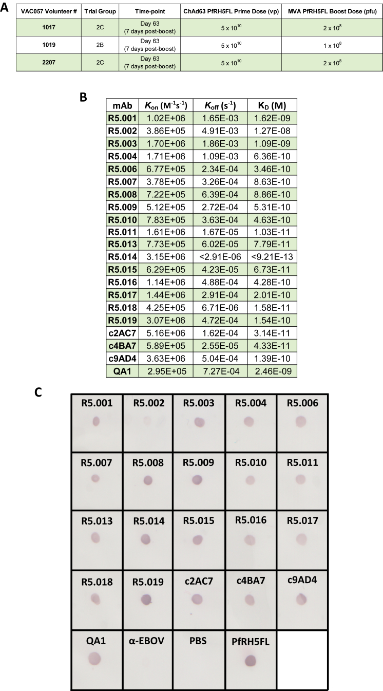
Figureu00a0S1
Clinical Trial (VAC057) Volunteer Information and Additional PfRH5 mAb Binding Data, Related to Figureu00a01 (A) Vaccination details of the three volunteers from which anti-PfRH5FL mAbs were isolated....
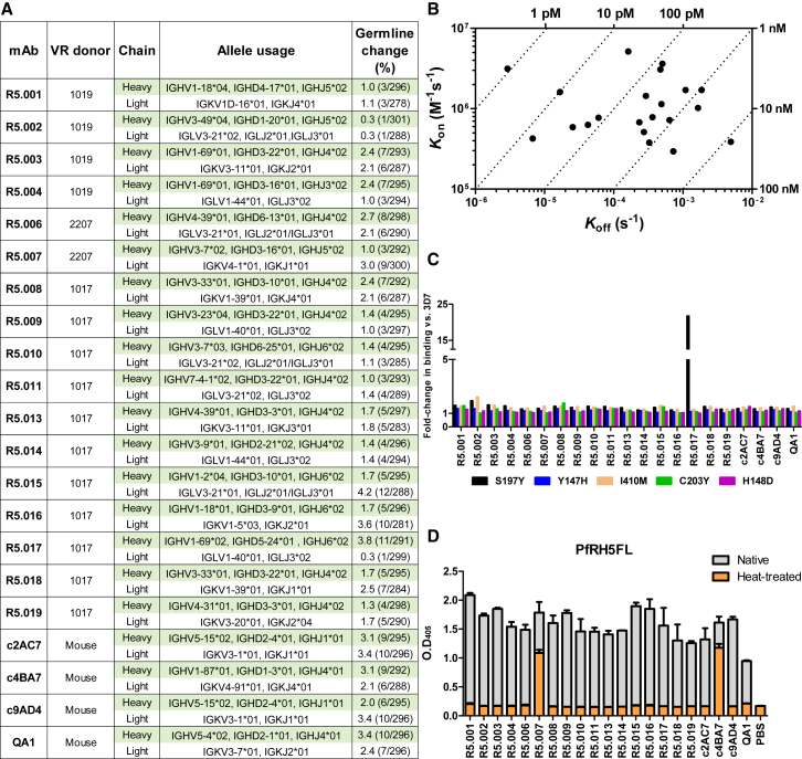
Figureu00a01
Description and Binding Characteristics of Anti-PfRH5 mAbs (A) Genetic lineage of variable regions from PfRH5-specific mAbs and their donor origin, showing percentage of nucleotide substitutions relat...
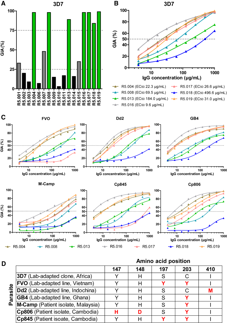
Figureu00a02
Growth Inhibitory Properties of Human PfRH5-Specific mAbs (A) Inu00a0vitro GIA of each mAb tested at 3u00a0mg/mL against 3D7 clone P.u00a0falciparum . Bars are color-coded to reflect potency (u201cGIA...
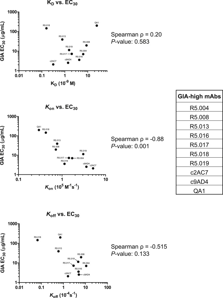
Figureu00a0S2
Correlation between nAb Kinetic Binding Parameters and GIA, Related to Figures 2 and S1 B The binding parameters of nAbs ( K on , K off and K D ) were correlated with growth inhibition using the GIA E...
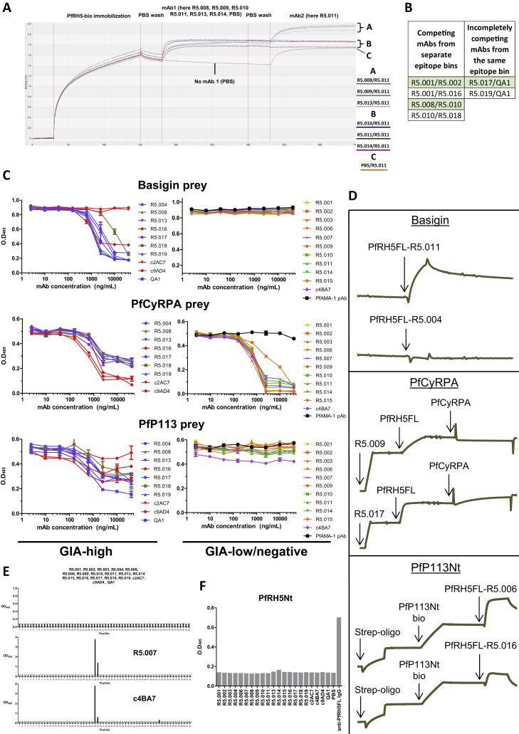
Figureu00a0S3
Further Investigations into Anti-PfRH5 mAb Binding Activity, Related to Figureu00a03 and Table S1 (A) Example graph of raw BLI data used to generate the epitope bins shown in Figureu00a03 A. (B) Table...
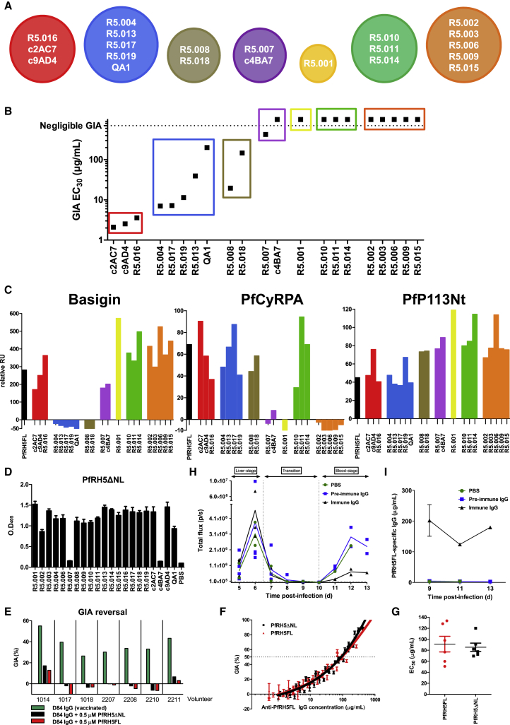
Figureu00a03
Epitope Binning Reveals that All Relevant Neutralizing Epitopes Lie within PfRH5u0394NL (A) Epitope bins determined by BLI from a matrix of sequential PfRH5FL-binding assays for different mAbs, with d...
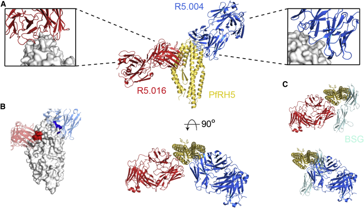
Figureu00a04
Structures of R5.004 and R5.016 Epitopes (A) Structure of PfRH5u0394NL bound to R5.004 and R5.016 Fab fragments. Insets show close-up views of epitopes. (B) The top PfRH5 peptides protected in HDX-MS ...
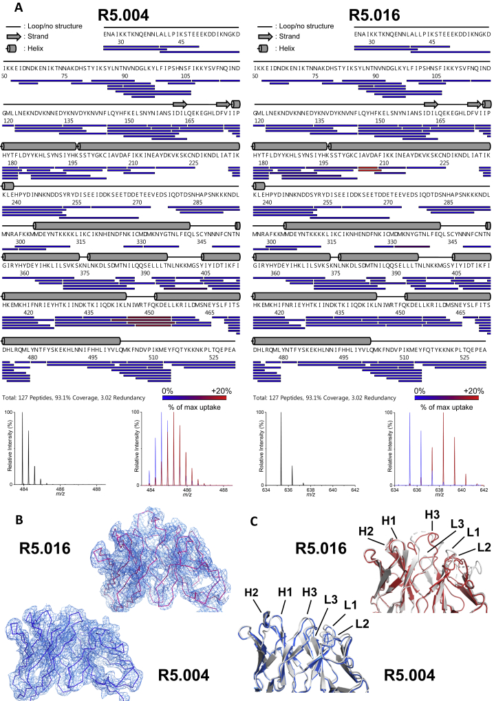
Figureu00a0S4
PfRH5 Peptide Protection in HDX-MS by R5.004 and R5.016 and Description of Bound R5.004 and R5.016 Fab Fragments, Related to Figureu00a04 (A) PfRH5 peptide map showing PfRH5FL protection from deuterat...
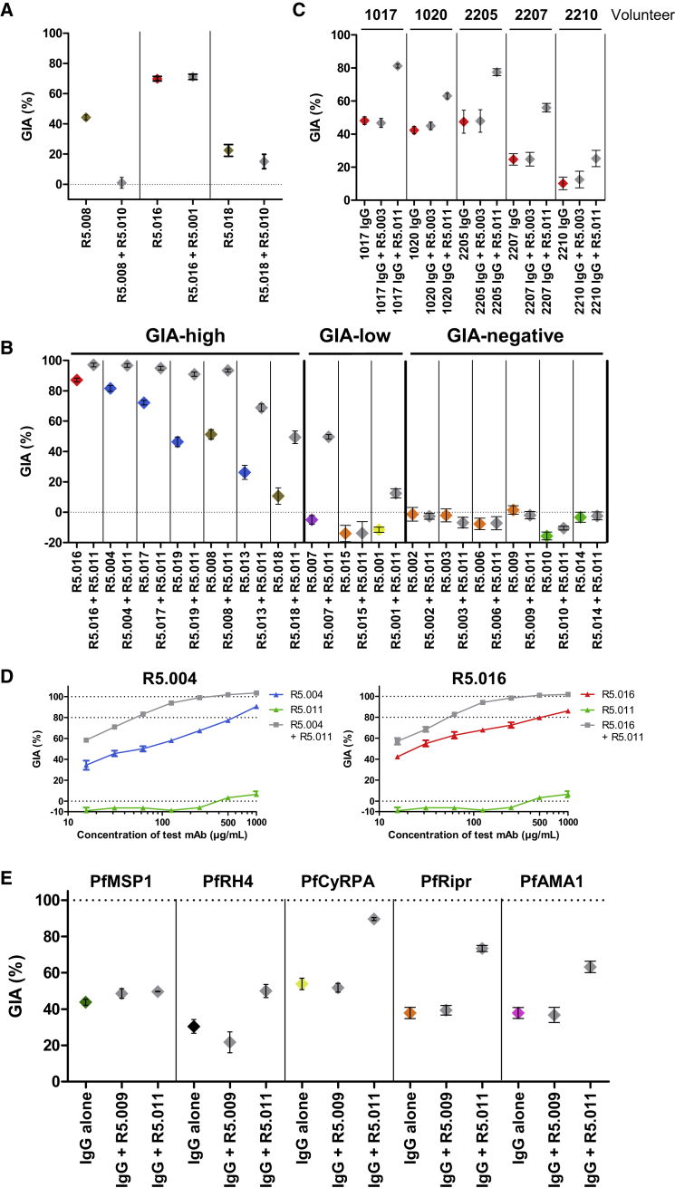
Figureu00a05
Non-neutralizing mAb R5.011 Potentiates the Growth Inhibitory Effect of Anti-PfRH5 nAbs (A) GIA of nAbs R5.008 (300u00a0u03bcg/mL), R5.016 (150u00a0u03bcg/mL), and R5.018 (400u00a0u03bcg/mL) alone (co...
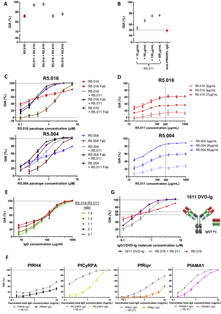
Figureu00a0S5
Investigations into the Effect of R5.011, Related to Figureu00a05 (A) GIA of 150u00a0u03bcg/mL of R5.016 alone or in combination with 150u00a0u03bcg/mL of each mAb from the green epitope bin (and R5.0...
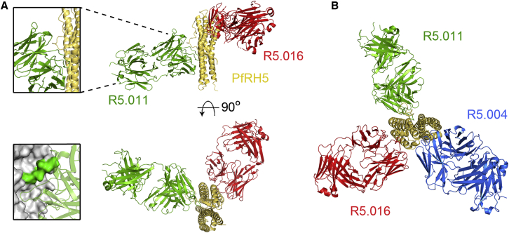
Figureu00a06
Structure of PfRH5u0394NL in Complex with R5.011 and R5.016 (A) Crystal structure of PfRH5u0394NL bound to Fab fragments from R5.011 and R5.016. The top left inset shows a close-up of the R5.011 epito...
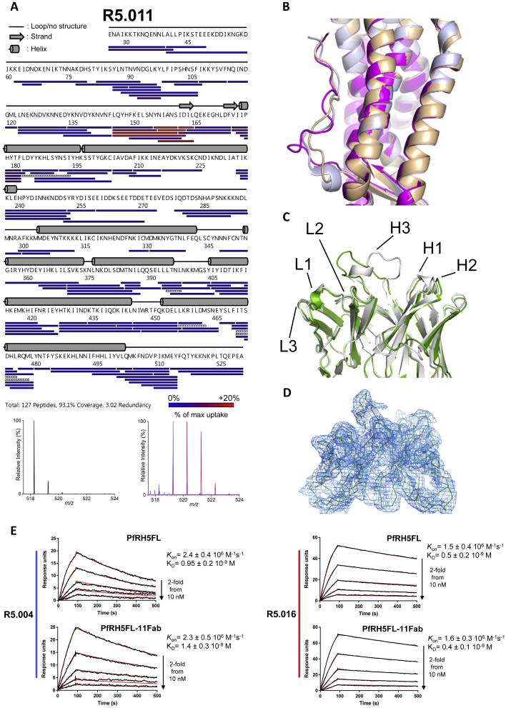
Figureu00a0S6
PfRH5 Peptide Protection in HDX-MS by R5.011 and Description of R5.011 Fab Fragment Interaction with PfRH5, Related to Figureu00a06 (A) Peptide map showing protection of PfRH5FL from deuteration durin...
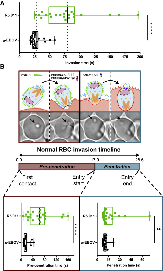
Figureu00a07
R5.011 Increases Parasite Invasion Time (A) Total time for RBC invasion in the presence of R5.011 or an irrelevant isotype-matched antibody control (u03b1-EBOV). (B) Time for early invasion (pre-penet...
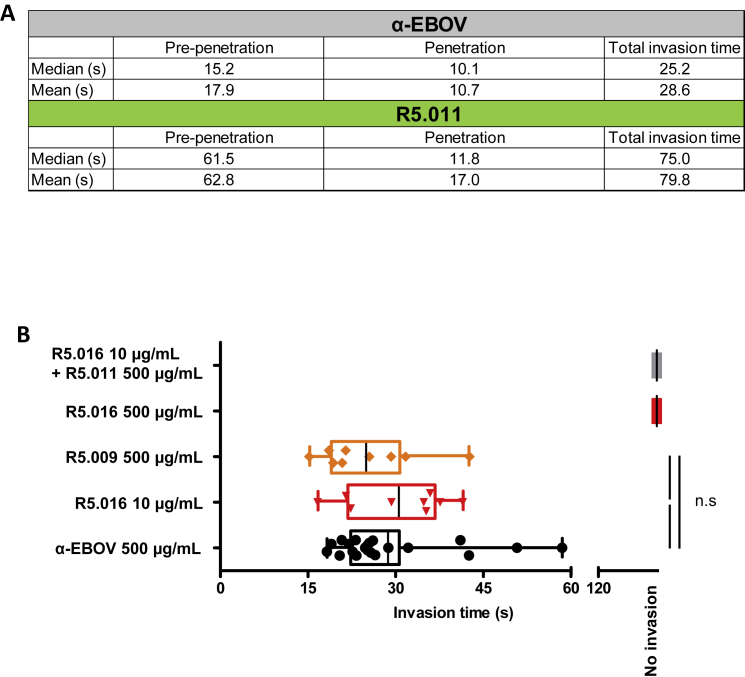
Figureu00a0S7
Additional Information from Live-Cell Microscopy Experiments, Related to Figureu00a07 and Videos S1 , S2 , and S3 (A) Mean and median early (pre-penetration), late (penetration) and total invasion tim...
Figure images are served from the NIH/NLM PubMed Central Open Access Subset or Europe PMC; copyright remains with the publishers and authors.
💬 Discussion
0 commentsNo comments yet. Be the first to start a discussion!
Leave a Comment