Abstract
While genetic screens have identified many genes essential for neurite outgrowth, they have been limited in their ability to identify neural genes that also have earlier critical roles in the gastrula, or neural genes for which maternally contributed RNA compensates for gene mutations in the zygote. To address this, we developed methods to screen the Drosophila genome using RNA-interference (RNAi) on primary neural cells and present the results of the first full-genome RNAi screen in neurons. We used live-cell imaging and quantitative image analysis to characterize the morphological phenotypes of fluorescently labelled primary neurons and glia in response to RNAi-mediated gene knockdown. From the full genome screen, we focused our analysis on 104 evolutionarily conserved genes that when downregulated by RNAi, have morphological defects such as reduced axon extension, excessive branching, loss of fasciculation, and blebbing. To assist in the phenotypic analysis of the large data sets, we generated image analysis algorithms that could assess the statistical significance of the mutant phenotypes. The algorithms were essential for the analysis of the thousands of images generated by the screening process and will become a valuable tool for future genome-wide screens in primary neurons. Our analysis revealed unexpected, essential roles in neurite outgrowth for genes representing a wide range of functional categories including signalling molecules, enzymes, channels, receptors, and cytoskeletal proteins. We also found that genes known to be involved in protein and vesicle trafficking showed similar RNAi phenotypes. We confirmed phenotypes of the protein trafficking genes Sec61alpha and Ran GTPase using Drosophila embryo and mouse embryonic cerebral cortical neurons, respectively. Collectively, our results showed that RNAi phenotypes in primary neural culture can parallel in vivo phenotypes, and the screening technique can be used to identify many new genes that have important functions in the nervous system.
🔬 Techniques
🧬 Organisms
💻 Software
✨ Fluorophores
🧪 Sample Preparation
🔬 Cell Lines
🏭 Microscope Brands
🧪 Reagent Suppliers
💻 Software Details
🏛️ Research Organizations (ROR)
Affiliated research institutions:
📋 Methods
Fly Genetics
The fly stock used to generating primary cell cultures for the full genome screen was w , elav C155 -GAL4 , UAS-mCD8-GFP/FM6;gcm-GAL4/CyO . The gcm-GAL4 line is a product of P-element exchange of the P[GAL4, w + ] with the P[lacZ, ry + ] element in the rA87 enhancer trap line [70] . Flies were raised in large populations and placed in population cages maintained at 25°C. For cell culture, embryos were collected on molasses plates streaked with paste made from autoclaved yeast mixed with water. For in vivo nervous system analysis, the Sec61α l(2)SH0190 [45] and Sec61α k04917 [71] alleles were analyzed. For unambiguous identification of homozygous mutant embryos, the Sec61α alleles were balanced over the CyO , wg en-lacZ chromosome.
Drosophila Cell Culture
Gastrula stage embryos ranging from 6 to 8 hours old were washed from molasses plates with 21°C water, and dechorionated in 50% household bleach in a Nytex-bottomed basket. Embryos were rinsed with sterile distilled water and transferred to Dounce homogenizers containing 100 mL Shields and Sang M3 media (Sigma), and homogenized using 10 pestle strokes. Homogenate was transferred to centrifuge tubes, and debris was pelleted twice with centrifugation at 40 g for 10 min and 5 min. Cells were pelleted by centrifugation at 380 g for 10 min. Cells were resuspended to 2×10 6 cells/mL using Shields and Sang M3 media (Sigma) supplemented with 10 U/mL penicillin, 10 ug/mL streptomycin, and 200 ng/mL insulin.
Full Genome RNAi Screen
At the Drosophila RNAi Screening Center at Harvard Medical School ( http://www.flyrnai.org ), dsRNAs were aliquoted in 5 µL volumes into plastic optical-bottomed, black 384-well plates (Corning), with each well containing 250 ng of dsRNAs ranging from approximately 200–500 bp. Specific details on dsRNA generation are previously reported [72] . 10 µL of primary cell preparation (at 2×10 6 cells/mL) were aliquoted to the dsRNA 384-well plates using a MultiDrop liquid dispenser, and incubated in a humidified chamber for 2 days at 18°C. Following serum-free incubation, cells were supplemented with 30 µL Shields and Sang M3 media (Gibco) containing 5% heat inactivated fetal bovine serum (JRH Biosciences), 10 U/mL penicillin, and 10 ug/mL streptomycin. Cells were incubated an additional 5 days at 18°C in serum-containing media. Following incubation, live, GFP expressing neurons and glia were scored visually, and 512×512 pixel images were collected with a Discovery1 automated widefield microscope (Universal Imaging). Raw images are available upon request. Control dsRNAs for Rho1, GFP, DIAP-1, and Neuroglian were included on each dsRNA plate. GFP dsRNA served effectively as a negative control, since GFP dsRNA wells were phenotypically indistinguishable from other negative controls such as lacZ dsRNA or water. The GFP dsRNA did not significantly knock down GFP signals in the cells due to the two very strong neuronal and glial promoters simultaneously driving GFP expression, in addition to protein perdurance effects. Except for Rho1, GFP, DIAP-1, and Neuroglian controls on each plate, all wells were scored blind. Importantly, dsRNAs for Rho1, DIAP-1, and Neuroglian were also represented in the dsRNA collection, and were detected as hits when scored blind. The primary screen was carried out on 1–2 independent replicates. For secondary screening, dsRNAs were resysnthesized as reported previously [73] . The secondary screen dsRNAs were aliquoted to 384-well plates in 2 different orientations to avoid plate location artifacts. The secondary validation screens were carried out on 12 independent replicates. For each gene 2 to 3 additional non-overlapping dsRNA amplicons were generated that had no 17–19 bp matches to other genes. Details of the dsRNAs used are publicly available at the DRSC website: www.flyrnai.org . Digital Image Analysis Image Feature Extraction of Cell Clusters From the automated widefield microscopy images, components of the cell phenotypes were quantified. First, neuron cell clusters sizes were determined using the Otsu method (Otsu, 1979), which chooses a global threshold to minimize the intraclass variance of the background and foreground pixels, to convert a raw intensity image to a binary image. The foreground was divided into three categories: small dots (area≤16 pixels), small cell clusters (16 pixels
Show full methods section
Fly Genetics
The fly stock used to generating primary cell cultures for the full genome screen was w , elav C155 -GAL4 , UAS-mCD8-GFP/FM6;gcm-GAL4/CyO . The gcm-GAL4 line is a product of P-element exchange of the P[GAL4, w + ] with the P[lacZ, ry + ] element in the rA87 enhancer trap line [70] . Flies were raised in large populations and placed in population cages maintained at 25°C. For cell culture, embryos were collected on molasses plates streaked with paste made from autoclaved yeast mixed with water. For in vivo nervous system analysis, the Sec61α l(2)SH0190 [45] and Sec61α k04917 [71] alleles were analyzed. For unambiguous identification of homozygous mutant embryos, the Sec61α alleles were balanced over the CyO , wg en-lacZ chromosome.
Drosophila Cell Culture
Gastrula stage embryos ranging from 6 to 8 hours old were washed from molasses plates with 21°C water, and dechorionated in 50% household bleach in a Nytex-bottomed basket. Embryos were rinsed with sterile distilled water and transferred to Dounce homogenizers containing 100 mL Shields and Sang M3 media (Sigma), and homogenized using 10 pestle strokes. Homogenate was transferred to centrifuge tubes, and debris was pelleted twice with centrifugation at 40 g for 10 min and 5 min. Cells were pelleted by centrifugation at 380 g for 10 min. Cells were resuspended to 2×10 6 cells/mL using Shields and Sang M3 media (Sigma) supplemented with 10 U/mL penicillin, 10 ug/mL streptomycin, and 200 ng/mL insulin.
Full Genome RNAi Screen
At the Drosophila RNAi Screening Center at Harvard Medical School ( http://www.flyrnai.org ), dsRNAs were aliquoted in 5 µL volumes into plastic optical-bottomed, black 384-well plates (Corning), with each well containing 250 ng of dsRNAs ranging from approximately 200–500 bp. Specific details on dsRNA generation are previously reported [72] . 10 µL of primary cell preparation (at 2×10 6 cells/mL) were aliquoted to the dsRNA 384-well plates using a MultiDrop liquid dispenser, and incubated in a humidified chamber for 2 days at 18°C. Following serum-free incubation, cells were supplemented with 30 µL Shields and Sang M3 media (Gibco) containing 5% heat inactivated fetal bovine serum (JRH Biosciences), 10 U/mL penicillin, and 10 ug/mL streptomycin. Cells were incubated an additional 5 days at 18°C in serum-containing media. Following incubation, live, GFP expressing neurons and glia were scored visually, and 512×512 pixel images were collected with a Discovery1 automated widefield microscope (Universal Imaging). Raw images are available upon request. Control dsRNAs for Rho1, GFP, DIAP-1, and Neuroglian were included on each dsRNA plate. GFP dsRNA served effectively as a negative control, since GFP dsRNA wells were phenotypically indistinguishable from other negative controls such as lacZ dsRNA or water. The GFP dsRNA did not significantly knock down GFP signals in the cells due to the two very strong neuronal and glial promoters simultaneously driving GFP expression, in addition to protein perdurance effects. Except for Rho1, GFP, DIAP-1, and Neuroglian controls on each plate, all wells were scored blind. Importantly, dsRNAs for Rho1, DIAP-1, and Neuroglian were also represented in the dsRNA collection, and were detected as hits when scored blind. The primary screen was carried out on 1–2 independent replicates. For secondary screening, dsRNAs were resysnthesized as reported previously [73] . The secondary screen dsRNAs were aliquoted to 384-well plates in 2 different orientations to avoid plate location artifacts. The secondary validation screens were carried out on 12 independent replicates. For each gene 2 to 3 additional non-overlapping dsRNA amplicons were generated that had no 17–19 bp matches to other genes. Details of the dsRNAs used are publicly available at the DRSC website: www.flyrnai.org . Digital Image Analysis Image Feature Extraction of Cell Clusters From the automated widefield microscopy images, components of the cell phenotypes were quantified. First, neuron cell clusters sizes were determined using the Otsu method (Otsu, 1979), which chooses a global threshold to minimize the intraclass variance of the background and foreground pixels, to convert a raw intensity image to a binary image. The foreground was divided into three categories: small dots (area≤16 pixels), small cell clusters (16 pixels
📊 Figures
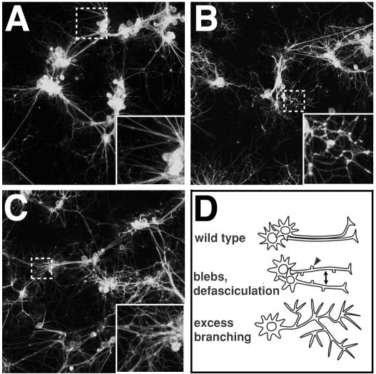
Figure 1
Morphological phenotype features and gene classes observed in RNAi screen.
(Au2013C) Confocal micrographs of live, GFP-labeled neurons and glia. Inset on bottom right hand corner is enlargement of dotted small box areas in same image. (A) Wild-type (no dsRNA) control culture...
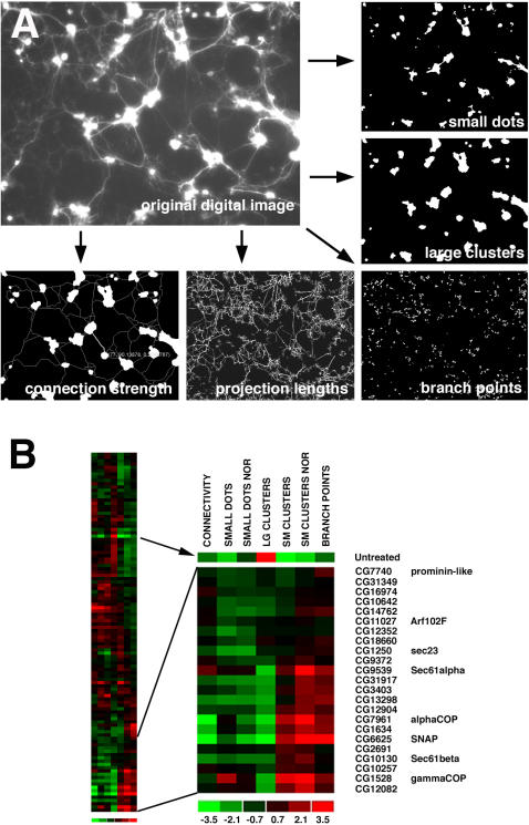
Figure 2
Quantification of RNAi phenotypes and cluster analysis.
(A) Screen images acquired on a robotic widefield fluorescence microscope were analyzed by custom image analysis software. Image features relating to the health of the cultures were extracted using fi...
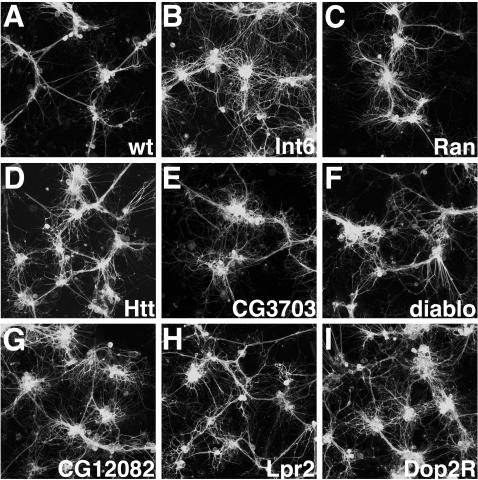
Figure 3
Confocal micrographs of live GFP-labeled primary neural cultures treated with dsRNAs showing altered cell morphologies.
All cultures were grown on glass coverslips coated with poly-L-lysine. (A) Wild type (negative control). Wild type primary neurons in a mature culture show cell body clusters interconnected by well-fa...
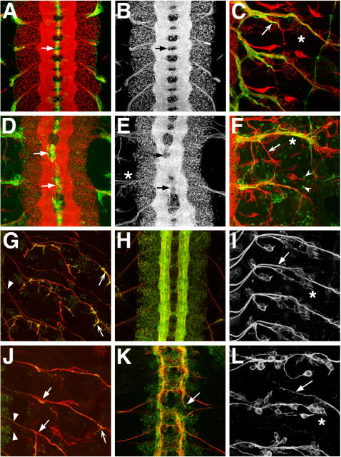
Figure 4
Phenotypes of wild type and Sec61u03b1 mutant embryonic nervous systems.
(Au2013C, Gu2013I) Wild type. (Du2013F) Sec61u03b1 SH190 homozygous mutant. (Ju2013L) Sec61u03b1 k04917 homozygous mutant. Anterior is to the top. PNS images have CNS off to the left in C,F,G,I,J,L. (...
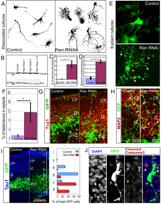
Figure 5
Ran knock down by RNAi results in abnormal neurite morphology in mouse neurons.
(A) Cortices from E14 embryos were transfected by electroporation with either a control GFP plasmid or Ran RNAi contructs, and then dissociated and cultured for 96 hours. Analysis of dissociated neuro...
Figure images are served from the NIH/NLM PubMed Central Open Access Subset or Europe PMC; copyright remains with the publishers and authors.
💬 Discussion
0 commentsNo comments yet. Be the first to start a discussion!
Leave a Comment