Abstract
The lipid bilayer of model membranes, liposomes reconstituted from cell lipids, and plasma membrane vesicles and spheres can separate into two distinct liquid phases to yield lipid domains with liquid-ordered and liquid-disordered properties. These observations are the basis of the lipid raft hypothesis that postulates the existence of cholesterol-enriched ordered-phase lipid domains in cell membranes that could regulate protein mobility, localization and interaction. Here we review the evidence that nano-scaled lipid complexes and meso-scaled lipid domains exist in cell membranes and how new fluorescence microscopy techniques that overcome the diffraction limit provide new insights into lipid organization in cell membranes.
🔬 Techniques
✨ Fluorophores
🧪 Sample Preparation
🔬 Cell Lines
🧪 Reagent Suppliers
🏛️ Research Organizations (ROR)
Affiliated research institutions:
📊 Figures
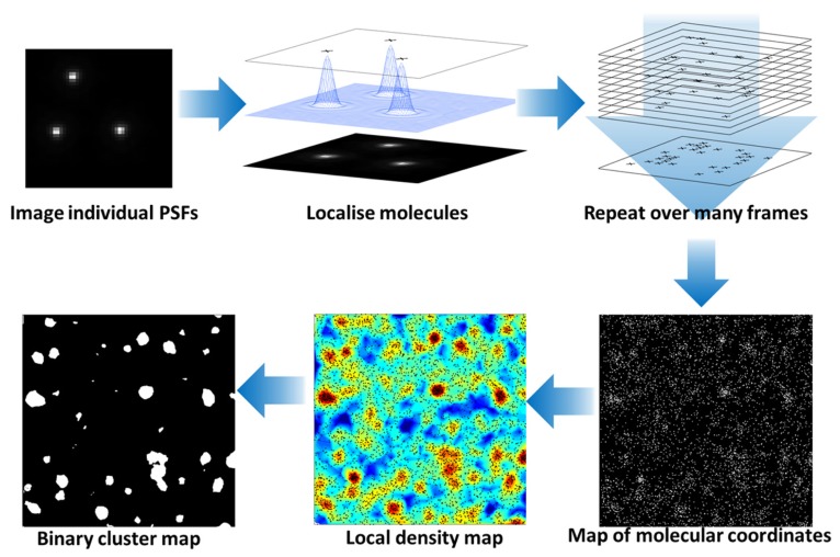
FIGURE 1
Photoactivated localization microscopy analysis of protein clustering at the cell surface. Photo-activation or stable dark states of fluorophores are exploited to limit the number of fluorescent molec...
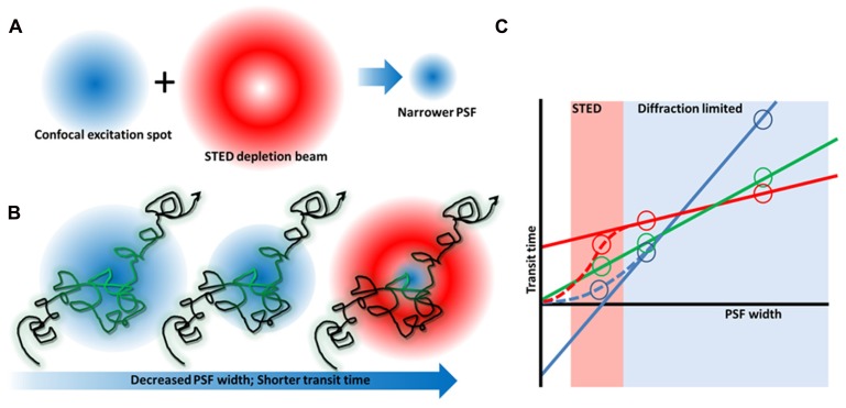
FIGURE 2
Stimulated emission depletion microscopy microscopy and FCS diffusion laws. (A) By combining a red-shifted, u201cdoughnutu201d-shaped depletion beam with a confocal laser beam, the excitation spot and...
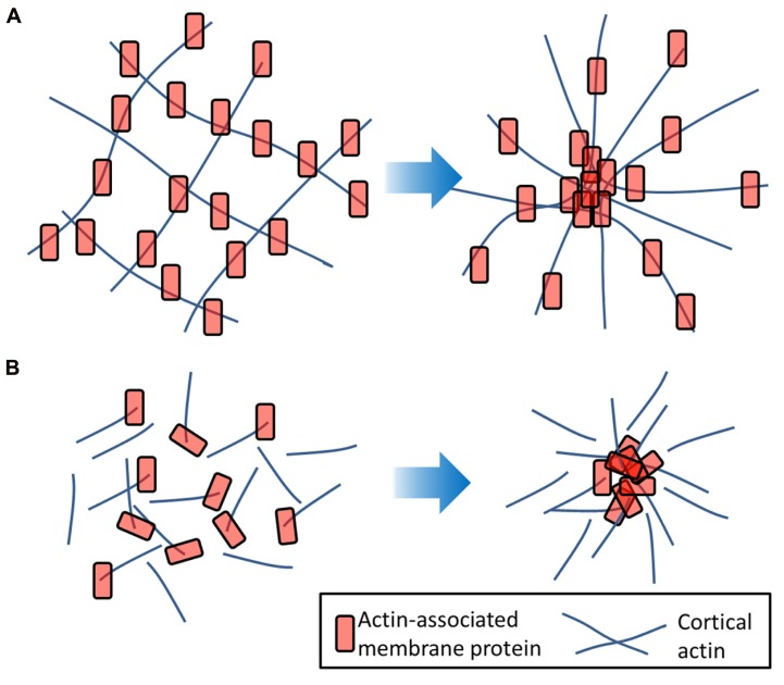
FIGURE 3
Actin-induced clustering of membrane proteins. (A) Membrane molecules (red) which are tethered to the underlying actin filaments (blue) may undergo transient clustering as the actin mesh flexes. (B) P...
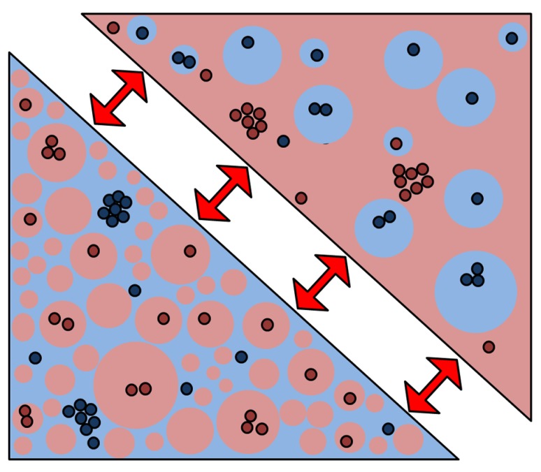
FIGURE 4
A new membrane model based on percolating phase switching. A new membrane model emerged from our data that indicates that the plasma membrane is indeed a mixture of ordered (red) and disordered (blue)...
Figure images are served from the NIH/NLM PubMed Central Open Access Subset or Europe PMC; copyright remains with the publishers and authors.
💬 Discussion
0 commentsNo comments yet. Be the first to start a discussion!
Leave a Comment