Abstract
Intravital microscopy is increasingly used to visualize and quantitate dynamic biological processes at the (sub)cellular level in live animals. By visualizing tissues through imaging windows, individual cells (e.g., cancer, host, or stem cells) can be tracked and studied over a time-span of days to months. Several imaging windows have been developed to access tissues including the brain, superficial fascia, mammary glands, liver, kidney, pancreas, and small intestine among others. Here, we review the development of imaging windows and compare the most commonly used long-term imaging windows for cancer biology: the cranial imaging window, the dorsal skin fold chamber, the mammary imaging window, and the abdominal imaging window. Moreover, we provide technical details, considerations, and trouble-shooting tips on the surgical procedures and microscopy setups for each imaging window and explain different strategies to assure imaging of the same area over multiple imaging sessions. This review aims to be a useful resource for establishing the long-term intravital imaging procedure.
🔬 Techniques
🔭 Microscopes
💻 Software
✨ Fluorophores
🧪 Sample Preparation
🔬 Cell Lines
🏭 Microscope Brands
🧪 Reagent Suppliers
💻 Software Details
📋 Protocols
🏛️ Research Organizations (ROR)
Affiliated research institutions:
📊 Figures
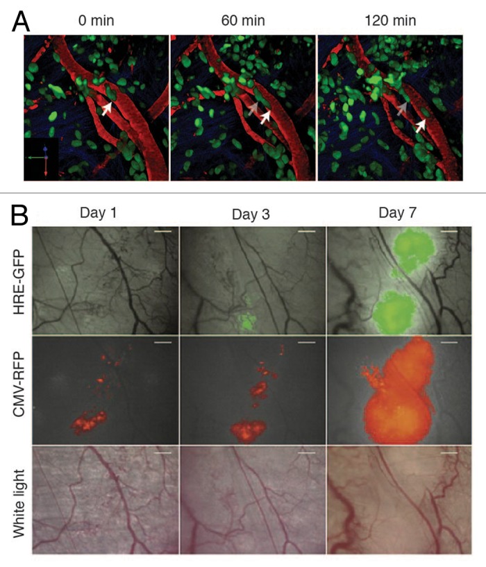
Figure 1
Figureu00a01. Intravital microscopy of tumors through imaging windows. ( A ) A 3D reconstruction of a murine orthotopic glioma (GL261-H2B Dendra2). The time-series show single cells migrating along th...
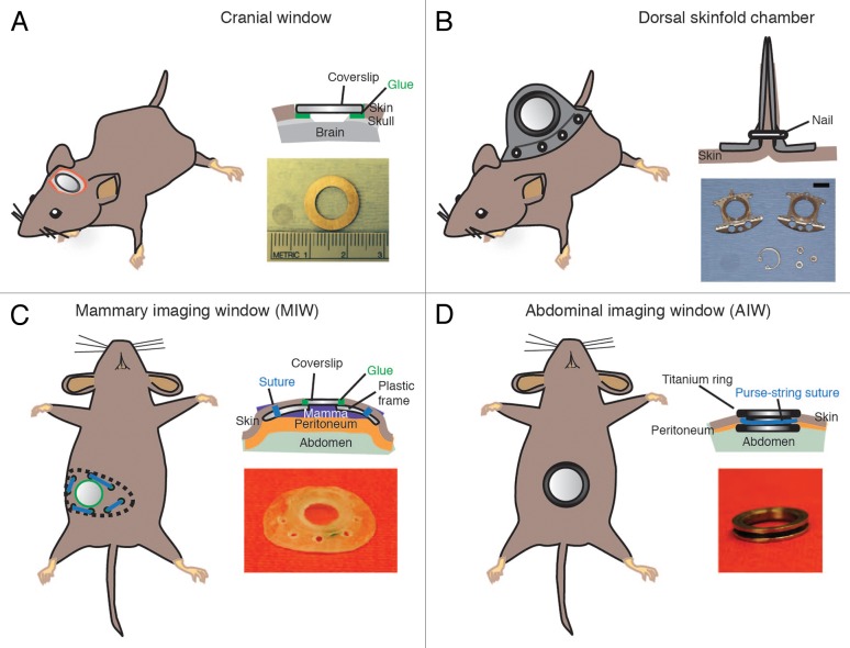
Figure 2
Figureu00a02. Most frequently used imaging windows in cancer research. ( A ) The CIW: the coverslip is glued onto the skull. The image depicts a coverslip and a metal ring. ( B ) The DSC: the metal fr...
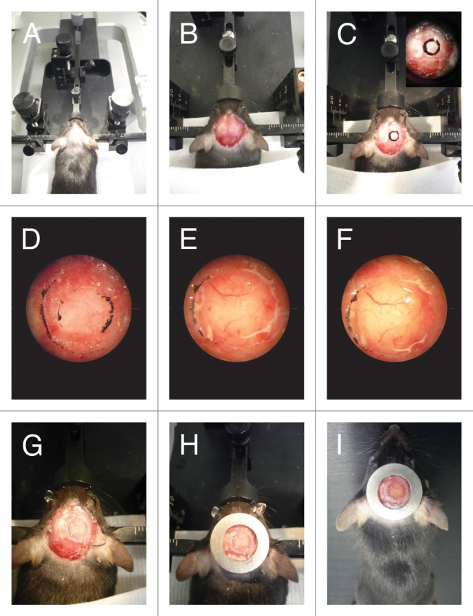
Figure 3
Figureu00a03. CIW surgery. ( A ) The mouse head is shaved and fixed on a stereotaxic frame. ( B ) The skin is cut in a circular manner around the skull and the periosteum is scraped. ( C ) 5mm diamete...
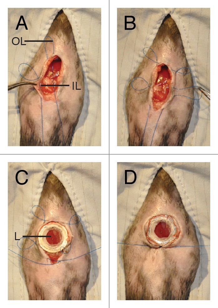
Figure 4
Figureu00a04. Purse-string suture fixes the AIW into the animal. ( A ) A purse-string suture is placed. The suture is first placed through skin and then through opposing abdominal wall. The outer loop...
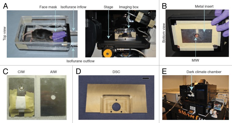
Figure 5
Figureu00a05. Fixation of the window during imaging. (A) A custom-designed imaging box that can be mounted on top of the stage of an inverted microscope. Reprinted from 31 by permission from Macmillan...
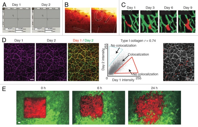
Figure 6
Figureu00a06. Retracing of the exact same field of view over multiple days. ( A ) A microscope with a motorized stage allows storage of regions of interest that can be imported at request. Reprinted f...
Figure images are served from the NIH/NLM PubMed Central Open Access Subset or Europe PMC; copyright remains with the publishers and authors.
💬 Discussion
0 commentsNo comments yet. Be the first to start a discussion!
Leave a Comment