Abstract
Malaria is initiated when the mosquito introduces sporozoites into the skin of a mammalian host. To successfully continue the infection, sporozoites must invade blood vessels in the dermis and be transported to the liver. A significant number of sporozoites, however, may enter lymphatic vessels in the skin or remain in the skin long after the mosquito bite. We have used fluorescence microscopy of Plasmodium berghei sporozoites expressing a fluorescent protein to evaluate the kinetics of sporozoite disappearance from the skin. Sporozoites injected into immunized mice were rapidly immobilized, did not appear to invade dermal blood vessels and became morphologically degraded within several hours. Strikingly, mosquitoes introduced significantly fewer sporozoites into immunized than into non-immunized mice, presumably by formation of an immune complex between soluble sporozoite antigens in the mosquito saliva and homologous host antibodies at the proboscis tip. These results indicate that protective antibodies directed against sporozoites may function both by reducing the numbers of sporozoites injected into immunized hosts and by inhibiting the movement of injected sporozoites into dermal blood vessels.
🔬 Techniques
🔭 Microscopes
🧬 Organisms
✨ Fluorophores
🧪 Sample Preparation
🏭 Microscope Brands
🧪 Reagent Suppliers
💻 Software Details
🏛️ Research Organizations (ROR)
Affiliated research institutions:
📋 Methods
Sporozoites Anopheles stephensi mosquitoes were infected with a clone of the rodent malaria parasite, P. berghei , whose sporozoites constitutively express RedStar, an improved red fluorescent protein [21] . For some studies we used mosquitoes infected with wild-type P. berghei (strain NK65) or P. yoelii (strain 17NXL), neither of whose sporozoites express fluorescent protein. We used standard protocols for infecting and maintaining mosquitoes [22] , which were infected by feeding upon gametocyte-carrying 6–8 wk-old Swiss-Webster mice (Taconic Farms Inc., Germantown, NY). Our protocols for maintenance and use of experimental animals were approved by the Institutional Animal Care and Use Committee at New York University School of Medicine, and our animal facility is accredited by the Association for Assessment and Accreditation of Laboratory Animal Care International (Rockville, MD). Mosquitoes were used for sporozoite transmission studies 18 days after the infective blood meal. Prior to use of infected mosquitoes for feedings observed by intravital microscopy, live, intact mosquitoes were examined by fluorescence microscopy to establish that they had salivary gland infections [23] , [24] ; mosquitoes found to be negative were discarded. Mosquito Feeding on Ear Pinnae For these and other sporozoite transmission studies, mosquitoes fed on BALB/c mice anesthetized by IP injection of ketamine (50 mg/kg) plus xylazine (10 mg/kg) and acepromazine (1.7 mg/kg), and placed on a warming tray. To restrict the area of sporozoite deposition for more efficient counting of sporozoites, the dorsal aspect of one ear pinna was partially masked with tape so that only its edge (8–10 mm long and 2–3 mm wide) was accessible to a feeding mosquito. Mosquitoes, previously selected for having positive salivary gland infections, were kept individually in plastic feeding tubes 2.5 cm in length and with an inside diameter of 1.5 cm; one end of the tube was covered with netting through which the mosquito was able to feed and the other end was closed with a screw-cap. Each mosquito was allowed to probe and feed on the ear through the netting for 3 min from the time that probing was first observed. At appropriate times after each feeding, the fed-upon region of the ear plus the taped adjacent area ∼2.5 mm beyond this was excised. This biopsy specimen was separated into dorsal and ventral leaflets with fine forceps [25] , after which each leaflet was mounted under a coverslip and examined by fluorescence microscopy to count sporozoites and record their distribution [8] . Biopsy specimens were taken either immediately after feeding or at intervals of 1, 2, 3 or 6 h after feeding. Parallel studies were done with mice that had been actively or passively immunized against sporozoites. Fed-upon mice were kept for up to 14 days to obtain blood smears from the tip of the tail; smears were stained with Giemsa and observed by bright field microscopy to detect patent blood infections. This is an extremely sensitive way to establish whether even a single sporozoite has left the skin to develop further in the liver and establish a blood infection. Mosquito Feeding on Ventral Abdomen Mosquitoes were allowed to feed on mice anesthetized as above. Hair was removed from an area of the ventral abdomen with a razor blade. Anesthetized mice were placed on a warming tray, ventral side facing up, and a portion of the abdominal skin was masked with tape that had a 4 mm-diameter hole punched into it. Mosquitoes placed individually in plastic feeding tubes, as above, were allowed to probe and feed through the hole in the tape for 3 min from the time that probing was first observed. Immediately after feeding, the periphery of the feeding circle was marked and the tape was removed. At appropriate times after each feeding, the full depth of skin of a circle 6 mm in diameter, centered around the bite site, was removed with a skin punch device (6 mm Miltex Biopsy Punch) while mice were under deep anesthesia. Then, an underlying circle of peritoneal muscle wall was removed and both incisions were closed [8] . Both biopsy specimens were mounted under cover-slips and viewed through a fluorescence stereoscopic microscope to count sporozoites, as above. We added the numbers found in the skin to the numbers found in peritoneal musculature to obtain the total numbers of sporozoites remaining in the ventral abdomen after mosquito feedings [8] . Biopsy specimens were taken either immediately after feeding or at intervals of 1, 2, 3 or 6 h after feeding. Parallel studies were done with mice that had been actively or passively immunized against sporozoites. For follow-up information, mice, were maintained for blood smears, as above, to detect patent blood infections.
Show full methods section
Sporozoites Anopheles stephensi mosquitoes were infected with a clone of the rodent malaria parasite, P. berghei , whose sporozoites constitutively express RedStar, an improved red fluorescent protein [21] . For some studies we used mosquitoes infected with wild-type P. berghei (strain NK65) or P. yoelii (strain 17NXL), neither of whose sporozoites express fluorescent protein. We used standard protocols for infecting and maintaining mosquitoes [22] , which were infected by feeding upon gametocyte-carrying 6–8 wk-old Swiss-Webster mice (Taconic Farms Inc., Germantown, NY). Our protocols for maintenance and use of experimental animals were approved by the Institutional Animal Care and Use Committee at New York University School of Medicine, and our animal facility is accredited by the Association for Assessment and Accreditation of Laboratory Animal Care International (Rockville, MD). Mosquitoes were used for sporozoite transmission studies 18 days after the infective blood meal. Prior to use of infected mosquitoes for feedings observed by intravital microscopy, live, intact mosquitoes were examined by fluorescence microscopy to establish that they had salivary gland infections [23] , [24] ; mosquitoes found to be negative were discarded. Mosquito Feeding on Ear Pinnae For these and other sporozoite transmission studies, mosquitoes fed on BALB/c mice anesthetized by IP injection of ketamine (50 mg/kg) plus xylazine (10 mg/kg) and acepromazine (1.7 mg/kg), and placed on a warming tray. To restrict the area of sporozoite deposition for more efficient counting of sporozoites, the dorsal aspect of one ear pinna was partially masked with tape so that only its edge (8–10 mm long and 2–3 mm wide) was accessible to a feeding mosquito. Mosquitoes, previously selected for having positive salivary gland infections, were kept individually in plastic feeding tubes 2.5 cm in length and with an inside diameter of 1.5 cm; one end of the tube was covered with netting through which the mosquito was able to feed and the other end was closed with a screw-cap. Each mosquito was allowed to probe and feed on the ear through the netting for 3 min from the time that probing was first observed. At appropriate times after each feeding, the fed-upon region of the ear plus the taped adjacent area ∼2.5 mm beyond this was excised. This biopsy specimen was separated into dorsal and ventral leaflets with fine forceps [25] , after which each leaflet was mounted under a coverslip and examined by fluorescence microscopy to count sporozoites and record their distribution [8] . Biopsy specimens were taken either immediately after feeding or at intervals of 1, 2, 3 or 6 h after feeding. Parallel studies were done with mice that had been actively or passively immunized against sporozoites. Fed-upon mice were kept for up to 14 days to obtain blood smears from the tip of the tail; smears were stained with Giemsa and observed by bright field microscopy to detect patent blood infections. This is an extremely sensitive way to establish whether even a single sporozoite has left the skin to develop further in the liver and establish a blood infection. Mosquito Feeding on Ventral Abdomen Mosquitoes were allowed to feed on mice anesthetized as above. Hair was removed from an area of the ventral abdomen with a razor blade. Anesthetized mice were placed on a warming tray, ventral side facing up, and a portion of the abdominal skin was masked with tape that had a 4 mm-diameter hole punched into it. Mosquitoes placed individually in plastic feeding tubes, as above, were allowed to probe and feed through the hole in the tape for 3 min from the time that probing was first observed. Immediately after feeding, the periphery of the feeding circle was marked and the tape was removed. At appropriate times after each feeding, the full depth of skin of a circle 6 mm in diameter, centered around the bite site, was removed with a skin punch device (6 mm Miltex Biopsy Punch) while mice were under deep anesthesia. Then, an underlying circle of peritoneal muscle wall was removed and both incisions were closed [8] . Both biopsy specimens were mounted under cover-slips and viewed through a fluorescence stereoscopic microscope to count sporozoites, as above. We added the numbers found in the skin to the numbers found in peritoneal musculature to obtain the total numbers of sporozoites remaining in the ventral abdomen after mosquito feedings [8] . Biopsy specimens were taken either immediately after feeding or at intervals of 1, 2, 3 or 6 h after feeding. Parallel studies were done with mice that had been actively or passively immunized against sporozoites. For follow-up information, mice, were maintained for blood smears, as above, to detect patent blood infections.
Immunization
Sporozoites for immunization were first purified on a DEAE ion-exchange column [26] in order to distinguish between effects due to anti-sporozoite vs. anti-saliva immunity. Purified sporozoites were irradiated within a gamma irradiator (MDS Nordion Gammacell® 1000 Elite) to a central dose of ±12049 cGy and a minimum dose of ±10266 cGy. Mice received an IV injection of 50,000 irradiated sporozoites, with 2 subsequent booster injections of 10,000 irradiated sporozoites, each, at 15 days intervals. They were challenged by mosquito bite 15 days after the second boost. Serum from immunized mice was taken the day prior to challenge to assess levels of anti-sporozoite antibodies. Some mice were passively immunized by IV injection of 320 µg per mouse of MoAb 3D11, directed against the repeat region of P. berghei CS protein [3] or with MoAb NYS1, directed against the repeat region of P. yoelii CS protein [9] . All mice were challenged by bite of infected mosquitoes 24 h after antibody transfer. Parallel challenges were done on non-immunized control mice.
Visualisation of Immuno-Complexes Formation at the Injection Site
For these studies MoAb 3D11 was conjugated with FITC prior to injection. For this, 150 µl of 1 mg/ml FITC solution (Fluorescein Isothiocyanate; Sigma, St. Louis, MO) was added to a 2 mg/ml 3D11 solution (or BSA for negative controls) and incubated at room temperature for 1 h. Unbound FITC was then separated from the conjugate with a PD-10 gel filtration column loaded with Sephadex G-25 M (Amersham Biosciences, Piscataway, NJ, USA). Mice were passively immunized by IV-injection of 150 µg per mouse of the conjugates. On the next day, the ear pinnae of these mice were fed upon by mosquitoes infected with P. berghei or P. yoelii sporozoites (none of these sporozoites expressing fluorescent protein) or by non-infected mosquitoes, while intravital fluorescence videomicroscopy observations were made of the feedings, with particular focus on the site of injection of saliva from the distal end of the proboscis. Subsequent to these observations, biopsy specimens were taken from the fed-upon ears and probed with FITC-conjugated Protein A or Protein A/G (Pierce Biotechnology IL, USA; 20 µg/mL). Sporozoite Injection into Drops of Media To study ejection of saliva and sporozoites by mosquitoes into media, we used a modification of the method of Frischknecht et al. (2004) [27] . The feeding stylets of individual mosquitoes immobilized on a microscope slide were positioned under a 13×13 mm coverslip and the mosquito was allowed to salivate into 5 µl of RPMI medium containing 1 mg/µl of either FITC-conjugated BSA (control) or FITC-conjugated MoAb 3D11. Salivation was observed for 10 min by videomicroscopy with the Leica MZ16FA fluorescence stereoscopic microscope, after which counts were made of the sporozoites released into the media.
ELISA Assay
Antibody titers were determined by enzyme-linked immunosorbent assay (ELISA), using the P. berghei -specific B4 multiple Ag peptide (MAP) as antigen (4 branch MAPS, each branch with 3 repeats of DPPPPNPN) from AnaSpec, Inc., San Jose, CA) [28] . Peroxidase-labeled anti-mouse IgG was used as a secondary antibody and 2,2′-azinobis(3-ethylbenthiazolinesulfonic acid) (ABTS) as the substrate. The end point was measured as the highest dilution of serum having a delta O.D. greater than the mean+3 standard deviations obtained with non-immune sera. Results were expressed as geometric mean titers.
Microscopy
For counting of sporozoites, we used a Leica MZ16FA fluorescence stereoscopic microscope with a 2.0× stereoscopic objective lens. Illumination for fluorescence studies was with an EXFO X-Cite 120 F1 illumination system and with a DsRED filter set, restricting illumination to 515–556 nm (peak = 545) and signal emission to 590 nm. Observations of remnant sporozoites and Protein A or A/G staining in the skin of control and immunized animals were performed using a Leica Inverted Laser Scanning Confocal Microscope (Model Number TCS SP2 AOBS) and Leica LCS Software. Each image is the average projection superimposed stacks of individual focal plane images. The total thickness of the stack varies depending upon the position of the sporozoites in the biopsy specimen. Intravital videomicroscopy was done with a Leica DMI 4000B inverted fluorescence microscope with a 10× objective lens. Illumination for fluorescence studies was with a CTR4000 illumination system and with a dual Green/Red filter set, restricting illumination to 480–500 nm (peak = 490) and 560–590 nm (peak = 575) and signal emission to 505 and 600 nm. Images were acquired with a Leica DFC300 FX digital camera and saved as digital files for further analysis and processing. We used Leica Application Suite software (LAS V2.7.1) for documentation and analysis. For 3D reconstruction and volume rendering, raw 3D data set were processed using Imaris 6.1.5 (BitPlane, 2008) software. A Gaussian filter was used for noise reduction on the average projection and Iso-surface objects were created on an intensity value on a per channel basis. Surfaces were colored in green for the green channel (FITC-conjugated Protein A) and red for the red channel ( P. berghei Red Star). Green surfaces were attributed a 50% transparency in order to visualize double staining.
Statistics
The numbers of sporozoites injected by mosquitoes did not follow a Normal distribution but were highly skewed with a clear floor effect. However, when log-transformed [ln (spz count+1)], the data sufficiently approximated a Normal distribution that allowed the use of parametric tests.
Analysis of variance
(ANOVA) examined the effects of site (abdomen vs. ear), immunization status (control vs. immunized) and time (0 to 6 h), as appropriate; Student's t-test (unpaired, 2-tailed) compared means when only two experimental groups were considered. The analyses were performed using SPSS 15.0 for Windows (SPSS Inc., 2006) and GraphPad Prism Version 5 software (San Diego, California.
Supporting Information Video S1 Intravital fluorescence video showing mosquito proboscis introducing saliva into mouse ear pinna after passive immunization with fluorescent antibodies (homologous antigen and antibodies). Mosquitoes had salivary gland infection of Plasmodium berghei sporozoites. Mouse had received passive IV transfer of anti- P. berghei sporozoite monoclonal antibodies conjugated with FITC. Saliva contained sporozoites in addition to secreted, soluble P. berghei CS protein. Green density (left arrow at start of video) shows apparent immune complex formed by interaction between soluble CS protein and homologous antibodies. (Proboscis had been observed at this site just prior to initiation of time-lapse video.) Another green density (right arrow) is seen resulting from probe of another proboscis. Proboscis is visualized as orange due to autofluorescence. During 3-min, real-time duration of video, several additional green densities are formed from probosci that are out of the field of focus. Because of background density caused by FITC-labeled antibodies in tissue, relatively long exposure times of 1 sec per frame were required. Bar = 100 µm for all videos. (2.26 MB MOV) Click here for additional data file. Video S2 Intravital fluorescence video showing mosquito proboscis introducing saliva into mouse ear pinna after passive immunization with fluorescent antibodies (heterologous antigen and antibodies). Mosquitoes had salivary gland infection of Plasmodium yoelii sporozoites. Mouse had received passive IV transfer of anti- P. berghei sporozoite monoclonal antibodies conjugated with FITC. Saliva contained sporozoites in addition to secreted, soluble P. yoelii CS protein. Probing proboscis is visualized as orange due to autofluorescence. No proboscis-associated green densities were ever seen during our observations. (1.30 MB MOV) Click here for additional data file. Video S3 Intravital fluorescence video showing mosquito proboscis introducing saliva into mouse ear pinna after passive immunization with antibodies (negative control). Mosquitoes had no salivary gland infections with sporozoites. Mouse had received passive IV transfer of anti- P. berghei sporozoite monoclonal antibodies conjugated with FITC. Saliva contained neither sporozoites nor sporozoite antigen. Probing proboscis is visualized as orange due to autofluorescence. No proboscis-associated green densities were ever seen during our observations, although secreted droplets of saliva (arrows) can be seen associated with two of the three probes within this sequence. (2.31 MB MOV) Click here for additional data file. Video S4 Secretion of saliva and P. berghei sporozoites from proboscis of immobilized mosquito into medium on microscope slide: Medium contained FITC-conjugated BSA. Sporozoites were freely released into medium. Secretion of saliva can be seen as a dark globule at the tip of proboscis. Green streak running along middle of distal end of proboscis indicates that some medium was being sucked back by mosquito. (Real-time duration of video was 5 min, 32 sec; each frame was captured for 1 sec.) (5.52 MB AVI) Click here for additional data file. Video S5 Secretion of saliva and P. berghei sporozoites from proboscis of immobilized mosquito into medium on microscope slide: Medium contained FITC-conjugated 3D11. Relatively few sporozoites were released into medium. Some of these have become coated with antibody and fluoresce green. Green plug at end of proboscis is associated with stasis of unreleased, red-fluorescing sporozoites within proboscis, suggesting formation of an immune complex that inhibits sporozoite release. (Real-time duration of Video S5 was 2 min, 22 sec; each frame was captured for 1 sec.) (4.31 MB AVI) Click here for additional data file. Video S6 Secretion of saliva and P. berghei sporozoites from proboscis of immobilized mosquito into medium on microscope slide: Medium contained FITC-conjugated 3D11. Relatively few sporozoites were released into medium. Some of these have become coated with antibody and fluoresce green. Green plug at end of proboscis is associated with stasis of unreleased, red-fluorescing sporozoites within proboscis, suggesting formation of an immune complex that inhibits sporozoite release. (Real-time duration of Video S6 was 3 min, 28 sec; each frame was captured for 1 sec.) (3.44 MB AVI) Click here for additional data file.
📊 Figures
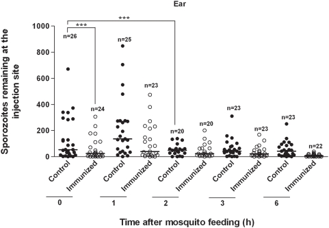
Figure 1
Mosquito injection of Plasmodium berghei sporozoites into mouse ear pinna.
Scatter plot shows numbers of sporozoites remaining at bite site on ear at various times following injection of sporozoites by mosquitoes feeding on non-immunized (control) mice vs. mice actively immu...
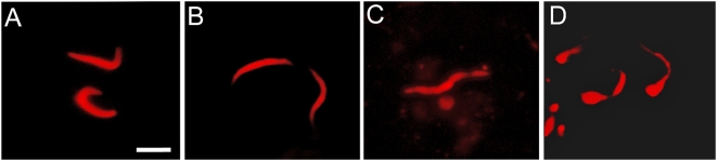
Figure 2
Confocal micrographs of remnant Plasmodium berghei sporozoites in the skin of control vs. immunized mice.
A & B show typical presentation of sporozoites 2 and 4 h, respectively after mosquito inoculation into ear pinnae. C & D show typical presentation of sporozoites 2 and 4 h, respectively, after mosquit...
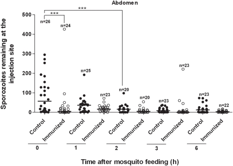
Figure 3
Mosquito injection of Plasmodium berghei sporozoites into mouse ventral abdomen.
Scatter plot shows numbers of sporozoites remaining at bite site in ventral abdomen at various times following injection of sporozoites by mosquitoes feeding on non-immunized (control) mice vs. mice a...
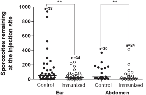
Figure 4
Mosquito injection of Plasmodium berghei sporozoites into mouse ear pinna or ventral abdomen (controls vs. mice passively immunized with anti-sporozoite antibodies).
Scatter plot shows numbers of sporozoites deposited at bite site from biopsy specimens taken immediately after mosquito feeding. Each point shows number of sporozoites left by a single mosquito. (Nu20...
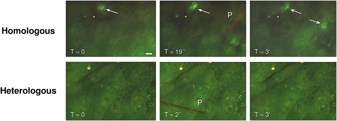
Figure 5
Intravital fluorescence micrographs showing proboscis introducing saliva into mouse ear pinnae after passive immunization with antibodies.
Antibodies directed against repeat region of Plasmodium berghei CS protein had been conjugated with FITC prior to IV injection into mice. Upper Panel: Mosquitoes with salivary gland infection of P. be...
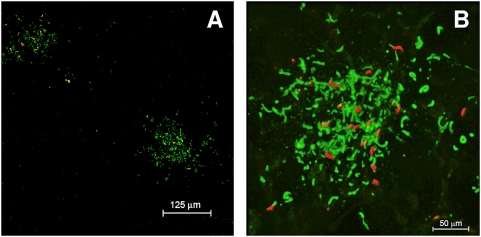
Figure 6
Fluorescence micrographs showing formation of apparent immune complexes after mosquito introduction of saliva and sporozoites into ear pinna of mouse that had been passively immunized with MoAb 3D11.
Following mosquito bite, biopsy specimen of ear was fixed with ice-cold acetone and probed with fluorescein-conjugated Protein A, which specifically binds to the Fc component of antibodies. Fig. 6A sh...
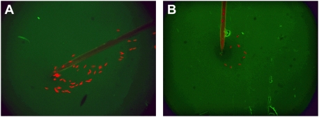
Figure 7
Fluorescence micrographs showing release of sporozoites from proboscis in drops of media on a glass slide.
Fig. 7A shows free release of Plasmodium berghei sporozoites into medium containing BSA conjugated with FITC. Fig. 7B shows release of smaller numbers of sporozoites into medium containing MoAb 3D11(d...
Figure images are served from the NIH/NLM PubMed Central Open Access Subset or Europe PMC; copyright remains with the publishers and authors.
💬 Discussion
0 commentsNo comments yet. Be the first to start a discussion!
Leave a Comment