Abstract
AbstractOptogenetics is increasingly used to map brain activation using techniques that rely on functional hyperaemia, such as opto-fMRI. Here we test whether light stimulation protocols similar to those commonly used in opto-fMRI or to study neurovascular coupling modulate blood flow in mice that do not express light sensitive proteins. Combining two-photon laser scanning microscopy and ultrafast functional ultrasound imaging, we report that in the naive mouse brain, light per se causes a calcium decrease in arteriolar smooth muscle cells, leading to pronounced vasodilation, without excitation of neurons and astrocytes. This photodilation is reversible, reproducible and energy-dependent, appearing at about 0.5 mJ. These results impose careful consideration on the use of photo-activation in studies involving blood flow regulation, as well as in studies requiring prolonged and repetitive stimulations to correct cellular defects in pathological models. They also suggest that light could be used to locally increase blood flow in a controlled fashion.
🔬 Techniques
💻 Software
✨ Fluorophores
🧪 Sample Preparation
🔬 Cell Lines
🏭 Microscope Brands
🧪 Reagent Suppliers
🔴 Lasers
📷 Detectors
🔎 Objectives
💻 Software Details
🏛️ Research Organizations (ROR)
Affiliated research institutions:
📋 Methods
Animal preparation and surgery
All animal care and experimentation was performed in accordance with the INSERM Animal Care and Use Committee guidelines (protocol numbers CEEA34.SC.122.12 and CEEA34.SC.123.12). Adult mice (2–6 months old, 20–35 g, both males and female, housed in 12-h light-dark cycle) were used in this study. Mice strains were obtained from the following suppliers; C57BL/6 , Janvier Labs; Thy1-GCaMP6f (GP5.11) , Jackson laboratory, Ai95(RCL-GCaMP6f) were donated from Hongkui Zeng (Allan Institute), NG2-CreERT2 were donated from Frank Kirchhoff (ULM University). All mice were bread on a C57BL/6 background. To generate mice with conditional GCaMP6f expression in mural cells, NG2-CreERT2 were crossed with Ai95(RCL-GCaMP6f) mice. NG2-CreERT2, Ai95(RCL-GCaMP6f) double transgenic mice were administered 2 mg of tamoxifen for 1–3 consecutive days and imaging was done 2–8 weeks later. The same strategy was used to generate mice with specific expression in astrocytes except that a Connexin30-CreERT2 (donated from Frank W Pfrieger), was crossed with a Ai95(RCL-GCaMP6f) mouse. Chronic craniotomies were performed as previously described 51 . In brief, mice were initially anesthetized with an intraperitoneal (IP) bolus of ketamine-xylazine (100 mg kg −1 and 10 mg kg −1 body mass, respectively). Further 10–20% of the same mixture was injected IP as necessary to maintain surgical plane anaesthesia. During surgery, the mice breathed a mixture of air and supplementary oxygen and the body temperature was monitored by a rectal probe and maintained at ∼36.5 °C by a feedback-controlled heating pad. A craniotomy was performed with a dental drill and care taken not to apply pressure to the bone and the area was regularly flushed with cool aqueous buffer solution to avoid damage or heating of the underlying tissue. Either a cover glass (100 μm thick) or Polymethylpentene (PMP) (250 μm thick) was used for the window and sealed in place with photopolymerizable dental cement, which was also used to form a head-cap in which a titanium head-bar was also embedded. The following veterinarian medications were used pre-, during and post-surgery; the anti-inflammatory, dexamethasone (Dexazone, 5 mg kg −1 body mass), administered once daily by subcutaneous injection pre-surgically and one day post-surgery; the analgesic, buprenorphine (Buprecare, 0.1 mg kg −1 body mass), administered by subcutaneous injection after the surgery and the following post-surgical day if necessary; The antibiotic enrofloxacine (Baytril, 5 mg kg −1 body mass), administered by subcutaneous injection pre-surgically and for two days post-surgery. Mice were permitted to recover for at least 1 week, before the experimental sessions began. For experiments mice were anesthetized with ketamine-xylazine (100 mg and 10 mg kg −1 body mass, respectively) injected IP. Experiments were performed within 20–120 min following injection of anaesthetics. Depth of anaesthesia was monitored with breathing rate (2–3 Hz) recorded by a pneumogram transducer (Biopac Systems) and toe pinch reflex. Body temperature was maintained at ∼36.5 °C using a heating pad. In a subset of experiments performed under isoflurane anaesthesia ( Fig. 5 ), mice were induced with 3% isoflurane for 1.5 min. For experiments, total flow to the nose cone was (1 L of air/min); (700 ml air/min) from the isoflurane apparatus set between 1 and 1.5% isoflurane, and an additional (300 ml air per min) from the olfactometer to deliver odour, thereby diluting the isoflurane concentration arriving at the nose cone to 0.7–1.1%. For acute kidney experiments, the same anaesthetics were used as for the chronic window implantation. A ∼2 cm incision was made on the skin to expose the kidney. A plastic palette was gently inserted in between the kidney and the diaphragm to prevent movement artefacts during the fUS experiments. The optic fibre was placed directly on the surface of the kidney and ultrasound gel was gently placed between the ultrasound probe and the kidney.
Show full methods section
Animal preparation and surgery
All animal care and experimentation was performed in accordance with the INSERM Animal Care and Use Committee guidelines (protocol numbers CEEA34.SC.122.12 and CEEA34.SC.123.12). Adult mice (2–6 months old, 20–35 g, both males and female, housed in 12-h light-dark cycle) were used in this study. Mice strains were obtained from the following suppliers; C57BL/6 , Janvier Labs; Thy1-GCaMP6f (GP5.11) , Jackson laboratory, Ai95(RCL-GCaMP6f) were donated from Hongkui Zeng (Allan Institute), NG2-CreERT2 were donated from Frank Kirchhoff (ULM University). All mice were bread on a C57BL/6 background. To generate mice with conditional GCaMP6f expression in mural cells, NG2-CreERT2 were crossed with Ai95(RCL-GCaMP6f) mice. NG2-CreERT2, Ai95(RCL-GCaMP6f) double transgenic mice were administered 2 mg of tamoxifen for 1–3 consecutive days and imaging was done 2–8 weeks later. The same strategy was used to generate mice with specific expression in astrocytes except that a Connexin30-CreERT2 (donated from Frank W Pfrieger), was crossed with a Ai95(RCL-GCaMP6f) mouse. Chronic craniotomies were performed as previously described 51 . In brief, mice were initially anesthetized with an intraperitoneal (IP) bolus of ketamine-xylazine (100 mg kg −1 and 10 mg kg −1 body mass, respectively). Further 10–20% of the same mixture was injected IP as necessary to maintain surgical plane anaesthesia. During surgery, the mice breathed a mixture of air and supplementary oxygen and the body temperature was monitored by a rectal probe and maintained at ∼36.5 °C by a feedback-controlled heating pad. A craniotomy was performed with a dental drill and care taken not to apply pressure to the bone and the area was regularly flushed with cool aqueous buffer solution to avoid damage or heating of the underlying tissue. Either a cover glass (100 μm thick) or Polymethylpentene (PMP) (250 μm thick) was used for the window and sealed in place with photopolymerizable dental cement, which was also used to form a head-cap in which a titanium head-bar was also embedded. The following veterinarian medications were used pre-, during and post-surgery; the anti-inflammatory, dexamethasone (Dexazone, 5 mg kg −1 body mass), administered once daily by subcutaneous injection pre-surgically and one day post-surgery; the analgesic, buprenorphine (Buprecare, 0.1 mg kg −1 body mass), administered by subcutaneous injection after the surgery and the following post-surgical day if necessary; The antibiotic enrofloxacine (Baytril, 5 mg kg −1 body mass), administered by subcutaneous injection pre-surgically and for two days post-surgery. Mice were permitted to recover for at least 1 week, before the experimental sessions began. For experiments mice were anesthetized with ketamine-xylazine (100 mg and 10 mg kg −1 body mass, respectively) injected IP. Experiments were performed within 20–120 min following injection of anaesthetics. Depth of anaesthesia was monitored with breathing rate (2–3 Hz) recorded by a pneumogram transducer (Biopac Systems) and toe pinch reflex. Body temperature was maintained at ∼36.5 °C using a heating pad. In a subset of experiments performed under isoflurane anaesthesia ( Fig. 5 ), mice were induced with 3% isoflurane for 1.5 min. For experiments, total flow to the nose cone was (1 L of air/min); (700 ml air/min) from the isoflurane apparatus set between 1 and 1.5% isoflurane, and an additional (300 ml air per min) from the olfactometer to deliver odour, thereby diluting the isoflurane concentration arriving at the nose cone to 0.7–1.1%. For acute kidney experiments, the same anaesthetics were used as for the chronic window implantation. A ∼2 cm incision was made on the skin to expose the kidney. A plastic palette was gently inserted in between the kidney and the diaphragm to prevent movement artefacts during the fUS experiments. The optic fibre was placed directly on the surface of the kidney and ultrasound gel was gently placed between the ultrasound probe and the kidney.
Imaging the mouse brain with fUS
Following anaesthesia the cranial PMP window was rinsed with sterile saline and 1 cm 3 of ultrasound coupling gel was placed between the window and the linear ultrasound probe (15 MHz central frequency, 128 elements; Vermon; Tours, France). The transducer was connected to an ultrafast ultrasound scanner (AixplorerT.M, SuperSonic Imagine; Aix-en-Provence, France). Programming of custom transmit/receive ultrasound sequences was done in Matlab (MathWorks; Natick, Massachusetts, USA), using software-based architecture of the scanner. Ultrasound sequences The concept of ultrafast Doppler relies on compounded plane-wave transmissions 52 . The mouse brain was insonified with a succession of ultrasound plane waves and the backscattered echoes were recorded and beamformed to produce an echographic image for each transmission. Although the frame rate of ultrafast ultrasound can reach more than 10 kHz, a 500 Hz frame rate was used as it allows correct sampling of the ultrasound signals backscattered by the red blood cells without aliasing in the mouse brain 53 . To increase the SNR of each echographic image taken at 500 Hz, the echographic images were compounded by transmitting several tilted plane waves and added their backscattered echoes. The compounded sequence resulted in enhanced echographic images, thereby increasing the sensitivity of the Doppler measurement 54 . In this study, the ultrasound sequence consisted of transmitting eleven different tilted plane waves (−10, −8, −6, −4, −2, 0, 2, 4, 6, 8, 10° tilted angle) with a 5,500 Hz pulse repetition frequency (PRF). The backscattered echoes were added to produce enhanced echographic images at a 500 Hz frame rate.
Power Doppler data treatment
As the backscattered signals from the mouse brain are composed of both tissue and blood signals, the following steps were performed to remove signals from the tissue. First, a singular value decomposition (SVD) was applied on the stack of the fUS images and the largest Eigenvalues were eliminated to filter out the slowest variations in the Power Doppler signal which represented the tissue signal 55 . Next, the backscattered signals were filtered with a fourth order Butterworth high-pass filter with a cut-off frequency of 50 Hz to further remove any tissue or motion artefacts. The Doppler signal of each spatial pixel was obtained by the incoherent temporal mean of the blood signal. The increase in Power Doppler signal (proportional to the cerebral blood volume 54 ) evoked by the light stimulations was measured in each pixel, which were 100 × 100 μm 2 in plane size with a slice thickness of 200 μm. Building activation maps Activation maps were made using average power Doppler signals from 3 to 5 trials. Activated pixels were found using a Pearson correlation coefficient r between the local power Doppler temporal signal computed from each spatial pixel of the fUS acquisition and a temporal binary step pattern from 0 to 1 for a duration of 3 and 4 s (for the brain and the kidney respectively) starting 1.5 s after the start of the light train stimulus. Activations were considered significant for a correlation r >2σ, where σ is the spatial standard deviation of the correlation map. Once activated areas were found, the activation maps were displayed as the percentage increase from baseline of the power Doppler signal, measured for 1 s and 2 s (for the brain and the kidney respectively) following the end of the light train. Power Doppler time course variations for individual pixels are shown in Fig. 1b . Averaged time course plots were calculated by averaging the temporal signal of the 10 most activated pixels contained within the activated region ( r >2σ). In a few cases (particularly in the kidney) 5–10 pixels were significantly activated. Signals were never observed using the same criteria in the absence of light.
Two-photon laser scanning microscopy
Imaging was performed using a femtosecond laser (Mai Tai eHP; SpectraPhysics) with a dispersion compensation module (Deepsee; Spectraphysics) emitted ∼70-fs pulses at 80MHz. Laser power was attenuated by an acoustic optical modulator (AA Optoelectronic, MT110-B50-A1.5-IR-Hk). XY scanning was performed with Galvanometric scanner (GS) mirrors (VM500; GSI Lumonics). GCaMP6 and Texas Red were excited at 920nm. Emitted light was collected with a 40X/0.8NA objective (Leica) and was sent to a pair of lenses, coupled into a 2-mm diameter core polymethyl methacrylate optical fibre as previously described 56 . Collected light was split using a dichroic mirror at 580 nm and the signals were each detected with a dedicated GaAsP photomultiplier tube (Hamamatsu) after passing through an appropriate emission filter (GCaMP6: 525 nm, 50 nm bp; Texas Red: 620 nm, 60 nm bp). Customized Labview software was used to control imaging paramaters. A mechanical shutter was placed directly before the 580 nm dichroic mirror to shield the PMTs during the photostimulation period. The laser light was blocked for a few additional milliseconds before and after the photostimulation period. Texas Red dextran (70 kDa, Molecular Probes) was administered intravenously by retro-orbital or tail vein injection. Analysis of vessel dilations represents maximum absolute value compared with baseline. Smooth muscle cell calcium decreases were averaged over 1 s surrounding the maximum absolute value compared with baseline. For mean values in pericyte and astrocyte experiments, Ca 2+ measurements were averaged during 2 s after light delivery, relative to the 2 s time period before light.
Light and Sensory stimulation
Optical stimulation, was performed with a 473 nm laser (Coblot MLD, Sweden), a dual laser 488 nm/561 nm (Oxxius, France), or 594 nm and 638 nm lasers (Oxxius, France) with FC/PC coupler to deliver the light pulse. The light pulse was triggered through an analogue module to deliver optical stimulations: Trains (20 ms, 20 Hz, 2 s duration) or single continuous pulses (100 ms duration). The multimode optical fibre was 62.5 μm (GIF625; ThorLabs, Germany). The light power delivered from the fibre tip was calibrated using optical power metres (Gentec-eo, Canada) and was measured during continuous mode. For spectrum comparison of 473, 594 and 638 nm lasers, the input to the optical fibre was manually moved between lasers without moving the position of the tip. Un-connecting and reconnecting the fibre from the laser resulted in power changes of
📊 Figures
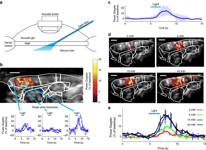
Figure 1
Blue light generates a rapid increase of cerebral blood flow (CBF) in the naive mouse brain.
( a ) Schematic, light emitted by the optic fibre diffuses through a PMP chronic window into the brain. ( b ) A single train of blue light pulses (20u2009ms, 20u2009Hz, 5u2009mW, 2u2009s) reliably gen...
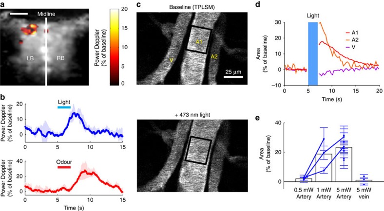
Figure 2
Light-evoked CBF increase results from artery dilation.
( a ) In the olfactory bulb (OB) of the naive mouse, a single train of blue light (20u2009ms, 20u2009Hz, 5u2009mW, 2u2009s) generates a focal increase of the power Doppler signal. Scale bar, 1u2009mm....
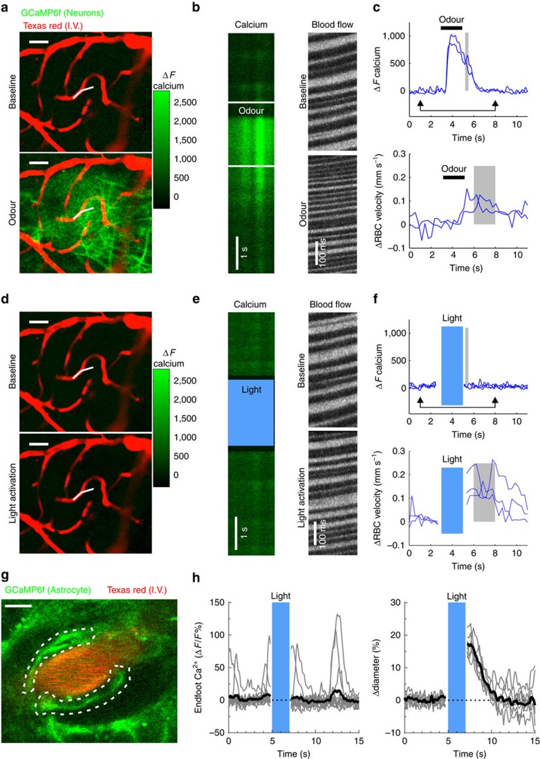
Figure 3
Light increases CBF independently of neuronal or astrocyte Ca 2+ dependent mechanisms.
( a ) Odour causes a large calcium increase in the glomerular layer of a mouse expressing GCaMP6f under the Thy1 promoter. Top, Fluorescence increases robustly in the dendritic tufts of mitral cells d...
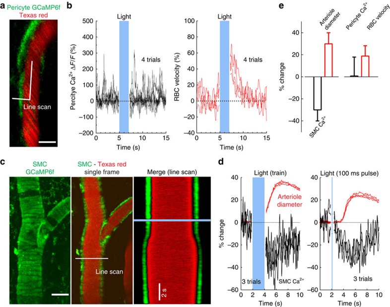
Figure 4
Light triggers dilation via a decrease in SMC calcium.
( a , b ) A glomerulus layer capillary, whose lumen is labelled with Texas red and contacted by the process of a longitudinal-type pericyte expressing GCaMP6f. A broken line scan acquisition to simult...
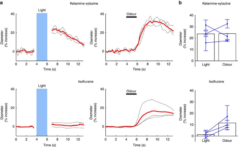
Figure 5
Isoflurane blocks light-triggered dilations.
( a ) The same vessel that dilated to light under ketamine-xylazine anaesthesia (top left) no longer dilated to light when the mouse was anesthetized with isoflurane (bottom left). Right, postive inte...
Figure images are served from the NIH/NLM PubMed Central Open Access Subset or Europe PMC; copyright remains with the publishers and authors.
💬 Discussion
0 commentsNo comments yet. Be the first to start a discussion!
Leave a Comment