Abstract
AbstractLight-sheet fluorescence microscopy has transformed our ability to visualize and quantitatively measure biological processes rapidly and over long time periods. In this review, we discuss current and future developments in light-sheet fluorescence microscopy that we expect to further expand its capabilities. This includes smart and adaptive imaging schemes to overcome traditional imaging trade-offs, i.e., spatiotemporal resolution, field of view and sample health. In smart microscopy, a microscope will autonomously decide where, when, what and how to image. We further assess how image restoration techniques provide avenues to overcome these tradeoffs and how “open top” light-sheet microscopes may enable multi-modal imaging with high throughput. As such, we predict that light-sheet microscopy will fulfill an important role in biomedical and clinical imaging in the future.
🔬 Techniques
🧬 Organisms
✨ Fluorophores
🧪 Sample Preparation
💻 Software Details
🏛️ Research Organizations (ROR)
Affiliated research institutions:
📋 Methods
Methods to overcome scattering and absorption processes To push the volumetric imaging barrier, multi-photon excitation 49 – 51 , wave-front shaping 52 , tissue clearing 53 and expansion microscopy 54 , 55 have been developed to overcome the physical limitations for imaging due to light-scattering and absorption processes. Physical scattering and absorption arises due to tissue-inherent absorbing chromophores such as blood, melanin, water or pigments, and small-and large-scale scatterers in the structure of cells and tissues 56 , 57 . This results in reduced penetration of optical microscopy into tissue, limiting imaging to few tens to hundreds of micrometers from the tissue surface 58 (i.e. one optical mean free path). Multi-photon excitation 49 – 51 has increased the optical penetration depth to more than a mm in some tissues 49 , 59 . Importantly though, light-sheet microscopy does not benefit as strongly from multiphoton excitation than raster scanning techniques do. This is because a light-sheet microscope still needs to form a widefield image, which is severely limited by light-scattering in the visible wavelength. In contrast, multi-photon raster scanning microscopes do not need to form a sharp image with the returning fluorescence photons and as such can go much deeper. Thus, intravital light-sheet microscopy is currently limited to a depth of less than 100 microns in most tissues. As an alternative, a judicious choice of a reasonably translucent model organism, such as zebrafish lines with no pigmentation 60 , has enabled light-sheet imaging in situ and in vivo. Furthermore, tuning the refractive index of the immersion medium better to the sample 61 , 62 reduces scattering and thus improves penetration depth. Also, a shift to fluorescent probes in the near infrared II window (900–1700 nm) has shown promise to increase the reach of light-sheet microscopes in tissues 63 . Longer wavelengths have intrinsically a longer scattering mean free path, and can overlap with the absorption window of biological tissues 64 . Development of probes for this optical window, however, has remained challenging as absorbing and emitting at longer wavelengths necessitates increased electronic conjugation, which is often accompanied with reduced molecular rigidity, increased sources for non-radiative decay, and low quantum yields 65 , 66 . Quantum dots 67 and carbon nanotubes 68 have been used as alternative to fluorescent proteins/dye molecules, but complicate labeling specificity and biocompatibility. Therefore, the future of near infrared light-sheet imaging strongly depends on future breakthroughs in probe development. Additionally, progress in wavefront correction schemes, in particular multi-conjugate adaptive optics (MCAO) 69 , 70 , may increase the optical penetration depth further. MCAO addresses the issue that conventional adaptive optics can only correct a small area, the so called isoplanatic patch 71 , 72 . In tissues, this patch can be smaller than the field of view of the camera, negating the benefits of parallelized detection of light-sheet microscopy. By correcting different regions of tissue separately, MCAO has the potential to increase the isoplanatic patch size 69 , 70 and may enable effective light-sheet imaging in tissues. Conventional adaptive optics for light-sheet microscopy has been demonstrated 73 , 74 , but the setup featured a high complexity. To rapidly correct spatially varying aberrations, dedicated wavefront sensors and deformable mirrors were employed both in the excitation and emission path of the light-sheet microscope. As such, it may seem at first far-fetched to add even more components for MCAO, making such a system overly complex. However, we envision that through machine learning, aberrations can be sensed without dedicated wavefront sensors 75 , 76 significantly easing the equipment constraints. Further, instead of deformable mirrors, transmissive deformable waveplates have shown promise for wavefront correction 77 . In principle, such devices can be stacked in an image space of the microscope to perform MCAO, or a dedicated, integrated 3D wavefront shaping device might be devised. For fixed tissues, sample preparation through tissue clearing 53 can largely overcome the depth limitations associated with light-scattering. Particularly interesting in this context is expansion microscopy 54 , 55 , which can physically magnify a sample tenfold or larger 78 , 79 (Fig. 1d ). This effectively increases the resolving power of any microscope by the expansion factor, and thus enables light-sheet microscopes to reach resolution levels that where hitherto limited to super-resolution microscopy (Fig. 2a ). Therefore, expansion microscopy is a way to overcome the volumetric imaging barrier by modifying the sample, with the caveat that the expansion process might not always preserve the ultrastructure and careful validation is needed 80 . The challenge is now to image the thousandfold larger volumes effectively, which will even test the most efficient volumetric light-sheet microscopes. As expansion microscopy progresses, we see further need to engineer novel light-sheet microscopes with ever larger field of view, larger cameras and working distances. Also, techniques that rapidly tile 81 , 82 or scan 39 the light-sheet to cover large field of views might become more necessary in this quest.
Show full methods section
Methods to overcome scattering and absorption processes To push the volumetric imaging barrier, multi-photon excitation 49 – 51 , wave-front shaping 52 , tissue clearing 53 and expansion microscopy 54 , 55 have been developed to overcome the physical limitations for imaging due to light-scattering and absorption processes. Physical scattering and absorption arises due to tissue-inherent absorbing chromophores such as blood, melanin, water or pigments, and small-and large-scale scatterers in the structure of cells and tissues 56 , 57 . This results in reduced penetration of optical microscopy into tissue, limiting imaging to few tens to hundreds of micrometers from the tissue surface 58 (i.e. one optical mean free path). Multi-photon excitation 49 – 51 has increased the optical penetration depth to more than a mm in some tissues 49 , 59 . Importantly though, light-sheet microscopy does not benefit as strongly from multiphoton excitation than raster scanning techniques do. This is because a light-sheet microscope still needs to form a widefield image, which is severely limited by light-scattering in the visible wavelength. In contrast, multi-photon raster scanning microscopes do not need to form a sharp image with the returning fluorescence photons and as such can go much deeper. Thus, intravital light-sheet microscopy is currently limited to a depth of less than 100 microns in most tissues. As an alternative, a judicious choice of a reasonably translucent model organism, such as zebrafish lines with no pigmentation 60 , has enabled light-sheet imaging in situ and in vivo. Furthermore, tuning the refractive index of the immersion medium better to the sample 61 , 62 reduces scattering and thus improves penetration depth. Also, a shift to fluorescent probes in the near infrared II window (900–1700 nm) has shown promise to increase the reach of light-sheet microscopes in tissues 63 . Longer wavelengths have intrinsically a longer scattering mean free path, and can overlap with the absorption window of biological tissues 64 . Development of probes for this optical window, however, has remained challenging as absorbing and emitting at longer wavelengths necessitates increased electronic conjugation, which is often accompanied with reduced molecular rigidity, increased sources for non-radiative decay, and low quantum yields 65 , 66 . Quantum dots 67 and carbon nanotubes 68 have been used as alternative to fluorescent proteins/dye molecules, but complicate labeling specificity and biocompatibility. Therefore, the future of near infrared light-sheet imaging strongly depends on future breakthroughs in probe development. Additionally, progress in wavefront correction schemes, in particular multi-conjugate adaptive optics (MCAO) 69 , 70 , may increase the optical penetration depth further. MCAO addresses the issue that conventional adaptive optics can only correct a small area, the so called isoplanatic patch 71 , 72 . In tissues, this patch can be smaller than the field of view of the camera, negating the benefits of parallelized detection of light-sheet microscopy. By correcting different regions of tissue separately, MCAO has the potential to increase the isoplanatic patch size 69 , 70 and may enable effective light-sheet imaging in tissues. Conventional adaptive optics for light-sheet microscopy has been demonstrated 73 , 74 , but the setup featured a high complexity. To rapidly correct spatially varying aberrations, dedicated wavefront sensors and deformable mirrors were employed both in the excitation and emission path of the light-sheet microscope. As such, it may seem at first far-fetched to add even more components for MCAO, making such a system overly complex. However, we envision that through machine learning, aberrations can be sensed without dedicated wavefront sensors 75 , 76 significantly easing the equipment constraints. Further, instead of deformable mirrors, transmissive deformable waveplates have shown promise for wavefront correction 77 . In principle, such devices can be stacked in an image space of the microscope to perform MCAO, or a dedicated, integrated 3D wavefront shaping device might be devised. For fixed tissues, sample preparation through tissue clearing 53 can largely overcome the depth limitations associated with light-scattering. Particularly interesting in this context is expansion microscopy 54 , 55 , which can physically magnify a sample tenfold or larger 78 , 79 (Fig. 1d ). This effectively increases the resolving power of any microscope by the expansion factor, and thus enables light-sheet microscopes to reach resolution levels that where hitherto limited to super-resolution microscopy (Fig. 2a ). Therefore, expansion microscopy is a way to overcome the volumetric imaging barrier by modifying the sample, with the caveat that the expansion process might not always preserve the ultrastructure and careful validation is needed 80 . The challenge is now to image the thousandfold larger volumes effectively, which will even test the most efficient volumetric light-sheet microscopes. As expansion microscopy progresses, we see further need to engineer novel light-sheet microscopes with ever larger field of view, larger cameras and working distances. Also, techniques that rapidly tile 81 , 82 or scan 39 the light-sheet to cover large field of views might become more necessary in this quest.
Supplementary information Reporting Summary
📊 Figures
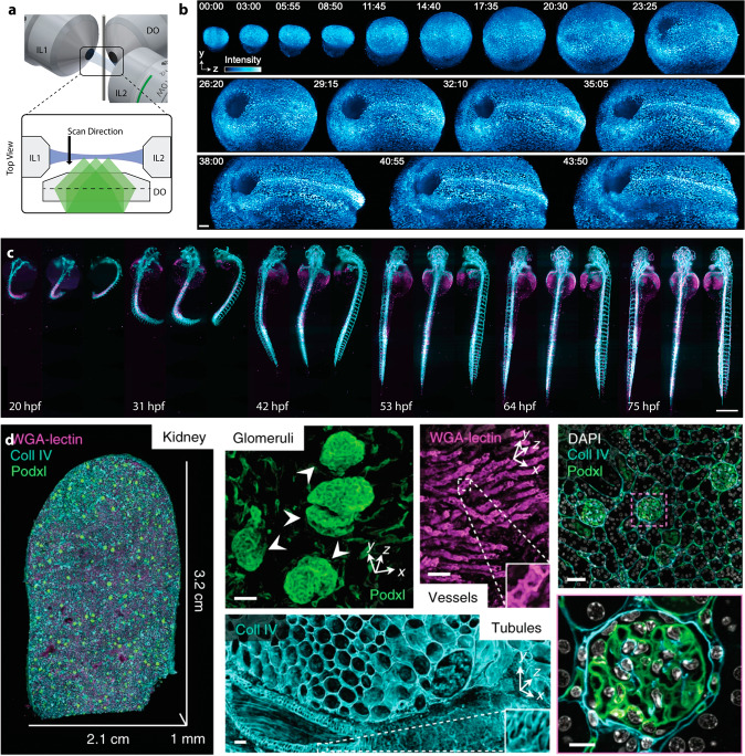
Fig. 1
Light-sheet microscopy provides fast imaging with minimized photo-toxicity and photo-bleaching, enabling diverse applications from imaging developmental processes to imaging of large, cleared tissues.
a Traditional light-sheet microscopy such as three-objective Selective Plane Illumination Microscopy (SPIM) relies on an orthogonal arrangement of the illumination (blue; illumination objectives IL1 a...
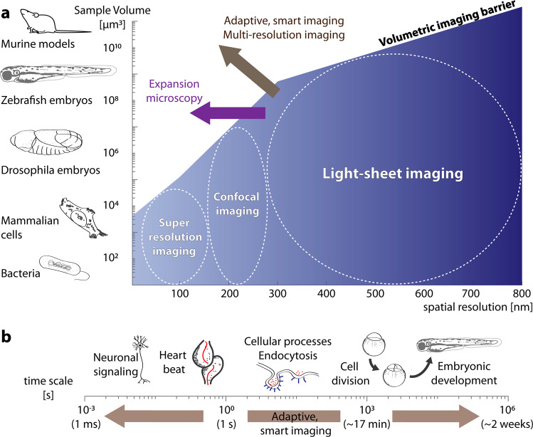
Fig. 2
Expansion microscopy, and novel adaptive, smart imaging methods combined with multi-resolution imaging will expand the available imaging capabilities.
a Current imaging techniques such as light-sheet, confocal and super-resolution microscopy are limited in the volume they can image due to technical and practical limitations (blue gradient: from low ...
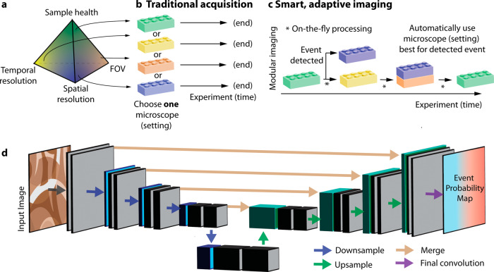
Fig. 3
Inu00a0the future, we expect novel smart and adaptive imaging schemes to overcome the traditional trade-offs and limitations of imaging.
a Traditionally, an acquisition is governed by a limited photon budget of the sample. Therefore, improved spatial and temporal resolution is typically antagonistic to sample health and the field of vi...
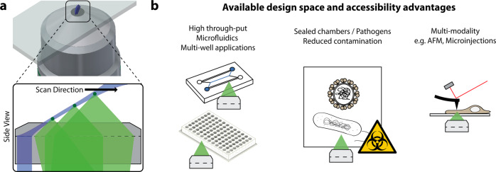
Fig. 4
Novel engineering solutions for light-sheet microscopy provide improved accessibility to enable new applications and integration with other modalities.
a Oblique Plane Light-Sheet Microscopy (OPM) is one example of open top geometries, where a high NA primary objective provides both, the illumination (blue) and detection (three fluorescent emitters a...
Figure images are served from the NIH/NLM PubMed Central Open Access Subset or Europe PMC; copyright remains with the publishers and authors.
💬 Discussion
0 commentsNo comments yet. Be the first to start a discussion!
Leave a Comment