Abstract
The αβ-tubulin subunits of microtubules can undergo a variety of evolutionarily-conserved post-translational modifications (PTMs) that provide functional specialization to subsets of cellular microtubules. Acetylation of α-tubulin residue Lysine-40 (K40) has been correlated with increased microtubule stability, intracellular transport, and ciliary assembly, yet a mechanistic understanding of how acetylation influences these events is lacking. Using the anti-acetylated tubulin antibody 6-11B-1 and electron cryo-microscopy, we demonstrate that the K40 acetylation site is located inside the microtubule lumen and thus cannot directly influence events on the microtubule surface, including kinesin-1 binding. Surprisingly, the monoclonal 6-11B-1 antibody recognizes both acetylated and deacetylated microtubules. These results suggest that acetylation induces structural changes in the K40-containing loop that could have important functional consequences on microtubule stability, bending, and subunit interactions. This work has important implications for acetylation and deacetylation reaction mechanisms as well as for interpreting experiments based on 6-11B-1 labeling.
🔬 Techniques
🧪 Sample Preparation
🔬 Cell Lines
🏭 Microscope Brands
🧪 Reagent Suppliers
📷 Detectors
🔎 Objectives
💻 Software Details
🏛️ Research Organizations (ROR)
Affiliated research institutions:
📋 Methods
Antibodies and plasmids
Polyclonal antibody production was carried out by ProteinTech Group and the entire study was approved by their Institutional Animal Care and Use Committee (IACUC). All animals were observed on a regular basis for tissue necrosis and abscess formation at the inoculation sites and for the animal's activity, food consumption and body condition. Euthanasia was done under anesthetics with ether with cardiac puncture. Rabbits were immunized with a synthetic peptide (amino acids QMPSD[AcK]TIGG common to all mouse α-tubulin isotypes) coupled to keyhole limpet hemocyanin and boosted at separate locations with the same peptide coupled to BSA. Production bleeds were obtained from the ear vein of sedated rabbits with a 21 gauge needle. Specific antibodies were affinity purified by adsorption to the same peptide coupled to a Sulfolink column (Pierce). The following monoclonal antibodies were purchased: anti-acetylated tubulin clone 6-11B-1 ( [5] Sigma T7451), anti-α-tubulin clone DM1A (Sigma T6199), and anti-β-tubulin clone E7 (Developmental Studies Hybridoma Bank). Secondary antibodies conjugated to fluorophores were purchased from Jackson ImmunoResearch Laboratories. Plasmids for expression of GST-MEC-17 ( [23] , gift of Jacek Gaertig, University of Georgia), pHEX-His-SIRT2 ( [26] , gift of Eric Verdin, UCSF) and HA-HDAC6 ( [45] , gift of Xiang-Jiao Yang, McGill University) have been described. MEC-17, HDAC6 and SIRT2 were sub-cloned by PCR into pmCitrine-C1 for mammalian expression.
Mammalian cell culture and Immunofluorescence
COS7 (monkey kidney fibroblast, ATCC) cells were grown in DMEM+10% fetal bovine serum (FBS) and 2 mM L-glutamine at 37°C with 5% CO 2 . PtK2 (rat kangaroo kidney epithelial, ATCC) cells were grown in EMEM+10% FBS and 2 mM L-glutamine at 37°C with 5% CO 2 . COS7 and PtK2 cells were transfected using Expressfect (Danville Scientific) and TransIT-LT1 (Mirus-Bio), respectively. The next day, coverslips were rinsed with PBS, fixed in 4% paraformaldehyde in PBS for 10 min, quenched with 50 mM ammonium chloride in PBS for 5 min, and permeabilized in 0.2% Triton-X-100 in PBS for 5 min. Subsequently, coverslips were blocked with 0.2% FSG (Fish Skin Gelatin) in PBS for 5 min and incubated in primary antibody for 1 h. After three washes with 0.2% FSG in PBS, cells were incubated in secondary antibody for 1 h, washed three times in PBS, and mounted with Prolong Gold. Images were obtained on an inverted epifluorescence microscope Nikon TE2000E, equipped with 60X 1.40 NA objective and a Photometrics CoolSnap HQ camera.
Show full methods section
Antibodies and plasmids
Polyclonal antibody production was carried out by ProteinTech Group and the entire study was approved by their Institutional Animal Care and Use Committee (IACUC). All animals were observed on a regular basis for tissue necrosis and abscess formation at the inoculation sites and for the animal's activity, food consumption and body condition. Euthanasia was done under anesthetics with ether with cardiac puncture. Rabbits were immunized with a synthetic peptide (amino acids QMPSD[AcK]TIGG common to all mouse α-tubulin isotypes) coupled to keyhole limpet hemocyanin and boosted at separate locations with the same peptide coupled to BSA. Production bleeds were obtained from the ear vein of sedated rabbits with a 21 gauge needle. Specific antibodies were affinity purified by adsorption to the same peptide coupled to a Sulfolink column (Pierce). The following monoclonal antibodies were purchased: anti-acetylated tubulin clone 6-11B-1 ( [5] Sigma T7451), anti-α-tubulin clone DM1A (Sigma T6199), and anti-β-tubulin clone E7 (Developmental Studies Hybridoma Bank). Secondary antibodies conjugated to fluorophores were purchased from Jackson ImmunoResearch Laboratories. Plasmids for expression of GST-MEC-17 ( [23] , gift of Jacek Gaertig, University of Georgia), pHEX-His-SIRT2 ( [26] , gift of Eric Verdin, UCSF) and HA-HDAC6 ( [45] , gift of Xiang-Jiao Yang, McGill University) have been described. MEC-17, HDAC6 and SIRT2 were sub-cloned by PCR into pmCitrine-C1 for mammalian expression.
Mammalian cell culture and Immunofluorescence
COS7 (monkey kidney fibroblast, ATCC) cells were grown in DMEM+10% fetal bovine serum (FBS) and 2 mM L-glutamine at 37°C with 5% CO 2 . PtK2 (rat kangaroo kidney epithelial, ATCC) cells were grown in EMEM+10% FBS and 2 mM L-glutamine at 37°C with 5% CO 2 . COS7 and PtK2 cells were transfected using Expressfect (Danville Scientific) and TransIT-LT1 (Mirus-Bio), respectively. The next day, coverslips were rinsed with PBS, fixed in 4% paraformaldehyde in PBS for 10 min, quenched with 50 mM ammonium chloride in PBS for 5 min, and permeabilized in 0.2% Triton-X-100 in PBS for 5 min. Subsequently, coverslips were blocked with 0.2% FSG (Fish Skin Gelatin) in PBS for 5 min and incubated in primary antibody for 1 h. After three washes with 0.2% FSG in PBS, cells were incubated in secondary antibody for 1 h, washed three times in PBS, and mounted with Prolong Gold. Images were obtained on an inverted epifluorescence microscope Nikon TE2000E, equipped with 60X 1.40 NA objective and a Photometrics CoolSnap HQ camera.
Enzyme purification
His-tagged human SIRT2 protein was bacterially expressed in BL21 (DE3) cells by inducing with 1 mM IPTG (isopropyl-β-D-thiogalactopyranoside) at 37°C for 3 h and purified under native conditions at 4°C by Ni-NTA (Qiagen) as described [26] . GST-tagged human MEC-17 was bacterially expressed in Rosetta2 cells, adsorbed to Glutathione Sepharose beads (GE Healthcare Biosciences), and eluted with in 50 mM Tris-HCl pH-8.0, 0.2 mM EDTA, 10 mM reduced glutathione as described. Purified proteins were dialyzed against dialysis buffer (20 mM Tris-HCl pH-8.0, 0.2 mM DTT) overnight at 4°C, mixed with 10% glycerol, and snap frozen in liquid nitrogen prior to storage at −80°C. Tubulin modification and polymerization Purified bovine brain tubulin was purchased from Cytoskeleton (TL238). Acetylated tubulin was generated by incubating purified bovine brain tubulin with recombinant GST-MEC-17 in the presence of 10 µM Acetyl coenzyme A (Sigma A2056) for 2 h at 28°C with constant agitation. Deacetylated tubulin was prepared by incubating purified bovine brain tubulin with recombinant His-SIRT2 in the presence of 1 mM NAD (β-Nicotinamide adenine dinucleotide, Sigma N8285) for 2 h at 37°C with constant mixing. The resulting modified tubulins were cycled through one round of polymerization/depolymerization to remove the enzyme (verified by SDS-PAGE, data not shown) before flash-freezing in liquid nitrogen and storage at −80°C. Untreated, acetylated or deacetylated tubulins were polymerized for 20 min at 37°C in BRB80 at a concentration of 10 mg/ml in the presence of 20% DMSO (v/v), 2 mM GTP, 20 µM taxol, 0.5 mM PMSF and 4 mM MgCl 2 .
Kinesin binding assay
Constitutively active rat kinesin heavy chain (KIF5C) constructs were expressed in COS-7 cells. The cells were lysed in lysis buffer (25 mM HEPES/KOH pH-7.4, 115 mM KOAc, 5 mM NaOAc, 5 mM MgCl 2 , 0.5 mM EGTA and 1% Triton X-100) containing 0.1 mM ATP and clarified by centrifugation at 14,000 rpm for 10 min at 4°C. Taxol-stabilized acetylated or deacetylated microtubules were added to 0.1 mg/ml together with 20 µM taxol and 1 mM AMPPNP (a non-hydrolyzable ATP analog) and incubated for 30 min at room temperature with constant mixing. The motor-microtubule complexes were sedimented at 90,000 rpm at 18°C for 10 min through a glycerol cushion (BRB80 containing 60% glycerol and 20 µM taxol). The pellet was dissolved in SDS-PAGE sample buffer and the amount of tubulins and motors in the pellets was determined by immunoblotting. The scanned blots were used for quantification in ImageJ software. Fab fragment preparation and microtubule decoration Fab fragments of 6-11B-1 antibodies were generated using the Fab micro-preparation kit (Pierce). In brief, 6-11B-1 IgG was digested with immobilized papain at 37°C for 6 h with end-over-end mixing. The digested products were eluted by low speed centrifugation and the Fc fragments were bound to protein A beads at room temperature for 20 min. Fab fragments were collected by low speed spin and concentrated. Untreated microtubules were incubated with GST-KHC motor domain (KR01, Cytoskeleton), whereas acetylated or deacetylated microtubules were incubated with 6-11B-1 Fab fragment at a 1:2 ratio for 1 h at room temperature. Excess motors and Fab fragments were removed by centrifugation of microtubules through a glycerol cushion (BRB80 containing 60% glycerol and 20 µM taxol) in a TLA100 rotor at 90,000 rpm for 10 min at 35°C. Fab-decorated microtubules were resuspended in BRB80 containing 20 µM taxol.
Electron Microscopy and 3D Reconstruction
Samples were prepared for negative stain electron microscopy using 0.75% solution of uranyl formate and conventional negative staining protocols [46] . For cryo-EM, 2 µl of the microtubule samples were applied on glow-discharged Quantifoil R2/2 200 holey carbon grids and vitrified using a Vitrobot Mark IV (FEI Co.). Vitrified specimen was imaged on a Tecnai F20 Transmission Electron Microscope (FEI Co.) equipped with a field emission gun and operated at 200 kV. Images were recorded at a magnification of 66,964x on a Gatan US4000 CCD camera at a ∼2 µm defocus. The pixel size of images acquired under these conditions is 2.24 Å. Micrographs were screened for helical, 15-protofilament microtubules using the PHOELIX software package [47] . For 3D reconstructions of microtubule-Fab complexes, we selected filaments with strong signal at the 1/8 nm layerline in the 2D power spectra, indicative of high levels of Fab decoration. For the 3D reconstruction of the acetylated microtubule in complex with Fab, we selected and averaged layer-line data from 42 near and far sides. For the 3D recontruction of deacetylated with attached Fab and control microtubules, we selected and averaged layer-line data from 10 near and far sides.
Immunostaining of acetylated and deacetylated microtubules
Polymerized microtubules were adsorbed onto coverslips and stained with 6-11B-1 antibodies without fixation (live) or after fixation with 4% paraformaldehyde (PFA fixed) in PBS containing 20 µM taxol. All subsequent steps were carried out in BRB80+20 µM taxol. The cover slips were blocked with 5 mg/ml casein for 30 min, incubated with primary antibodies for 1 h, washed three times, incubated with secondary antibodies for 1 h, washed three times, and mounted with Prolong Gold. The images were obtained on an inverted epi-fluorescence microscope Nikon TE2000E, equipped with 60X 1.40 NA objective and a Photometrics CoolSnap HQ camera.
Supporting Information Figure S1 Purification of recombinant MEC-17 and SIRT2 enzymes and Fab fragment preparation. A,B) Coomassie-stained SDS-PAGE gels showing purification profile of recombinant A) GST-MEC-17 or B) His-SIRT2. C) Coomassie-stained SDS-PAGE gel showing preparation of Fab fragments from the monoclonal 6-11B-1 antibody. (TIF) Click here for additional data file. Figure S2 Raw cryo-EM images of representative microtubule segments. Filament sections have been excised from larger micrographs and enlarged to show detail. Shown are representative sections of A) control (no enzyme treatment, no Fab binding), B) MEC-17-acetylated and 6-11B-1 Fab-decorated, and C) SIRT2-deactylated and 6-11B-1 Fab-decorated microtubules. Scale bar, 25 nm. (TIF) Click here for additional data file. Figure S3 Representative power spectra. A) A representative power spectrum from a single vitrified control microtubule. B) A representative power spectrum from a single vitrified MEC-17-acetylated microtubule decorated with 6-11B-1 Fab. Regular Fab decoration is indicated by the presence of a 1/8 nm layer line, compared to the control microtubule (A). C) A representative power spectrum from a single vitrified SIRT2-deacetylated microtubule decorated with 6-11B-1 Fab. A weaker 1/8 nm signal is observed, corresponding to lower Fab occupancy. (TIF) Click here for additional data file. Figure S4 Monoclonal 6-11B-1 and polyclonal anti-acetyl-K40 antibodies recognize acetylated but not unacetylated microtubules in cells. A) COS7 and PtK2 cells were fixed and double stained with monoclonal 6-11B-1 and total tubulin antibodies (left panels) or polyclonal anti-acetyl-K40 and total tubulin antibodies (right panels). Neither antibody recognizes microtubule filaments in PtK2 cells which contain only unacetylated (never modified) α-tubulin. B) COS7 and PtK2 cells expressing the acetytransferase mCit-MEC-17 (green) were double stained with monoclonal 6-11B-1 (red) and total tubulin (magenta) antibodies (left panels) or with polyclonal anti-acetyl-K40 (red) and total tubulin (magenta) antibodies (right panels). Both antibodies recognize the highly acetylated microtubules induced by expression of mCit-MEC-17. Scale bars, 20 µm. (TIF) Click here for additional data file. Figure S5 The 6-11B-1 epitope is sensitive to the level of expression of the α-tubulin K40 deacetylases HDAC6 and SIRT2. COS7 cells transfected with A) mCit-HDAC6 or B) mCit-SIRT2 were fixed and stained with monoclonal 6-11B-1 (red) and total tubulin (magenta) antibodies. Scale bars, 20 µm. Transfected cells are indicated by a yellow dotted outline. Previous work showed that expression of HDAC6 or SIRT2 in mammalian cells resulted in a complete loss of 6-11B-1 staining [1] – [3] , suggesting that the 6-11B-1 antibody does not recognize deacetylated α-tubulin. In contrast, we show in Figure 4 that moderate expression of HDAC6 or SIRT2 results in deacetylated microtubules that can still be recognized by the 6-11B-1 antibody. To explain the difference between our results and the previous work, we looked at 6-11B-1 and anti-acetyl-K40 labeling at different levels of deacetylase expression. Figure 5 shows cells expressing moderate levels of HDAC6 and SIRT2 expression (based on fluorescence intensity) whereas this figure shows cells expressing high levels of HDAC6 and SIRT2. In agreement with previous work [1] – [3] , this figure shows that 6-11B-1 antigenicity is lost in cells expressing high levels of HDAC6 or SIRT2 enzymes. The fact that the polyclonal anti-acetyl-K40 antibody does not recognize any microtubules even in cells expressing moderate levels of HDAC6 or SIRT2 enzymes ( Figures 5B ), indicates that expression of these deacetylase enzymes results in microtubules that are fully non-acetylated (deacetylated and unacetylated). The fact that 6-11B-1 stains microtubules in cells expressing moderate levels of HDAC6 or SIRT2 ( Figure 5A ) but not high levels of the enzymes ( Figure S5 ) indicates that α-tubulin subunits undergo a structural conversion from the deacetylated (recognized by 6-11B-1) to non-acetylated (not recognized by 6-11B-1) state. Whether this conversion is due to increased levels or time of deacetylase expression is presently unclear. 1. North BJ, Marshall BL, Borra MT, Denu JM, Verdin E (2003) The human Sir2 ortholog, SIRT2, is an NAD(+)-dependent tubulin deacetylase. Mol Cell 11: 437-444. 2. Matsuyama A, Shimazu T, Sumida Y, Saito A, Yoshimatsu Y, et al. (2002) In vivo destabilization of dynamic microtubules by HDAC6-mediated deacetylation. EMBO J 21: 6820–6831. 3. Zhang Y, Li N, Caron C, Matthias G, Hess D, et al. (2003) HDAC-6 interacts with and deacetylates tubulin and microtubules in vivo. EMBO J 22: 1168–1179. (TIF) Click here for additional data file. Figure S6 HDAC6 or SIRT2 binding does not create an epitope for the 6-11B-1 antibody in PtK2 cells. PtK2 cells expressing the deacetylases mCit-HDAC6 or mCit-SIRT2 (green) were fixed and double stained using monoclonal 6-11B-1 anti-acetylated tubulin (red) and total tubulin (magenta) antibodies. Transfected cells are indicated by the yellow dotted outline. Scale bars, 20 µm. (TIF) Click here for additional data file.
📊 Figures
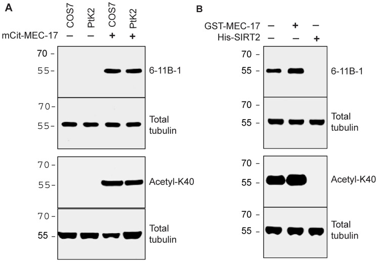
Figure 1
Generation of acetylated and deacetylated tubulins.
A ) Lysates of COS7 and PtK2 cells either untransfected (lanes 1 and 2) or expressing the acetyltransferase MEC-17 (lanes 3 and 4) were immunoblotted for K40 acetylation of u03b1-tubulin using monoclo...
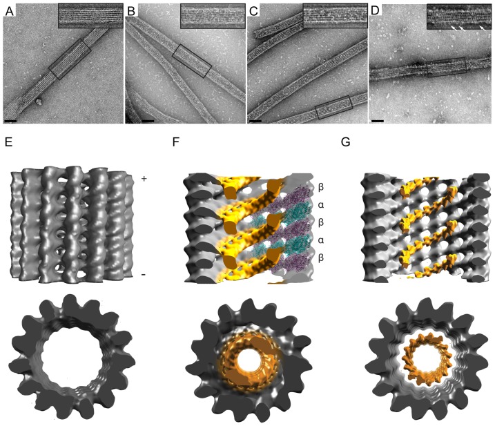
Figure 2
2D and 3D EM visualization of the 6-11B-1 Fab within the microtubule lumen.
A-D ) Microtubules polymerized from A,D) untreated B) MEC-17-treated (acetylated), or C) SIRT2-treated (deacetylated) tubulins were incubated with A-C) 6-11B-1 Fab fragments or D) GST-KHC motor domain...
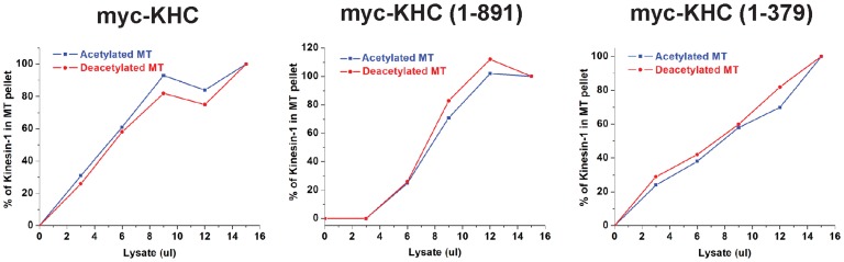
Figure 3
K40 acetylation does not directly influence the binding of Kinesin-1 to microtubules.
Myc-tagged versions of full-length kinesin-1 heavy chain (myc-KHC) or truncated, constitutively active constructs (1u2013891 and 1u2013379) were expressed in COS7 cells. Increasing amounts of cell lys...
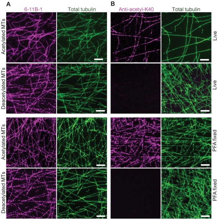
Figure 4
Monoclonal (6-11B-1) and polyclonal (anti-acetyl-K40) antibodies differ in their ability to recognize deacetylated microtubules in vitro .
Taxol-stabilized microtubules polymerized from acetylated or deacetylated tubulins were stained immediately (Live) or after fixation with paraformaldehyde (PFA fixed) with A) monoclonal 6-11B-1 antibo...
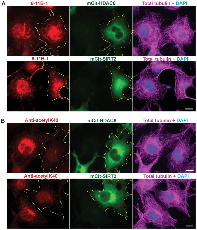
Figure 5
Monoclonal (6-11B-1) and polyclonal (anti-acetyl-K40) antibodies differ in their ability to recognize deacetylated microtubules in cells.
COS7 cells expressing the deacetylases mCit-HDAC6 or mCit-SIRT2 (green) were fixed and double stained using A) monoclonal 6-11B-1 (red) and total tubulin (magenta) antibodies or B) polyclonal anti-ace...
Figure images are served from the NIH/NLM PubMed Central Open Access Subset or Europe PMC; copyright remains with the publishers and authors.
💬 Discussion
0 commentsNo comments yet. Be the first to start a discussion!
Leave a Comment