Abstract
Mast cells (MCs) and dendritic cells (DCs) are essential innate sentinels populating host-environment interfaces. Using longitudinal intravital multiphoton microscopy of DCGFP/MCRFP reporter mice, we herein provide in vivo evidence that migratory DCs execute targeted cell-to-cell interactions with stationary MCs before leaving the inflamed skin to draining lymph nodes. During initial stages of skin inflammation, DCs dynamically scan MCs, whereas at a later stage, long-lasting interactions predominate. These innate-to-innate synapse-like contacts ultimately culminate in DC-to-MC molecule transfers including major histocompatibility complex class II (MHCII) proteins enabling subsequent ex vivo priming of allogeneic T cells with a specific cytokine signature. The extent of MHCII transfer to MCs correlates with their T cell priming efficiency. Importantly, preventing the cross talk by preceding DC depletion decreases MC antigen presenting capacity and T cell-driven inflammation. Consequently, we identify an innate intercellular communication arming resident MCs with key DC functions that might contribute to the acute defense potential during critical periods of migration-based DC absence.
🔬 Techniques
🔭 Microscopes
✨ Fluorophores
🧪 Sample Preparation
🔬 Cell Lines
🏭 Microscope Brands
🧪 Reagent Suppliers
🔴 Lasers
🎨 Filters
💻 Software Details
💾 Data Repositories
🏛️ Research Organizations (ROR)
Affiliated research institutions:
📋 Methods
Mice
Mcpt5-Cre mice crossed to the R26-tdRFP, R-DTA or CD11c-DTR/eGFP line, and CD11c-Cre mice crossed to the iDTR line, were bred and housed at the Experimental Centre at the Technische Universität Dresden, Medical Faculty Carl-Gustav Carus, under specific pathogen-free conditions. CD11c-DTR/eGFP and CD11c-Cre mice for breeding were purchased from Jackson Laboratories. Mcpt5-Cre mice were provided by A. Roers (Technische Universität, Dresden, Germany), R26-tdRFP by H.J. Fehling (Ulm University, Ulm, Germany), iDTR by A. Waisman (Johannes-Gutenberg University, Mainz, Germany), R-DTA by D. Voehringer (University Clinic, Erlangen, Germany), and MHCII −/− mice (B6.129S2-H2 dlAb1-Ea /J) by B. Schröder (Christian-Albrechts University, Kiel, Germany). CD45.2 + C57BL/6 and CD45.1 + SJL/J mice were purchased from Janvier and Charles River, respectively. All mice were of C57BL/6 background. In all experiments, mice were used at the age of 8 to 16 wk. All procedures were in accordance with institutional guidelines on animal welfare and were approved by the Landesdirektion Dresden (24-9168.11-1/2012-38). Chemicals and reagents DNFB (1-fluoro-2,4-dinitrobenzene) and hyaluronidase were obtained from Sigma-Aldrich. Liberase TM and DNase I were purchased from Roche. DT was obtained from Enzo Life Sciences. Recombinant murine IL-3 and recombinant rat SCF were obtained from Peprotech. The following mAbs directed against mouse antigens were obtained from eBiosciences: CD117 (clone 2B8), FcεRIα (MAR-1), CD45 (30-F11), CD11c (N418), F4/80 (BM8), MHCII I-A/I-E (M5/114.15.2), MHCII I-A b (AF6-120.1), CD45.1 (A20), CD45.2 (104), CD3 (eBio500A2), and CD4 (GK1.5). CFSE was obtained from Molecular Probes (Life Technologies). CHS Mice were sensitized with 100 µl 0.5% DNFB in acetone/oil (4:1) on the shaved back skin and challenged 6 d later with 20 µl 0.2% DNFB on the ear (10 µl/ear side). For imaging analysis, 20 µl 0.2% DNFB in acetone/olive oil (4:1) was epicutaneously applied on one side of the ear. Vehicle control mice were treated with the solvent only.
Show full methods section
Mice
Mcpt5-Cre mice crossed to the R26-tdRFP, R-DTA or CD11c-DTR/eGFP line, and CD11c-Cre mice crossed to the iDTR line, were bred and housed at the Experimental Centre at the Technische Universität Dresden, Medical Faculty Carl-Gustav Carus, under specific pathogen-free conditions. CD11c-DTR/eGFP and CD11c-Cre mice for breeding were purchased from Jackson Laboratories. Mcpt5-Cre mice were provided by A. Roers (Technische Universität, Dresden, Germany), R26-tdRFP by H.J. Fehling (Ulm University, Ulm, Germany), iDTR by A. Waisman (Johannes-Gutenberg University, Mainz, Germany), R-DTA by D. Voehringer (University Clinic, Erlangen, Germany), and MHCII −/− mice (B6.129S2-H2 dlAb1-Ea /J) by B. Schröder (Christian-Albrechts University, Kiel, Germany). CD45.2 + C57BL/6 and CD45.1 + SJL/J mice were purchased from Janvier and Charles River, respectively. All mice were of C57BL/6 background. In all experiments, mice were used at the age of 8 to 16 wk. All procedures were in accordance with institutional guidelines on animal welfare and were approved by the Landesdirektion Dresden (24-9168.11-1/2012-38). Chemicals and reagents DNFB (1-fluoro-2,4-dinitrobenzene) and hyaluronidase were obtained from Sigma-Aldrich. Liberase TM and DNase I were purchased from Roche. DT was obtained from Enzo Life Sciences. Recombinant murine IL-3 and recombinant rat SCF were obtained from Peprotech. The following mAbs directed against mouse antigens were obtained from eBiosciences: CD117 (clone 2B8), FcεRIα (MAR-1), CD45 (30-F11), CD11c (N418), F4/80 (BM8), MHCII I-A/I-E (M5/114.15.2), MHCII I-A b (AF6-120.1), CD45.1 (A20), CD45.2 (104), CD3 (eBio500A2), and CD4 (GK1.5). CFSE was obtained from Molecular Probes (Life Technologies). CHS Mice were sensitized with 100 µl 0.5% DNFB in acetone/oil (4:1) on the shaved back skin and challenged 6 d later with 20 µl 0.2% DNFB on the ear (10 µl/ear side). For imaging analysis, 20 µl 0.2% DNFB in acetone/olive oil (4:1) was epicutaneously applied on one side of the ear. Vehicle control mice were treated with the solvent only.
Flow cytometry analysis of skin cells
For preparation of skin cell suspensions, samples of ear skin were cut into small pieces and digested in 1 ml DMEM containing 20 mM Hepes, 0.025 mg/ml Liberase TM, 396 U/ml DNase I, and 0.5 mg/ml hyaluronidase at 37°C, 1400 rpm for 1 h. Samples were passed through a 40-µm sieve and the cell suspension was washed twice with PBS. For flow cytometry analysis, cells were resuspended in PBS/2% BSA and stained with mAbs for 30 min at 4°C. Cell suspensions were washed twice and resuspended in 200 µl PBS/2% BSA. Analysis was done using the Miltenyi MacsQuant flow cytometer with MacsQuant or FlowJo Analysis Software. MC reconstitution of MC-deficient mice Peritoneal MCs reflecting CTMCs were obtained from either C57BL/6 WT mice or MHCII −/− mice (B6.129S2- H2 dlAb1-Ea /J) as previously described ( Dudeck et al., 2011b ). In brief, cells from peritoneal lavage of respective donor mice were cultured in RPMI with 10% FCS, 1% penicillin/streptomycin, 10 ng/ml IL-3, and 30 ng/ml SCF. After 48 h, nonadherent cells were carefully removed and replaced by fresh culture medium to enhance the purity of resulting MCs. After 8 d of culture, these peritoneal cultured MCs (PCMCs) were enriched using mouse CD117 microbeads (Miltenyi) and cultured for further 48 h to avoid MC activation. MC-deficient Mcpt5-Cre + R-DTA + mice and Mcpt5-Cre + R-DTA + × CD11c-eGFP/DTR were reconstituted by i.d. injection with 1.25 × 10 6 PCMCs from C57BL/6 WT mice into one ear and the same number of PCMCs from MHCII −/− mice into the contralateral ear. DC depletion Dermal DCs were locally depleted in ear skin of CD11c-Cre iDTR mice or Mcpt5-Cre R-DTA x CD11c-eGFP/DTR mice by i.d. injection of DT (100 ng/20 µl/ear) into the ear pinnae at 48 h after DNFB administration onto the ear skin or at 4 h and 48 h after DNFB. Haplotype mismatch BM transfer Genetically engineered inbred mouse strains commonly used in biomedical research differ in their inherited haploid genotype (also referred to as haplotype). 6-wk-old SJL/J recipient mice (CD45.1 + MHCII haplotype H2 s ) were lethally irradiated (whole body irradiation, 9 Gy; x-ray source, MaxiShot, Yxlon) before retro-orbital i.v. injection of 20 × 10 6 whole BM cells isolated from age- and sex-matched C57BL/6 donor mice (CD45.2 + MHCII haplotype H2 b ). After BM transplantation, mice were given neomycin-containing drinking water for 3 wk. To determine the chimerism of recipient mice, a 50-µl blood sample was isolated 2 wk after transplantation, and surface expression of CD45.1/CD45.2 on blood leukocytes was determined using flow cytometry (Fig. S3 A). An additional week later, the DC-to-MC communication was initiated in SJL/J B6 mice by DNFB administration, and the expression of C57BL/B6 donor cell–restricted H2 b MHCII on MCs and DCs was analyzed by flow cytometry 24 h after DNFB administration in comparison to SJL/J mice without BM transplantation. The H2 b MHCII expression analysis was performed using the specific mAb clone AF6-120.1 reported to bind to C57BL/6-restricted H2 b MHCII but to not cross-react with SJL/J H2 s MHCII.
Analysis of skin
DCs from C57BL/6 and SJL/J control mice with the AF6-120.1 mAb demonstrated, as expected, that C57BL/6-restricted H2 b MHCII was labeled but not SJL/J H2 s MHCII (Fig. S3 B). Allogeneic T cell stimulation DCs (CD45.2 + CD11c + H2 b+ in SJL/J B6 ; CD45.1 + CD11c + in SJL/J) and MCs (CD45.1 + c-kit + FcεRI + ) were sorted from same samples of ear skin cell suspension of SJL/J B6 chimera mice 24 h after DNFB administration and untreated SJL/J mice. Spleen DCs as positive control for allogeneic T cell stimulation were isolated from C57BL/6 mice using CD11c microbeads (Miltenyi). 2 × 10 4 DCs or MCs were seeded to 96-well cell-culture plates in 50 µl complete RPMI medium supplemented with 10% FCS and 1% penicillin/streptomycin. T cells were negatively enriched from spleens of untreated SJL/J mice using the pan T cell isolation kit (Miltenyi) and labeled with CFSE. Allogeneic T cell proliferation was verified by co-culturing 10 5 CFSE-labeled T cells with preseeded DCs or MCs over 6 d. Subsequently, CD3 + CD4 + T cell proliferation was measured as CFSE dilution by flow cytometry and quantified as the fraction of proliferated CD4 + T cells bearing low CFSE staining and as mean fluorescence intensity. Cytokines were quantified in the co-culture supernatants using the bead based mouse essential Th1/Th2 cytokine 6-plex ProcartaPlex panel (ThermoFisher). T cell–induced skin inflammation C57BL/6 WT donor mice were sensitized with DNFB (0.5%, 100 µl) on the shaved back, and preactivated T cells were isolated from inguinal LNs 6 d later using a pan T cell enrichment kit (Miltenyi). CD11c-Cre + iDTR mice and Cre − littermates were treated with DNFB 0.2% on the ear skin (0.2%, 20 µl/ear) followed by DC depletion at 48 h after DNFB (group 1: CD11c-Cre − ; and group 2: CD11c-Cre + iDTR mice) or at 4 h and 48 h after DNFB (group 3: CD11c-Cre + iDTR). Skin inflammation was induced in CD11c-Cre iDTR recipient mice by i.d. injection of 1.25 × 10 6 preactivated T cells from sensitized WT mice into the ear 72 h after recipient mice DNFB treatment and quantified by measuring the ear thickness at the indicated time points using a caliper (Mitutoyo) in relation to basal ear thickness. Intravital 2-photon microscopy of mouse ear skin Mice were prepared for intravital microscopy as previously described ( Dudeck et al., 2011a ). In brief, animals were subjected to intubation narcosis with a mixture of Isofluran (1.0%) and oxygen (99%) with a mechanical ventilator (Mini-Vent, Hugo-Sachs Elektronik). Two-photon intravital microscopy was performed with a Zeiss LSM780 NLO microscope with simultaneous detection via four external non-descanned detectors. Illumination was performed at 920 nm with a Chameleon Vision II (Coherent, Inc.) laser (10–12% laser power, at 1,400 mW maximum power) via a 20× water-dipping lens with 1.0 NA. GFP-expressing DCs were detected with a 525/50 bandpass filter (BP), tdRFP-expressing MCs with a 600/20 BP, and blood vessels by i.v. injection of 12.5 µl Qtracker 705 nontargeted Qdots (Invitrogen) in 83 µl isotonic NaCl (BP 710/40). Collagen I structures were visualized by its SHG (
📊 Figures
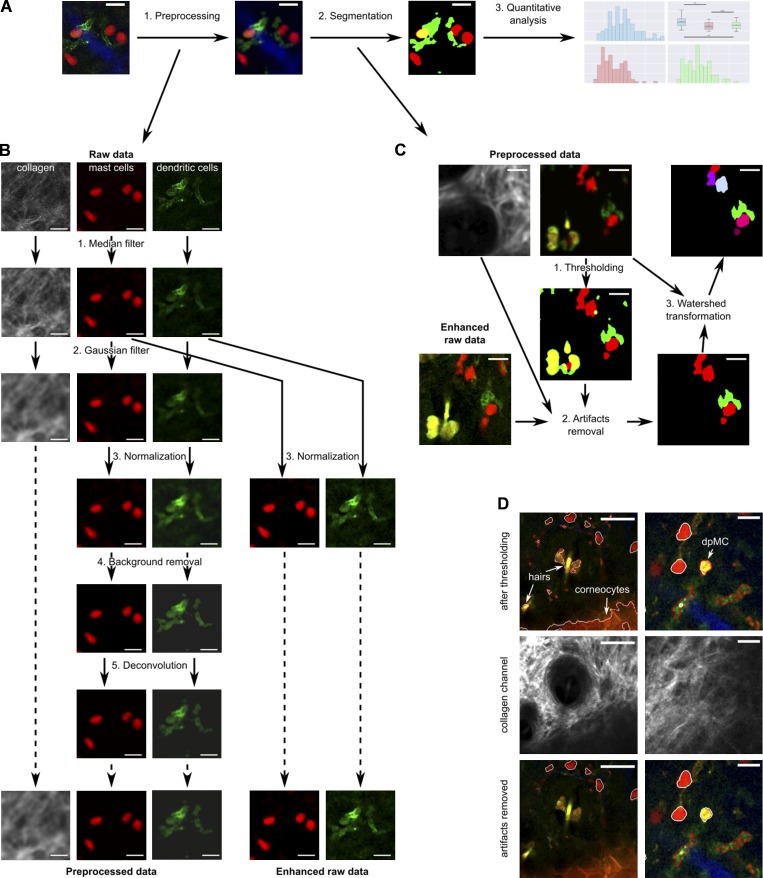
Figure 1.
Image analysis workflow. (A) Overview of image analysis. (B) Preprocessing workflow. (C) Segmentation workflow. (D) Using the collagen signal (second harmonic generation) for classification between au...
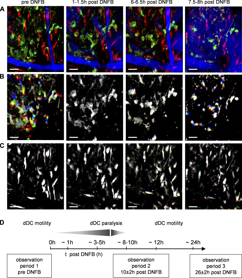
Figure 2.
Dermal DC paralysis and remobilization in responses to DNFB. (A) Maximum intensity projection DC GFP /MC RFP mouse ear skin representing DCs (green), MCs (red), and blood vessels (blue) before and aft...
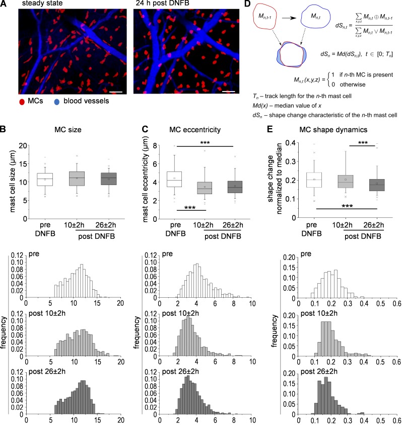
Figure 3.
DNFB-driven skin inflammation induced changes in MCs shape and dynamics. (A) Intravital multiphoton imaging of MC RFP reporter mouse ear skin before and 24 h after DNFB administration, MIP. Bars, 50 u...
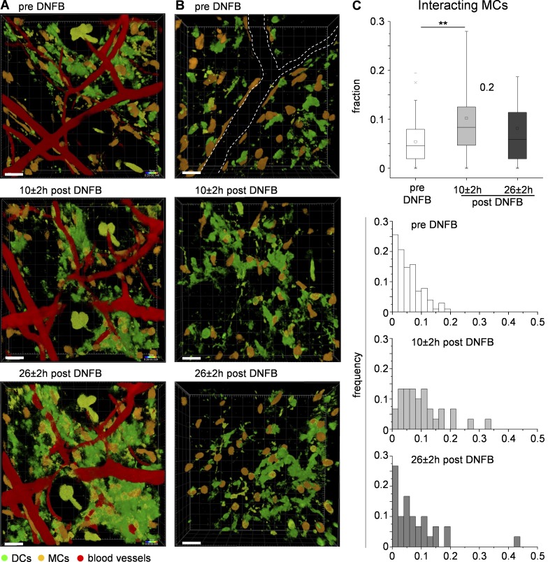
Figure 4.
DCs dynamically cross talk with MCs upon skin inflammation. Intravital multiphoton imaging of DC GFP /MC RFP mouse ear skin before and after DNFB administration, 3D rendered z-stack. Bars, 40 u00b5m. ...
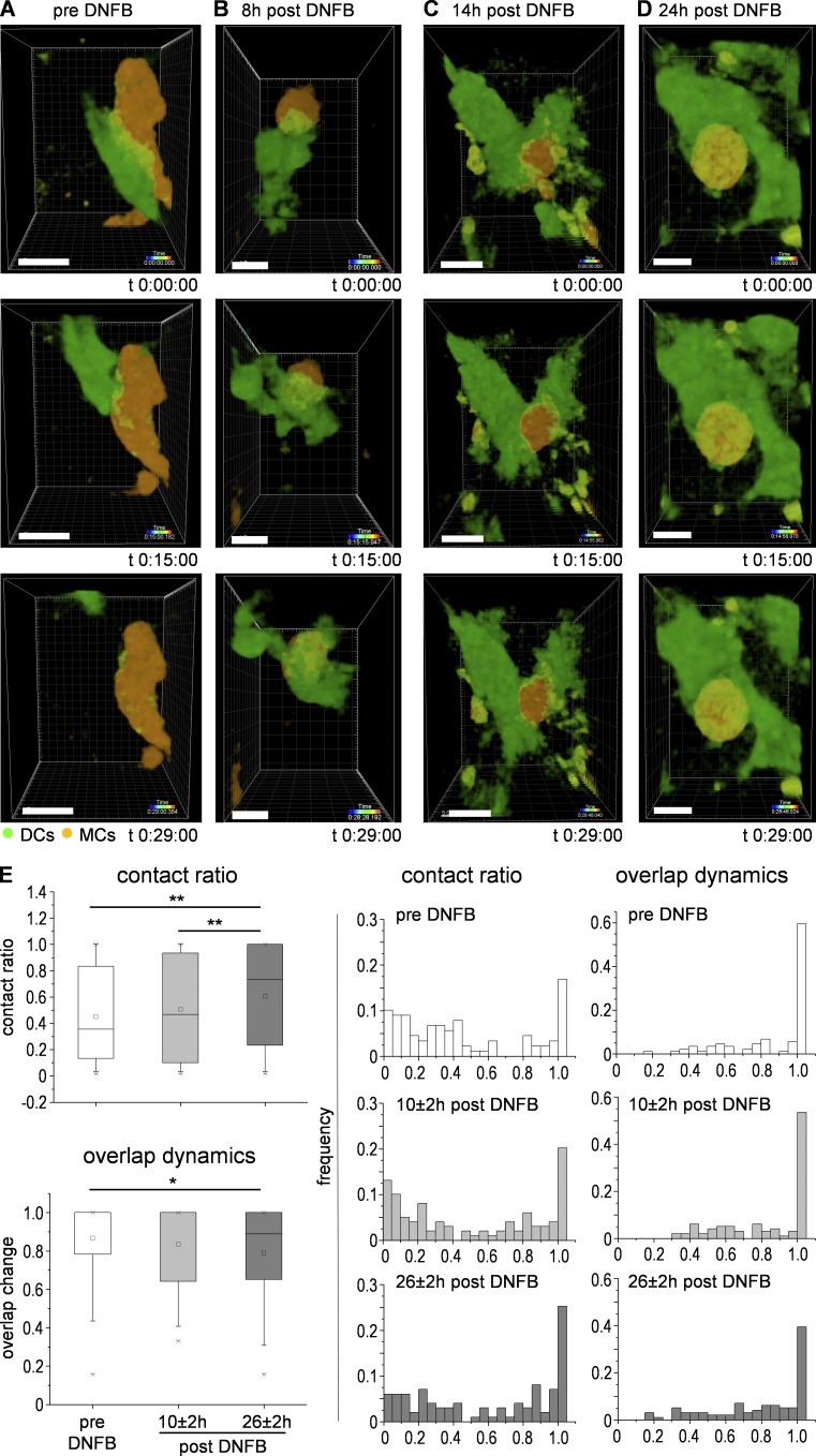
Figure 5.
DC-to-MC communication shifts from dynamic scanning to firm and long-lasting contacts in the progression of skin inflammation. (Au2013D) Sequences of intravital time-lapse series (3D-rendered z-stack)...
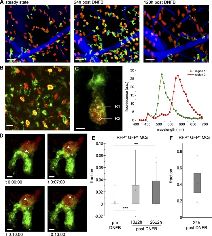
Figure 6.
DC-to-MC interactions culminate in DC protein transfer to MCs. Intravital imaging of DC GFP /MC RFP mouse ear skin (A) before and after DNFB, MIP. Bars, 50 u00b5m. (B) MIP, 24 h after DNFB. Bar, 20 u0...
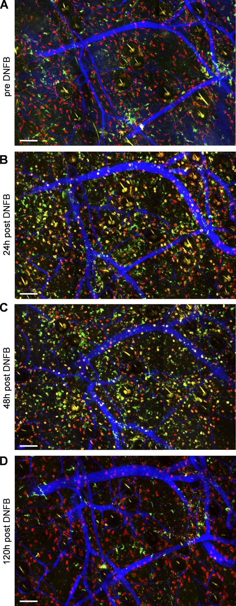
Figure 7.
Skin inflammation-induced DC-GFP transfer to MCs is followed by complete degradation of GFP inclusions on site. (Au2013D) Longitudinal and side-matched intravital imaging of DC GFP /MC RFP mouse ear s...
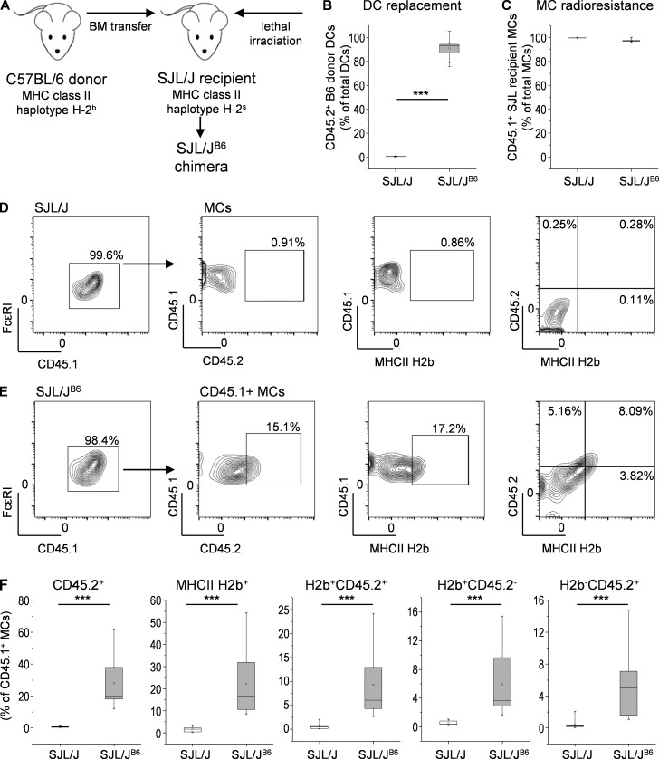
Figure 8.
MCs incorporate donor DC-derived H2 b MHCII protein complexes. (A) SJL/J B6 BM chimera were generated by BM transplantation from C57BL/6 donor mice (CD45.2 + H2 b MHCII) to SJL/J recipients (CD45.1 + ...
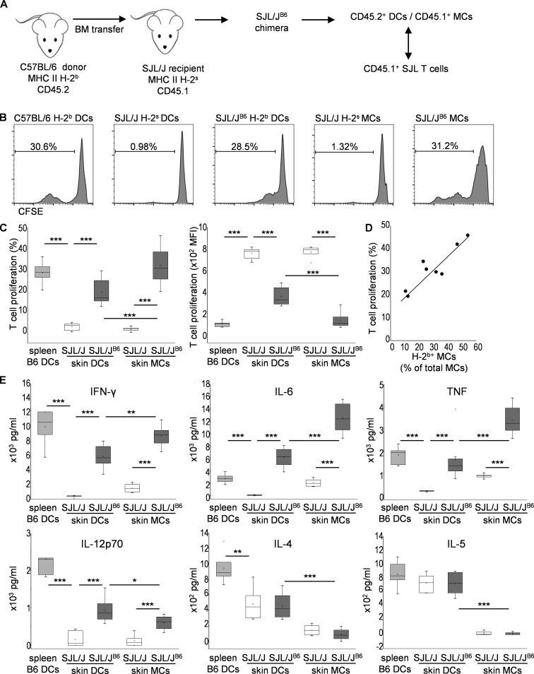
Figure 9.
DC-instructed MCs efficiently induce ex vivo allogeneic T cell priming. (A) SJL/J B6 chimera were generated by lethal irradiation of SJL/J recipient mice and transfer of BM cells from C57/BL6 donor mi...
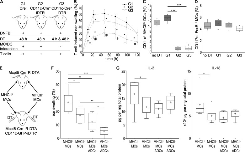
Figure 10.
DC-to-MC communication critically contributes to hapten-specific T cellu2013induced skin inflammation. (A) Skin inflammation was induced by i.d. injection of T cells from DNFB-sensitized WT donor mice...
Figure images are served from the NIH/NLM PubMed Central Open Access Subset or Europe PMC; copyright remains with the publishers and authors.
💬 Discussion
0 commentsNo comments yet. Be the first to start a discussion!
Leave a Comment