Abstract
The microtubule cytoskeleton is involved in many vital cellular processes. Microtubules act as tracks for molecular motors, and their polymerization and depolymerization can be harnessed to generate force. The structures of microtubules provide key information about the mechanisms by which their cellular roles are accomplished and the physiological context in which these roles are performed. Cryo-electron microscopy allows the visualization of in vitro-polymerized microtubules and has provided important insights into their overall morphology and the influence of a range of factors on their structure and dynamics. Cryo-electron tomography can be used to determine the unique three-dimensional structure of individual microtubules and their ends. Here, a previous cryo-electron tomography study of in vitro-polymerized GMPCPP-stabilized microtubules is revisited, the findings are compared with new tomograms of dynamic in vitro and cellular microtubules, and the information that can be extracted from such data is highlighted. The analysis shows the surprising structural heterogeneity of in vitro-polymerized microtubules. Lattice defects can be observed both in vitro and in cells. The shared ultrastructural properties in these different populations emphasize the relevance of three-dimensional structures of in vitro microtubules for understanding microtubule cellular functions.
🔬 Techniques
🧪 Sample Preparation
🔬 Cell Lines
🏭 Microscope Brands
🧪 Reagent Suppliers
💻 Software Details
💾 Data Repositories
🏛️ Research Organizations (ROR)
Affiliated research institutions:
📊 Figures
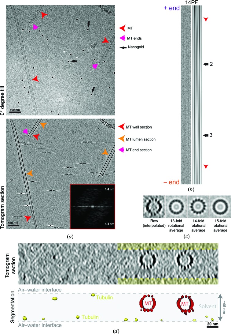
Figure 1
GMPCPP-stabilized in vitro -polymerized MTs visualized by cryo-electron tomography. ( a ) Top, 0u00b0 tilt image (motion-corrected movie sum) from a tilt series, showing a typical field of MTs that in...
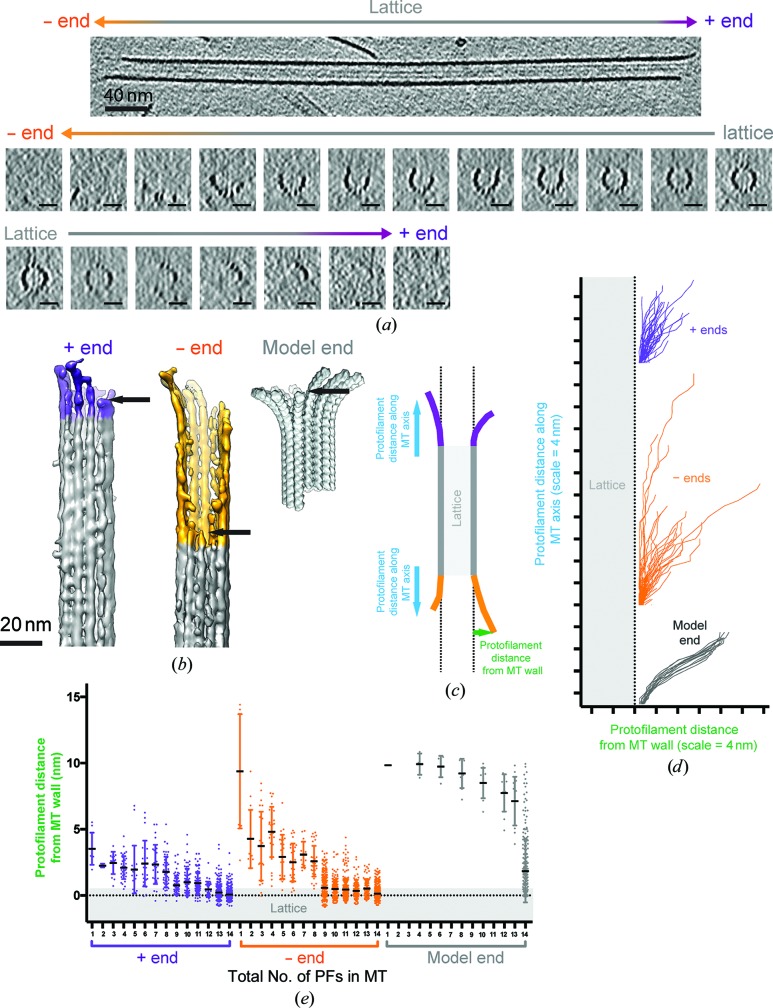
Figure 2
GMPCPP MT end structure visualized by cryo-electron tomography. ( a ) Top, longitudinal slice through the tomographic reconstruction, showing an MT in which both plus and minus ends (as indicated and ...
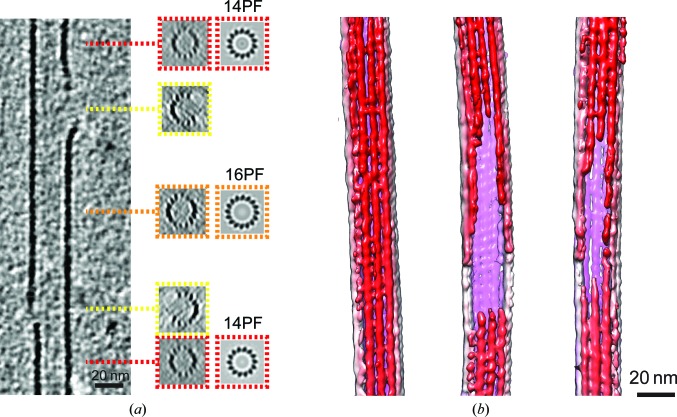
Figure 3
Cryo-electron tomography of GMPCPP MTs reveals lattice defects. ( a ) Longitudinal section through a three-dimensional tomographic reconstruction in which two different lattice defects are visible. In...
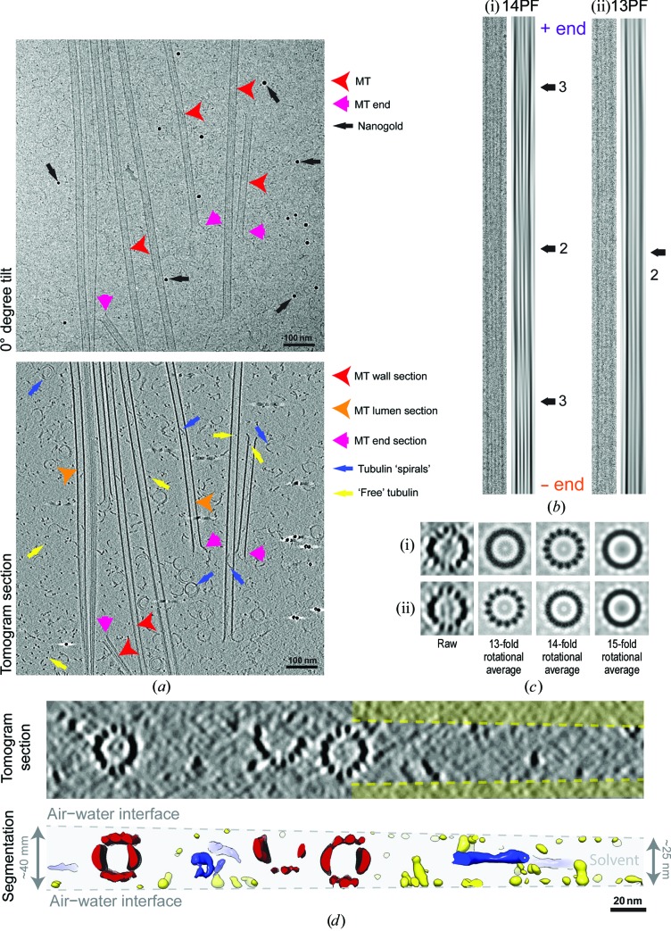
Figure 4
Dynamic in vitro -polymerized MTs visualized by cryo-electron tomography. ( a ) Top, 0u00b0 tilt image (motion-corrected movie sum; u223c2u2005e u2212 u00c5 u22122 total dose) from a tilt series, show...
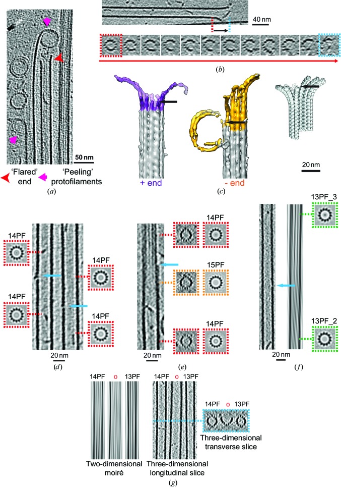
Figure 5
Dynamic MT end structure visualized in vitro by cryo-electron tomography. ( a ) Longitudinal slice through the tomographic reconstruction, showing a range of MT end morphologies including the highly c...
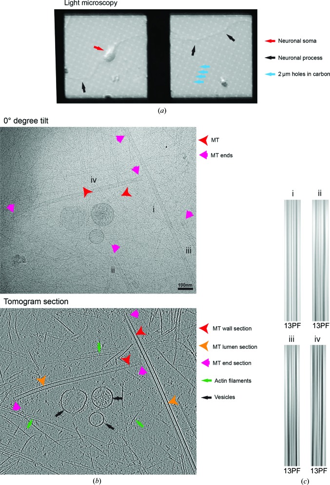
Figure 6
Characterization of MTs in cultured mouse neurons. ( a ) Phase-contrast light-microscope overview of mouse cortical neurons growing on cryo-EM grids. ( b ) Top, 0u00b0 tilt image (motion-corrected mov...
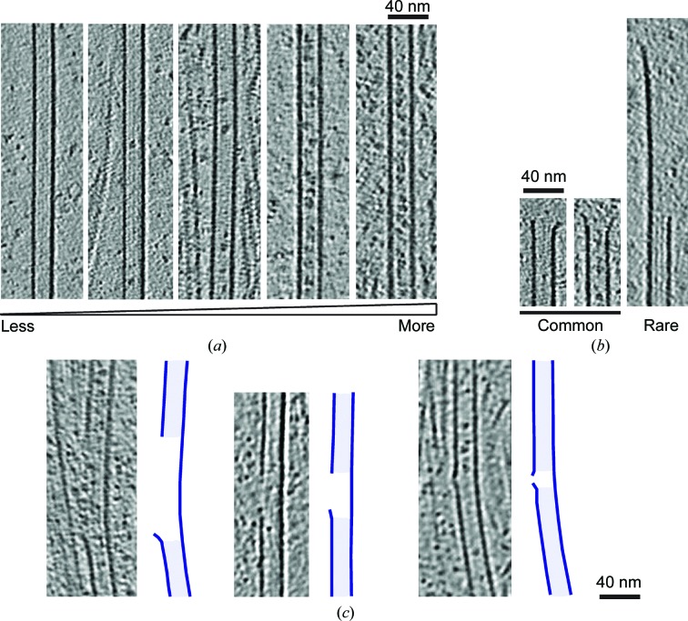
Figure 7
Diversity of MT ultrastructure in neurons. ( a ) A range of particle sizes and distributions are seen in the lumen of neuronal MTs. ( b ) The majority (24/25) of neuronal MT ends have relatively short...
Figure images are served from the NIH/NLM PubMed Central Open Access Subset or Europe PMC; copyright remains with the publishers and authors.
💬 Discussion
0 commentsNo comments yet. Be the first to start a discussion!
Leave a Comment