Abstract
A novel type of self-fluorescent unimolecular micelle nanoparticle (NP) formed by multi-arm star amphiphilic block copolymer, Boltron® H40 (H40, a 4th generation hyperbranched polymer)-biodegradable photo-luminescent polymer (BPLP)-poly(ethylene glycol) (PEG) conjugated with cRGD peptide (i.e., H40-BPLP-PEG-cRGD) was designed, synthesized, and characterized. The hydrophobic BPLP segment was self-fluorescent, thereby making the unimolecular micelle NP self-fluorescent. cRGD peptides, which can effectively target αvβ3 integrin-expressing tumor neovasculature and tumor cells, were selectively conjugated onto the surface of the micelles to offer active tumor-targeting ability. This unique self-fluorescent unimolecular micelle exhibited excellent photostability and low cytotoxicity, making it an attractive bioimaging probe for NP tracking for a variety of microscopy techniques including fluorescent microscopy, confocal laser scanning microscopy (CLSM), and two-photon microscopy. Moreover, this self-fluorescent unimolecular micelle NP also demonstrated excellent stability in aqueous solutions due to its covalent nature, high drug loading level, pH-controlled drug release, and passive and active tumor-targeting abilities, thereby making it a promising nanoplatform for targeted cancer theranostics.
🔬 Techniques
🔭 Microscopes
✨ Fluorophores
🧪 Sample Preparation
🔬 Cell Lines
🏭 Microscope Brands
🧪 Reagent Suppliers
🏛️ Research Organizations (ROR)
Affiliated research institutions:
📋 Methods
2.1. Materials Boltorn® H40 (a hyperbranched polyester with hydroxyl terminal groups) was provided by Perstorp Polyols Inc. The heterobifunctional poly(ethylene glycol) (PEG) derivatives, COOH-PEG-maleimide (Mw=5000 g/mol) and COOH-PEG-OCH 3 (Mw=3500 g/mol), were acquired from JenKem Technology. Citric acid, 1,8-octanediol, and L-cysteine were purchased from Sigma-Aldrich. Succinic anhydrous, 4-dimethylamino pyridine (DMAP), and 1,3-dicyclohexylcarbodiimide (DCC) were purchased from ACROS and used without further purification. Triethylamine (TEA), anhydrous dimethyl sulfoxide (DMSO), anhydrous dimethylformamide (DMF), and tris(2-carboxyethyl)phosphine (TCEP) were purchased from Sigma-Aldrich. All other chemicals and reagents used were of analytical reagent grade. The anticancer drug, doxorubicin hydrochloride (DOX HC1) was purchased from Beijing Mesochem Technology Co., Ltd. Cyclo (Arg-Gly-Asp-D-Phe-Cys) (cRGD) peptide was purchased from Peptides International. Ultrapure deionized water (DI water, Milli-Q Water Systems) was used for all buffer solutions and experiments. Dulbecco's phosphate-buffered saline (DPBS, pH 7.4), Dulbecco's Modified Eagle Medium (DMEM, high glucose, pyruvate), Trypsin-EDTA (0.25%), and Fetal Bovine Serum (FBS) were purchased from Invitrogen, USA. The U87MG human glioblastoma cells (expressing high levels of intergrin α v β 3 ) were purchased from ATCC and cultured in DMEM supplemented with 10% fetal bovine serum and 1% penicillin-streptomycin.
Synthesis of BPLP Polymers
BPLP polymers were synthesized following a literature protocol with slight modifications[ 32 ]. Briefly, citric acid, 1, 8-octanediol, and L-cysteine with a molar ratio of 1:1.2:0.2 were added into a 100 mL two-neck flask and dried under vacuum for 2 h. After the mixture was melted at 160 °C in an oil bath under continuous stirring, the temperature was lowered to 120 °C for another 75 min. Next, the polymer was dissolved in 1,4-dioxane and the resulting polymer solution was added dropwise into DI water under constant stirring. The BPLP polymer was obtained after lyophilization.
Show full methods section
2.1. Materials Boltorn® H40 (a hyperbranched polyester with hydroxyl terminal groups) was provided by Perstorp Polyols Inc. The heterobifunctional poly(ethylene glycol) (PEG) derivatives, COOH-PEG-maleimide (Mw=5000 g/mol) and COOH-PEG-OCH 3 (Mw=3500 g/mol), were acquired from JenKem Technology. Citric acid, 1,8-octanediol, and L-cysteine were purchased from Sigma-Aldrich. Succinic anhydrous, 4-dimethylamino pyridine (DMAP), and 1,3-dicyclohexylcarbodiimide (DCC) were purchased from ACROS and used without further purification. Triethylamine (TEA), anhydrous dimethyl sulfoxide (DMSO), anhydrous dimethylformamide (DMF), and tris(2-carboxyethyl)phosphine (TCEP) were purchased from Sigma-Aldrich. All other chemicals and reagents used were of analytical reagent grade. The anticancer drug, doxorubicin hydrochloride (DOX HC1) was purchased from Beijing Mesochem Technology Co., Ltd. Cyclo (Arg-Gly-Asp-D-Phe-Cys) (cRGD) peptide was purchased from Peptides International. Ultrapure deionized water (DI water, Milli-Q Water Systems) was used for all buffer solutions and experiments. Dulbecco's phosphate-buffered saline (DPBS, pH 7.4), Dulbecco's Modified Eagle Medium (DMEM, high glucose, pyruvate), Trypsin-EDTA (0.25%), and Fetal Bovine Serum (FBS) were purchased from Invitrogen, USA. The U87MG human glioblastoma cells (expressing high levels of intergrin α v β 3 ) were purchased from ATCC and cultured in DMEM supplemented with 10% fetal bovine serum and 1% penicillin-streptomycin.
Synthesis of BPLP Polymers
BPLP polymers were synthesized following a literature protocol with slight modifications[ 32 ]. Briefly, citric acid, 1, 8-octanediol, and L-cysteine with a molar ratio of 1:1.2:0.2 were added into a 100 mL two-neck flask and dried under vacuum for 2 h. After the mixture was melted at 160 °C in an oil bath under continuous stirring, the temperature was lowered to 120 °C for another 75 min. Next, the polymer was dissolved in 1,4-dioxane and the resulting polymer solution was added dropwise into DI water under constant stirring. The BPLP polymer was obtained after lyophilization.
Synthesis of COOH-PEG-cRGD
COOH-PEG-cRGD was prepared via a thiol-maleimide coupling reaction. Briefly, the COOH-PEG-Mal and cRGD-SH with a molar ratio of 1:1.2 were added in a 50 mL two-neck flask and dissolved in DMSO. TECP was added to avoid disulfide formation among cRGD peptides. The mixture was stirred under argon gas at room temperature for 24 h and followed by dialysis against DI water to remove impurities using a cellulose dialysis membrane (molecular weight cut-off, 2 kDa). After 48 h dialysis, the product was dried by lyophilization. Synthesis of BPLP-PEG-cRGD and BPLP-PEG-OCH 3 BPLP-PEG-cRGD was prepared via an esterification process in the presence of DCC and DMAP. Briefly, COOH-PEG-cRGD (5000 g/mol), BPLP, DCC, and DMAP with a molar ratio of 1.1:1:1.2:0.24 were dissolved in 10 mL of DMSO. The reaction was carried out at room temperature for 48 h. Next, the dicyclohexylurea (DCU) precipitated in the reaction solution was filtered out and the remaining solution was added dropwise into cold ethyl ether to obtain the crude polymers. The crude polymers were redissolved in DMSO and dialyzed against DI water for 48 h using a cellulose dialysis membrane (molecular weight cut-off, 5 kDa). The purified polymer was dried via lyophilization. BPLP-PEG-OCH 3 was prepared following a similar procedure by using COOH-PEG-OCH 3 (3500 g/mol) instead.
Synthesis of H40-COOH
To purify H40-OH, it was dissolved in acetone overnight and was then precipitated in cold ethyl ether. H40-COOH was prepared by converting the hydroxyl terminal groups into carboxyl terminal groups in the presence of succinic anhydrous. Briefly, H40-OH, succinic anhydrous, and DMAP with a molar ratio of 1:70:7 were dissolved into 10 mL of CH 2 Cl 2 . The reaction was carried out at room temperature for 48 h and the product was precipitated using diethyl ether and vacuum-dried. Synthesis of H40-BPLP-PEG-OCH 3 /cRGD H40-BPLP-PEG-OCH 3 /cRGD was synthesized by reacting H40-COOH with BPLP-PEG-cRGD and BPLP-PEG-OCH 3 in 10 mL of DMF in the presence of DCC and DMAP. The molar ratio of reactants (H40-COOH:BPLP-PEG-cRGD:BPLP-PEG-OCH 3 ) was 1:7:28. The reaction mixture was stirred at room temperature for 48 h and the by-product, DCU, was removed by filtration. The impurities were removed by dialysis against DMF for 12 h and against DI water for another 36 h using a cellulose dialysis membrane (molecular weight cut-off, 15 kDa). The resulting polymer H40-BPLP-PEG-OCH 3 /cRGD was obtained after lyophilization and used to prepare targeted unimolecular micelles. Multi-arm star amphiphilic block copolymer H40-BPLP-PEG-OCH 3 (without cRGD conjugation) was also prepared following a similar procedure and was used to prepare non-targeted unimolecular micelles. Preparation of DOX-Loaded Unimolecular Micelles DOX·HCl (4 mg) was dissolved in 4 mL of anhydrous DMSO and treated with 2 moles excess of TEA for 2 h. Subsequently, the multi-arm star amphiphilic block copolymer H40-BPLP-PEG-cRGD or H40-BPLP-PEG-OCH 3 (20 mg) was added to this solution. Thereafter, 12 ml of DI water was added dropwise into the solution under constant stirring. The resulting solution was stirred using a magnetic stirring bar for 4 h and then dialyzed against DI water using a cellulose dialysis membrane (molecular weight cut-off, 2 kDa) for 24 h followed by freeze-drying. Characterization 1 H NMR spectra of all intermediate and final polymer products were recorded on a Varian Mercury Plus 300 spectrometer using DMSO-d6 as a solvent at 25°C. Molecular weights (M n and M w ) and polydispersity indices (PDI) of the polymers were determined by gel permeation chromatography (GPC) equipped with a refractive index detector, a viscometer detector, and a light scattering detector (Viscotek, USA). DMF with 0.1 mmol of LiBr was used as a mobile phase with a flow rate of 1 mL/min. Fluorescent spectra of the unimolecular micelle solutions were acquired on a Nanolog FL3-2iHR spectrofluorometer (HORIBA Jobin Yvon Inc., USA). The sizes and morphologies of the unimolecular micelles were determined by dynamic light scattering (DLS, ZetaSizer Nano ZS90, Malvern Instrument, USA) and transmission electron microscopy (TEM, FEI Tecnai G 2 F30 TWIN 300 KV, E.A. Fischione Instruments, Inc. USA) at a polymer concentration of 0.05 mg/ml. The TEM sample was prepared by depositing a drop of the copolymer solution (0.05 mg/ml) containing 1 wt% of phosphotungstic acid onto a 200 mesh copper grid coated with carbon. The DOX loading level, defined as the weight percentage of DOX in the DOX-loaded unimolecular micelle NPs, was measured by a Cary 500 UV-Vis-NIR spectrophotometer based on a standard calibration curve of DOX at 485 nm. In Vitro Drug Release Study Drug release studies were carried out in a glass apparatus at 37 °C in either an acetate buffer (pH 5.3) or a phosphate buffer (pH 7.4) solution. Five mg of DOX-loaded non-targeted or targeted unimolecular micelles were dispersed uniformly in 5 ml of medium and then placed in a dialysis bag with a molecular weight cut-off of 2 kDa. The dialysis bag was immersed in 50 ml of the release medium and kept at 37 °C under a horizontal laboratory shaker at 100 rpm (Thermo Scientific MaxQ Shaker, USA). At specific time points, 3 ml of release media were collected and replaced by the same volume of fresh media. The amount of released DOX was analyzed by a UV-Vis-NIR spectrophotometer at 485 nm.
Cellular Uptake Study
The cellular internalization and intracellular distribution of the self-fluorescent unimolecular micelles in the U87MG cells were analyzed by fluorescence microscopy. U87MG cells (1.4×10 4 cells/cm 2 ) were seeded in the 8-well chamber slide and cultured overnight. When about 80% confluence was reached, the cells were treated with H40-BPLP-PEG (non-targeted) and H40-BPLP-PEG-cRGD (targeted) unimolecular micelles at two different concentrations (i.e., 0.5 mg/ml or 1.0 mg/ml) for 6 and 20 h. A blocking experiment with 2 µM of free cRGD was also carried out for targeted micelles. After a certain period of treatment, cells were washed with DPBS and fixed by 4% polyformaldehyde (PFA). Cell nuclei were stained by propidium iodide (PI, Sigma, USA). Cells were first treated by RNaseA at 37 °C for 30 min to eliminate any interference from RNA, which PI can also bind to, followed by treatment with PI solution (1 µg/mL) at room temperature for 1 h. Thereafter, cells were imaged under a fluorescence microscope (Nikon, Japan) using two different filters. To image the self-fluorescent unimolecular micelle NPs, a UV-2E/C filter with an excitation wavelength of 325 to 375 nm and an emission wavelength of 435 to 485 nm was used. For cell nuclei imaging, a Texas-red filter with an excitation wavelength of 533 to 588 nm, and an emitter wavelength of 608 to 683 nm, was used. Digital monochromatic images were acquired using NIS-Element AR software. Cell uptake studies of DOX-loaded unimolecular micelles (non-targeted and targeted NPs) were carried out using confocal laser scanning microscopy (CLSM), two-photon microscopy and flow cytometry. For CLSM and two-photon microscopy studies, after reaching 80% confluence, the cells (1.4×l0 4 cells/cm 2 ) were treated with free DOX, DOX-loaded non-targeted, and DOX-loaded targeted micelles at a DOX concentration of 20 µg/ml. After 5 h incubation, cells were washed by DPBS and images were taken under a CLSM (Nikon Eclipse Ti inverted microscope equipped with Nikon AIR confocal diode lasers, Japan) or two-photon microscope (Bruker Ultima IV, USA) to observe the intracellular locations of both the self-fluorescent unimolecular micelle NPs and DOX. For the CLSM imaging, a UV-2E/C filter was used for unimolecular micelle imaging and a Texas-red filter was used for cell nuclei imaging. For two-photon microscope imaging, the excitation wavelength was set at 700 nm, and emissions at 445 nm (for unimolecular micelle imaging) and 620 nm (for DOX imaging) were collected. For the flow cytometry study, U87MG cells (1.5×10 4 cells/cm 2 ) were seeded in 24-well plates and cultured overnight. When about 80%, confluence was reached, cells were treated with free DOX, DOX-loaded non-targeted micelles, and DOX-loaded targeted micelles at a DOX concentration of 20 µg/ml for 5 h. A blocking experiment with 2 µM of free cRGD was also carried out for the DOX-loaded targeted micelles. After 5 h of treatment, DOX uptake was analyzed by an Accuri™C6 flow cytometer system (BD Bioscience, USA). A minimum of 2×10 4 cells for each sample were analyzed for DOX fluorescence intensity on a decade log scale.
Cytotoxicity Assay
The cytotoxicity of free DOX, DOX-loaded non-targeted micelles, and DOX-loaded targeted micelles for U87MG cells was analyzed by MTT assay. U87MG cells (3×10 4 cells/cm 2 ) were seeded in a 96-well plate overnight. When about 80% confluence was reached, cells were treated with free DOX, DOX-loaded non-targeted micelles, DOX-loaded targeted micelles, and empty targeted and non-targeted micelles at two DOX concentrations (i.e., 2.5 and 5.0 µg/ml). After 48 h of treatment, the cells were incubated with media containing 500 µg/ml of MTT for another 4 h, followed by adding 100 µl of DMSO to dissolve the precipitates. An absorbance at 560 nm was measured by a GloMax®-Multi+ Detection System (Promega, USA) with an absorbance of 730 nm as the reference.
2.1. Materials Boltorn® H40 (a hyperbranched polyester with hydroxyl terminal groups) was provided by Perstorp Polyols Inc. The heterobifunctional poly(ethylene glycol) (PEG) derivatives, COOH-PEG-maleimide (Mw=5000 g/mol) and COOH-PEG-OCH 3 (Mw=3500 g/mol), were acquired from JenKem Technology. Citric acid, 1,8-octanediol, and L-cysteine were purchased from Sigma-Aldrich. Succinic anhydrous, 4-dimethylamino pyridine (DMAP), and 1,3-dicyclohexylcarbodiimide (DCC) were purchased from ACROS and used without further purification. Triethylamine (TEA), anhydrous dimethyl sulfoxide (DMSO), anhydrous dimethylformamide (DMF), and tris(2-carboxyethyl)phosphine (TCEP) were purchased from Sigma-Aldrich. All other chemicals and reagents used were of analytical reagent grade. The anticancer drug, doxorubicin hydrochloride (DOX HC1) was purchased from Beijing Mesochem Technology Co., Ltd. Cyclo (Arg-Gly-Asp-D-Phe-Cys) (cRGD) peptide was purchased from Peptides International. Ultrapure deionized water (DI water, Milli-Q Water Systems) was used for all buffer solutions and experiments. Dulbecco's phosphate-buffered saline (DPBS, pH 7.4), Dulbecco's Modified Eagle Medium (DMEM, high glucose, pyruvate), Trypsin-EDTA (0.25%), and Fetal Bovine Serum (FBS) were purchased from Invitrogen, USA. The U87MG human glioblastoma cells (expressing high levels of intergrin α v β 3 ) were purchased from ATCC and cultured in DMEM supplemented with 10% fetal bovine serum and 1% penicillin-streptomycin.
📊 Figures

Fig. 1
A schematic illustration of a self-fluorescent unimolecular micelle NP made of multi-arm star amphiphilic block copolymer, H40-BPLP-PEG-cRGD, for tumor-targeted drug delivery and bioimaging.

Fig. 2
(A) 1 H NMR and (B) FT-IR spectra of the BPLP polymers.

Fig. 3
1 H NMR spectra of (A) BPLP-PEG-cRGD and (B) H40-BPLP-PEG-OCH 3 /cRGD polymers.

Fig. 4
(A) DLS histogram and (B) TEM images of the unimolecular micelles.

Fig. 5
Fluorescence properties of the unimolecular micelles. (A) Fluorescent spectra of the unimolecular micelles in water at various excitations. (B) Digital photograph of the unimolecular micelles (in wate...

Fig. 6
Fluorescence microscopy images of U87MG cells incubated with H40-BPLP-PEG non-targeted micelles (NT) and H40-BPLP-PEG-cRGD targeted unimolecular micelles (T) at 37 u00b0C for 6 h at (A) 0.5 mg/ml and ...

Fig. 7
Fluorescence microscopy images of U87MG cells incubated with H40-BPLP-PEG non-targeted micelles (NT) and H40-BPLP-PEG-cRGD targeted micelles (T), or targeted micelles with a blocking dose of free cRGD...

Fig. 8
In vitro drug release profiles of DOX-loaded unimolecular micelles at a pH of 7.4 and 5.3.

Fig. 9
CLSM images of U87MG cells incubated with free DOX, DOX-loaded targeted micelles (DOX-T), and DOX-loaded non-targeted micelles (DOX-NT) at 37 u00b0C for 5 h with a DOX concentration of 20 u00b5g/ml. T...

Fig. 10
Flow cytometry analysis of U87MG cells incubated with free DOX (green line), DOX-T (blue line), DOX-NT micelles (red line), or DOX-T with a blocking dose of free cRGD (purple line) at 37 u00b0C for 5 ...

Fig. 11
Cytotoxicity of free DOX, DOX-loaded targeted micelles (DOX-T), DOX loaded non-targeted micelles (DOX-NT), empty (DOX-free) non-targeted micelles (NT), or empty targeted micelles (T) against U87MG cel...
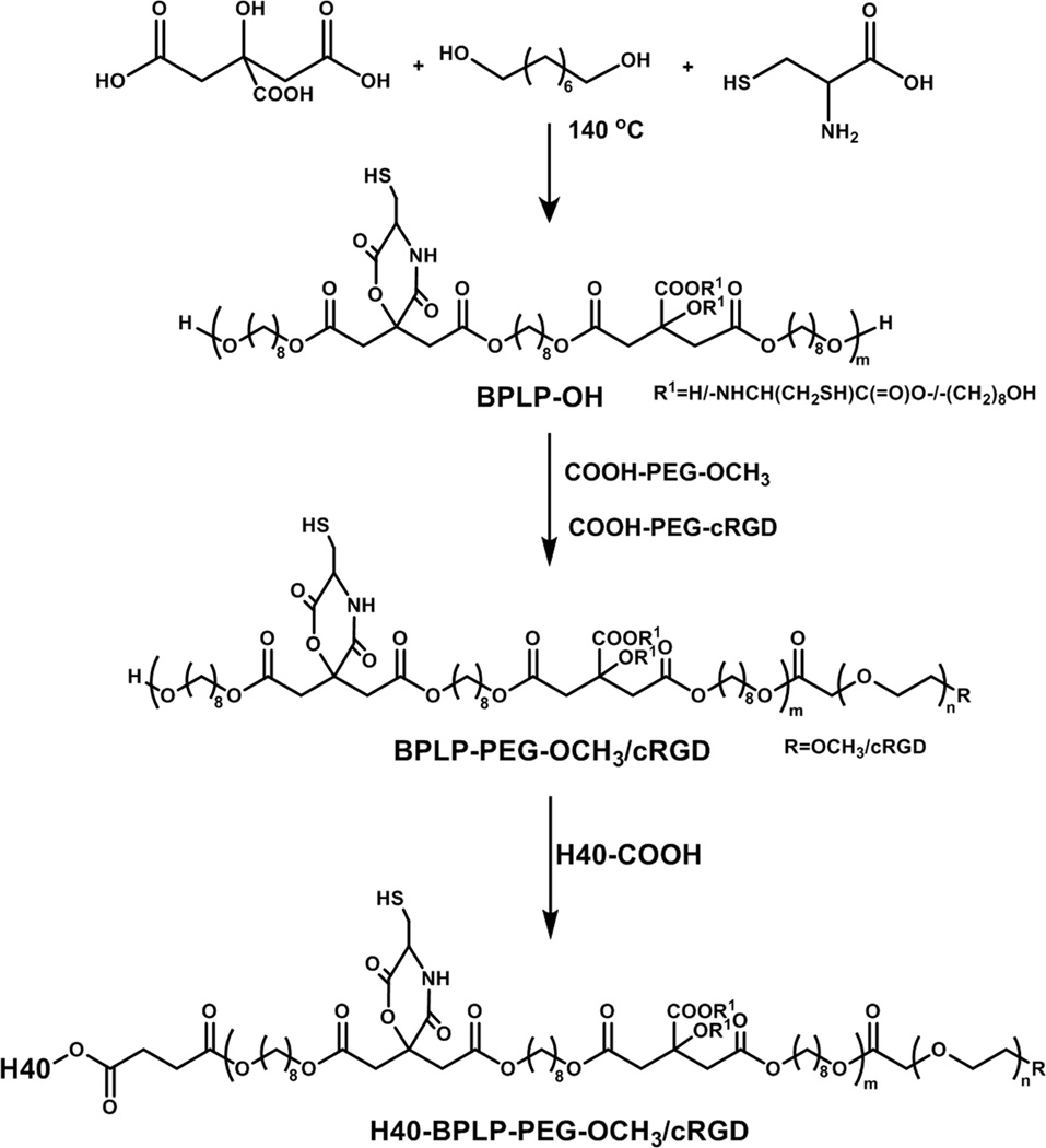
Scheme 1
Synthesis scheme of the self-fluorescent multi-arm star amphiphilic block copolymer H40-BPLP-PEG-OCH 3 /cRGD.
Figure images are served from the NIH/NLM PubMed Central Open Access Subset or Europe PMC; copyright remains with the publishers and authors.
💬 Discussion
0 commentsNo comments yet. Be the first to start a discussion!
Leave a Comment