Abstract
The mitochondrial contact site and cristae organizing system (MICOS) is a multisubunit protein complex that is essential for the proper architecture of the mitochondrial inner membrane. MICOS plays a key role in establishing and maintaining crista junctions, tubular or slit-like structures that connect the cristae membrane with the inner boundary membrane, thereby ensuring a contiguous inner membrane. MICOS is enriched at crista junctions, but the detailed distribution of its subunits around crista junctions is unclear because such small length scales are inaccessible with established fluorescence microscopy. By targeting individually activated fluorophores with an excitation beam featuring a central zero-intensity point, the nanoscopy method called MINFLUX delivers single-digit nanometer-scale three-dimensional (3D) resolution and localization precision. We employed MINFLUX nanoscopy to investigate the submitochondrial localization of the core MICOS subunit Mic60 in relation to two other MICOS proteins, Mic10 and Mic19. We demonstrate that dual-color 3D MINFLUX nanoscopy is applicable to the imaging of organellar substructures, yielding a 3D localization precision of ∼5 nm in human mitochondria. This isotropic precision facilitated the development of an analysis framework that assigns localization clouds to individual molecules, thus eliminating a source of bias when drawing quantitative conclusions from single-molecule localization microscopy data. MINFLUX recordings of Mic60 indicate ringlike arrangements of multiple molecules with a diameter of 40 to 50 nm, suggesting that Mic60 surrounds individual crista junctions. Statistical analysis of dual-color MINFLUX images demonstrates that Mic19 is generally in close proximity to Mic60, whereas the spatial coordination of Mic10 with Mic60 is less regular, suggesting structural heterogeneity of MICOS.
🔬 Techniques
🧬 Organisms
✨ Fluorophores
🔬 Cell Lines
💻 Software Details
🏛️ Research Organizations (ROR)
Affiliated research institutions:
📊 Figures
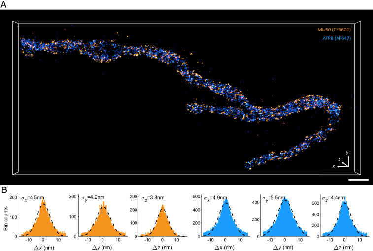
Fig. 1.
Two-color 3D MINFLUX acquisition of a mitochondrion in primary human dermal fibroblasts. ( A ) Three-dimensional MINFLUX recording of Mic60 (orange) and ATPB (blue) using directly labeled primary anti...
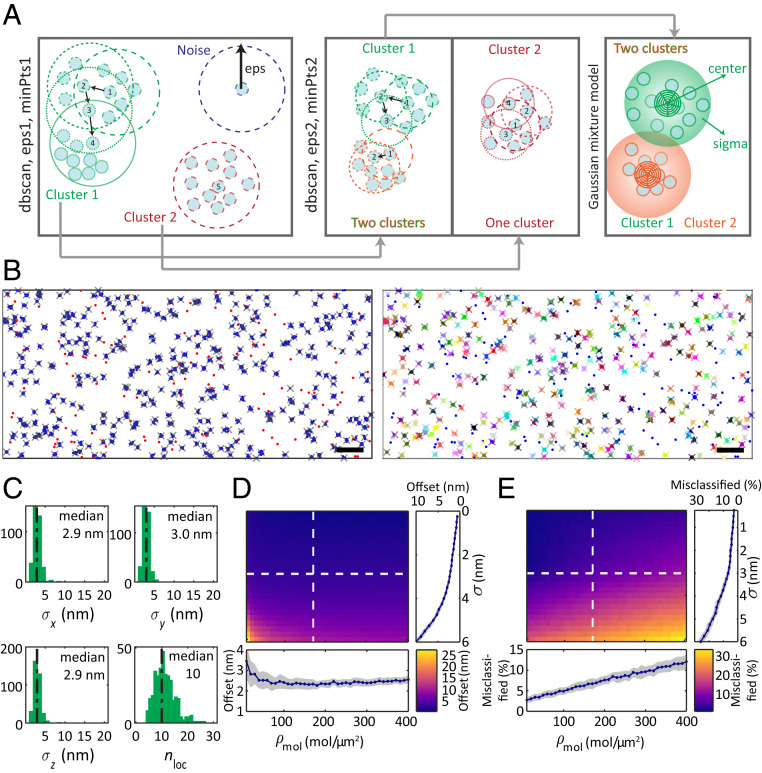
Fig. 2.
Molecular assignment of localization clouds with the modified dbscan algorithm. ( A ) Schematic of the clustering algorithm developed in this work. In the first dbscan step, noise (blue) and isolated ...
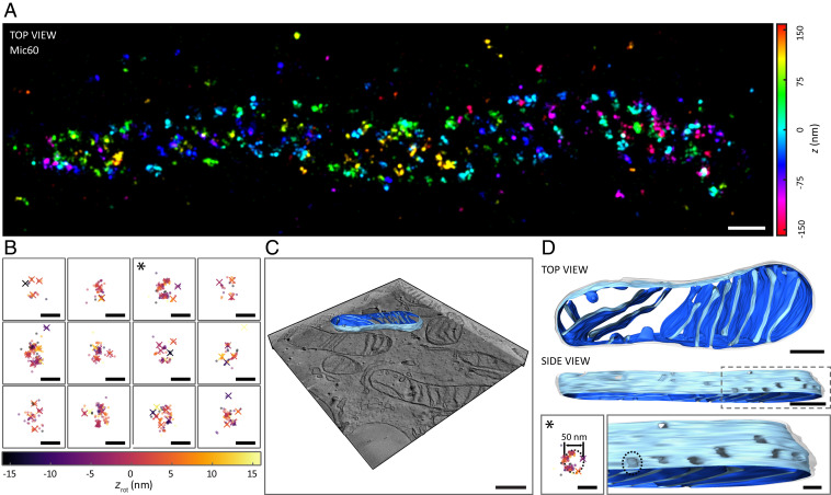
Fig. 3.
Distribution of Mic60 in mitochondria of human U-2 OS cells. ( A ) Three-dimensional MINFLUX image of Mic60 stained with primary labeled antibodies conjugated to Alexa Fluor 647. The axial position of...
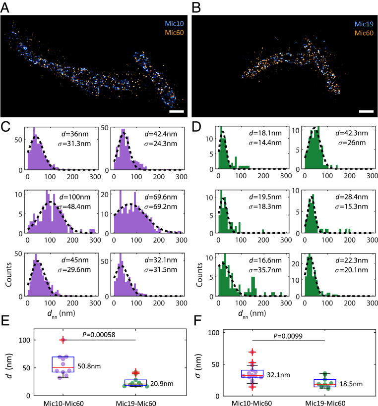
Fig. 4.
Measurement of distances between proteins of the MICOS complex. ( A ) Representative 3D two-color MINFLUX acquisition of Mic10 (CF680, blue) and Mic60 (Alexa Fluor 647, orange) in Mic10-TO HeLa cells....
Figure images are served from the NIH/NLM PubMed Central Open Access Subset or Europe PMC; copyright remains with the publishers and authors.
💬 Discussion
0 commentsNo comments yet. Be the first to start a discussion!
Leave a Comment