Abstract
Adult skeletal muscle is capable of complete regeneration after an acute injury. The main parameter studied to assess muscle regeneration efficacy is the cross-sectional area (CSA) of the myofibers as myofiber size correlates with muscle force. CSA analysis can be time-consuming and may trigger variability in the results when performed manually. This is why programs were developed to completely automate the analysis of the CSA, such as SMASH, MyoVision, or MuscleJ softwares. Although these softwares are efficient to measure CSA on normal or hypertrophic/atrophic muscle, they fail to efficiently measure CSA on regenerating muscles. We developed Open-CSAM, an ImageJ macro, to perform a high throughput semi-automated analysis of CSA on skeletal muscle from various experimental conditions. The macro allows the experimenter to adjust the analysis and correct the mistakes done by the automation, which is not possible with fully automated programs. We showed that Open-CSAM was more accurate to measure CSA in regenerating and dystrophic muscles as compared with SMASH, MyoVision, and MuscleJ softwares and that the inter-experimenter variability was negligible. We also showed that, to obtain a representative CSA measurement, it was necessary to analyze the whole muscle section and not randomly selected pictures, a process that was easily and accurately be performed using Open-CSAM. To conclude, we show here an easy and experimenter-controlled tool to measure CSA in muscles from any experimental condition, including regenerating muscle.
🔬 Techniques
🔭 Microscopes
💻 Software
✨ Fluorophores
🧪 Sample Preparation
🏭 Microscope Brands
🧪 Reagent Suppliers
📷 Detectors
💻 Software Details
🏛️ Research Organizations (ROR)
Affiliated research institutions:
📊 Figures
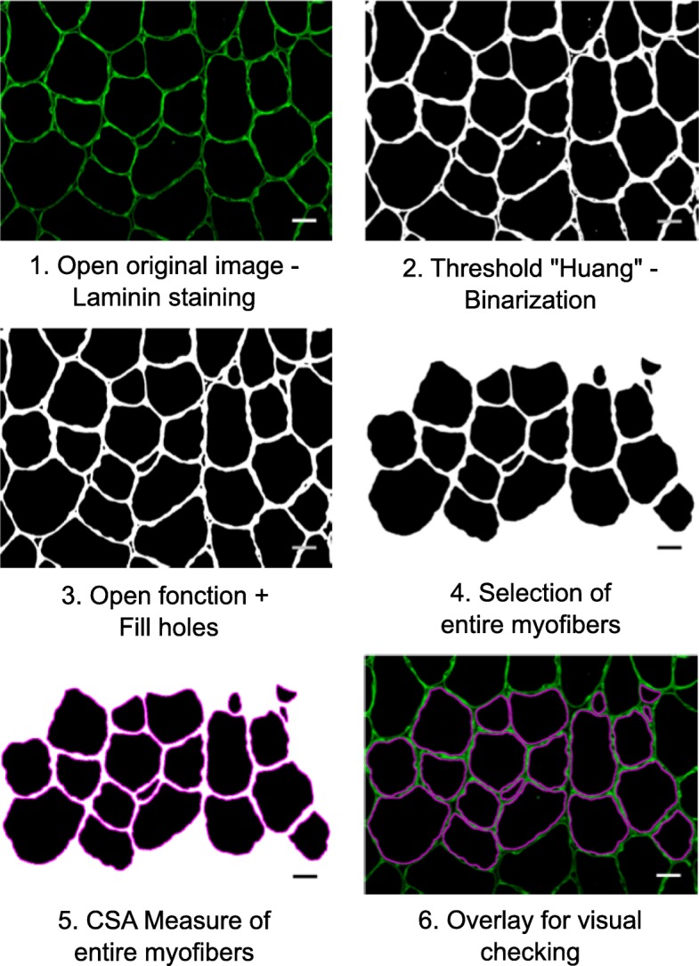
Fig. 1
Open-CSAM workflow. Step 1: When the macro starts, a window automatically opens to select the image to be analyzed (here muscle cryosections immunostained for laminin). Step 2: Open-CSAM applies the I...
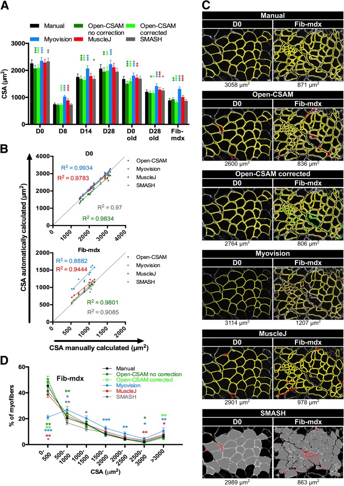
Fig. 2
Open-CSAM comparison with MyoVision, MuscleJ, and SMASH softwares. The same pictures were analyzed either by manual measurement or using Open-CSAM (with or without manual correction), MyoVision, Muscl...
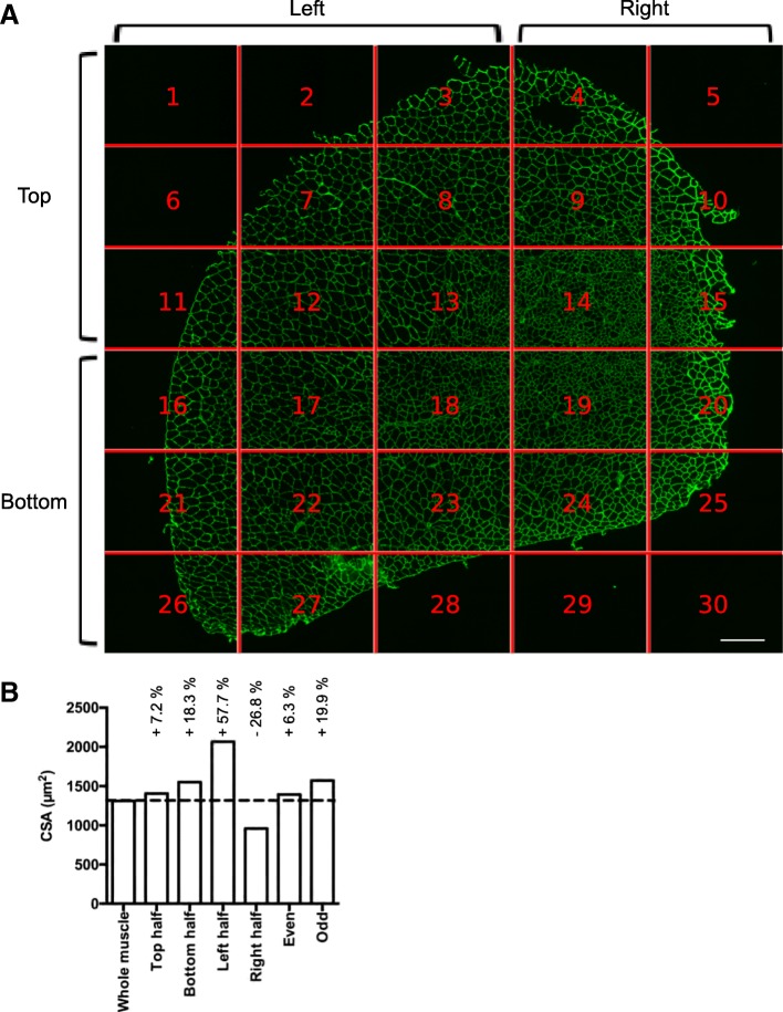
Fig. 3
Whole muscle analysis by Open-CSAM. a Whole reconstitution of a laminin-stained cryosection of a TA muscle 28u2009days post-CTX injury (30 pictures were automatically recorded and assembled by MetaMor...
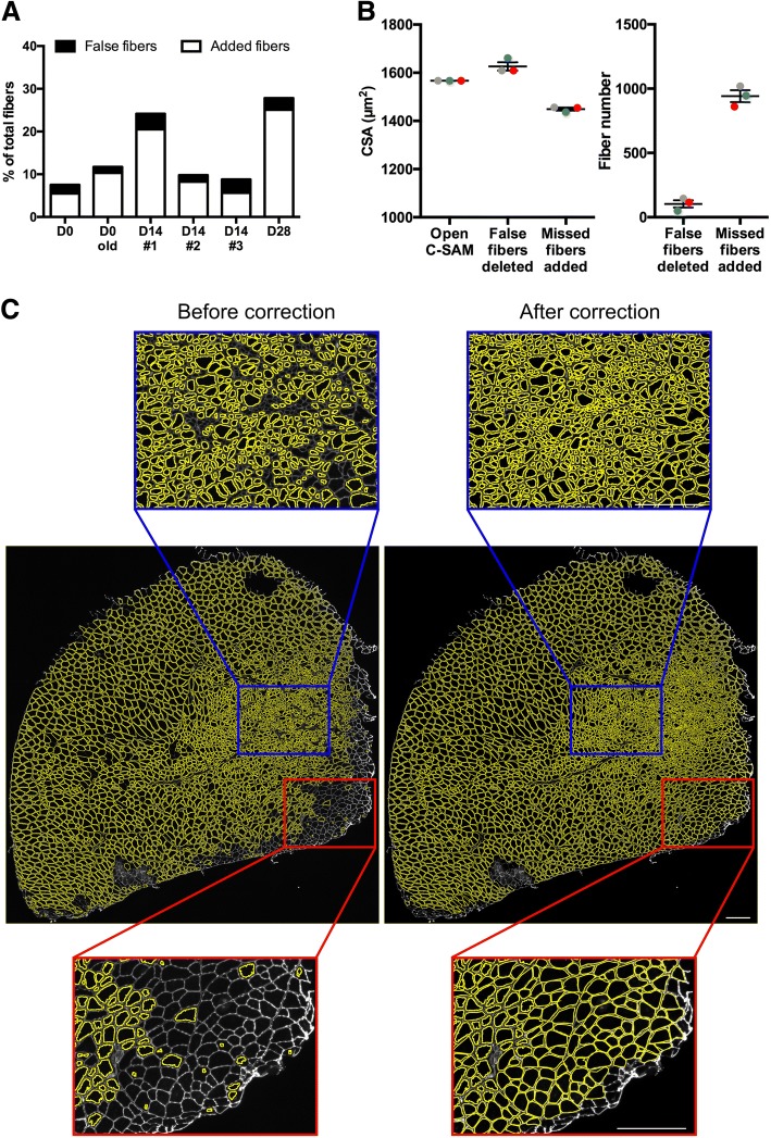
Fig. 4
Manual corrections after Open-CSAM analysis. a Percentage of false myofibers detected (black histograms) and missed myofibers (white histograms) by Open-CSAM that needed manual correction for TA muscl...
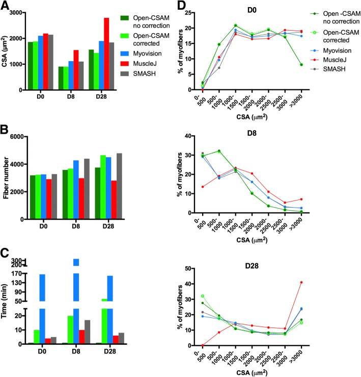
Fig. 5
Comparison of Open-CSAM, MyoVision, MuscleJ, and SMASH CSA quantification on whole muscle sections. CSA was measured using Open-CSAM (with or without manual correction), MyoVision, MuscleJ, or SMASH o...
Figure images are served from the NIH/NLM PubMed Central Open Access Subset or Europe PMC; copyright remains with the publishers and authors.
💬 Discussion
0 commentsNo comments yet. Be the first to start a discussion!
Leave a Comment