Abstract
AbstractStructured illumination microscopy relies on reconstruction algorithms to yield super-resolution images. Artifacts can arise in the reconstruction and affect the image quality. Current reconstruction methods involve a parametrized apodization function and a Wiener filter. Empirically tuning the parameters in these functions can minimize artifacts, but such an approach is subjective and produces volatile results. We present a robust and objective method that yields optimal results by two straightforward filtering steps with Richardson-Lucy-based deconvolutions. We provide a resource to identify artifacts in 2D-SIM images by analyzing two main reasons for artifacts, out-of-focus background and a fluctuating reconstruction spectrum. We show how the filtering steps improve images of test specimens, microtubules, yeast and mammalian cells.
🔬 Techniques
✨ Fluorophores
🧪 Sample Preparation
🔬 Cell Lines
🏭 Microscope Brands
🧪 Reagent Suppliers
🎨 Filters
💻 Software Details
🏛️ Research Organizations (ROR)
Affiliated research institutions:
📊 Figures
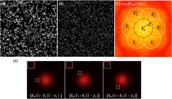
Figure 1
Simulation of 2D structured illumination with fluorescent beads.
( a ) Wide-field image. ( b ) I SIM reconstruction. ( c ) Logarithm of the I SIM power spectrum: . ( d ) Cross-correlations of extended domains with the central one, , produce a sharp peak due to thei...
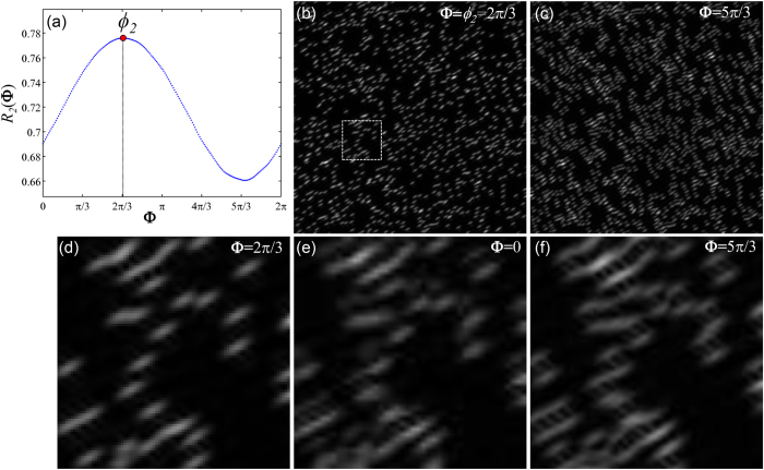
Figure 2
Estimation of initial phase u03d5 j by correlating the reconstruction I SIM,j and the wide-field.
( a ) The R 2 ( u03a6 ) curve provides the values of the normalized cross-correlation between the wide-field and the reconstruction I SIM,2 at different phases from 0 to 2u03c0. Arguments of the maxim...
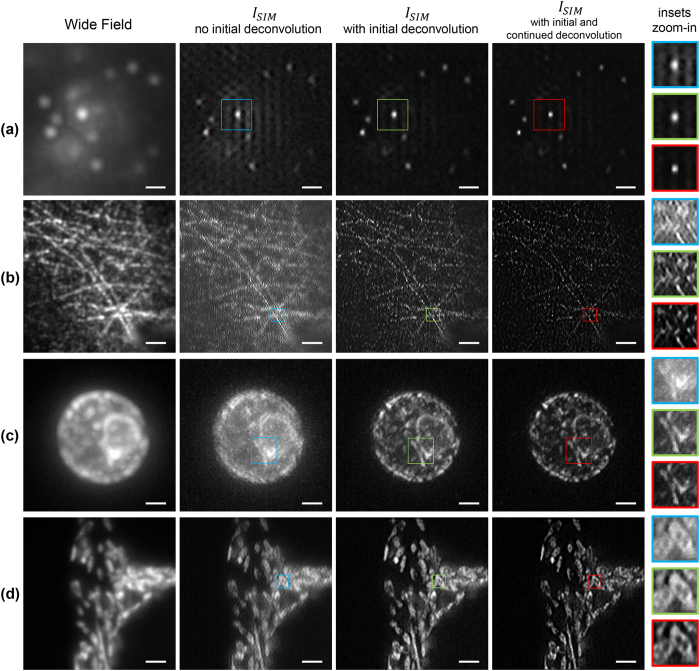
Figure 3
Comparison of SIM image reconstruction of different samples with/without the initial and continued deconvolution steps.
All samples are excited with a 488u2009nm laser, while a bandpass filter (FF02-525/50-25, Semrock) is used in the detection path. ( a ) Fluorescent beads are 40u2009nm (F8759, Invitrogen) in diameter....
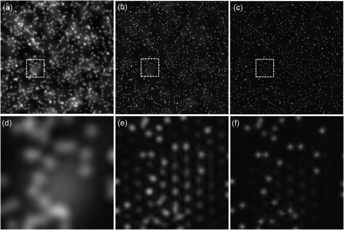
Figure 4
Using raw images O j,m with out-of-focus background yields I SIM reconstructions with artifacts in the background regions.
If the raw O j,m images are deconvolved prior to the reconstruction, no artifacts are visible in the reconstructed image. ( a ) Wide-field image of simulated beads with out-of-focus fluorescence backg...
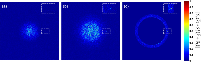
Figure 5
Initial deconvolution improves the estimate of the parameter by enhancing the peak occurrence in the correlation .
Correlation result for a sample ( a ) without and ( b ) with initially deconvolved raw data O j,m . In the first case, there is no peak in the region where is expected (white dashed inset). In the sec...
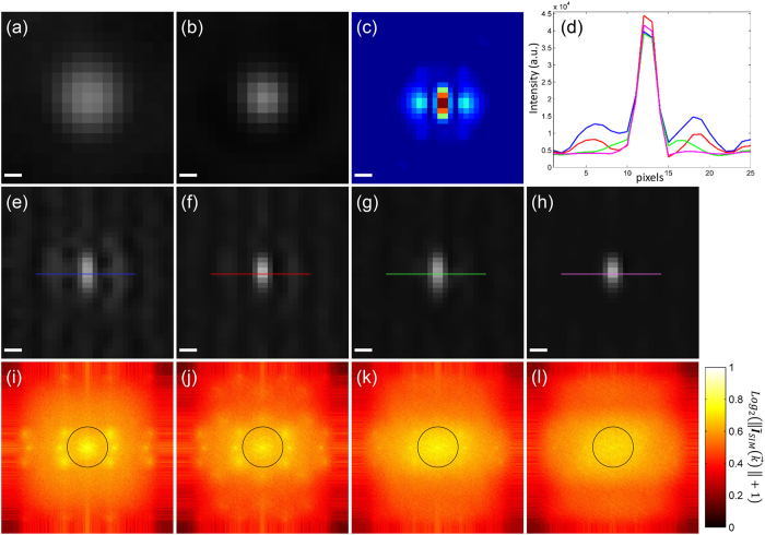
Figure 6
Continued deconvolution improves the reconstructed image I SIM .
A single bead taken from a larger image consisting of 40u2009nm fluorescent beads is selected to show the effects of the initial and continued deconvolution on the reconstruction. ( a ) Wide-field, ( ...
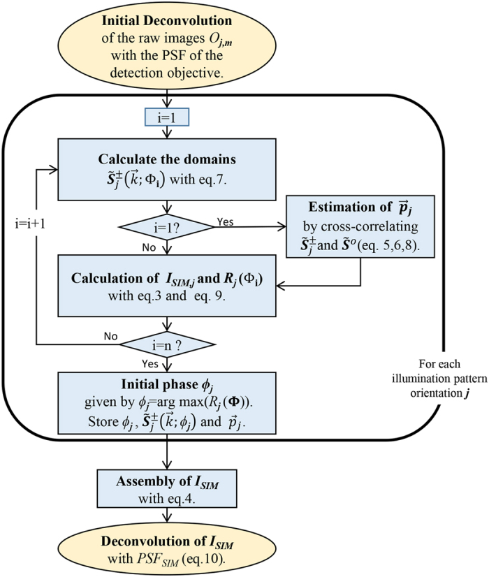
Figure 7
Flow chart of our reconstruction approach.
For the Richardson-Lucy deconvolutions applied in the first and final steps we use no more than 10 and 5 iterations respectively. To estimate the initial phase u03d5 j an interval u03a6 =u2009[0, 2u03...
Figure images are served from the NIH/NLM PubMed Central Open Access Subset or Europe PMC; copyright remains with the publishers and authors.
💬 Discussion
0 commentsNo comments yet. Be the first to start a discussion!
Leave a Comment