Abstract
BACKGROUND: Variation in the gene encoding zinc finger binding protein 804A (ZNF804A) is associated with schizophrenia and bipolar disorder. Evidence suggests that ZNF804A is a regulator of gene transcription and is present in nuclear and extranuclear compartments. However, a detailed examination of ZNF804A distribution and its neuronal functions has yet to be performed. METHODS: The localization of ZNF804A protein was examined in neurons derived from human neural progenitor cells, human induced pluripotent stem cells, or in primary rat cortical neurons. In addition, small interfering RNA-mediated knockdown of ZNF804A was conducted to determine its role in neurite formation, maintenance of dendritic spine morphology, and responses to activity-dependent stimulations. RESULTS: Endogenous ZNF804A protein localized to somatodendritic compartments and colocalized with the putative synaptic markers in young neurons derived from human neural progenitor cells and human induced pluripotent stem cells. In mature rat neurons, Zfp804A, the homolog of ZNF804A, was present in a subset of dendritic spines and colocalized with synaptic proteins in specific nanodomains, as determined by super-resolution microscopy. Interestingly, knockdown of ZNF804A attenuated neurite outgrowth in young neurons, an effect potentially mediated by reduced neuroligin-4 expression. Furthermore, knockdown of ZNF804A in mature neurons resulted in the loss of dendritic spine density and impaired responses to activity-dependent stimulation. CONCLUSIONS: These data reveal a novel subcellular distribution for ZNF804A within somatodendritic compartments and a nanoscopic organization at excitatory synapses. Moreover, our results suggest that ZNF804A plays an active role in neurite formation, maintenance of dendritic spines, and activity-dependent structural plasticity.
🔬 Techniques
🔭 Microscopes
💻 Software
✨ Fluorophores
🧪 Sample Preparation
🔬 Cell Lines
🏭 Microscope Brands
🧪 Reagent Suppliers
🔎 Objectives
💻 Software Details
🏛️ Research Organizations (ROR)
Affiliated research institutions:
📋 Methods
The localization of ZNF804A protein was examined in neurons derived from human neural progenitor cells, human induced pluripotent stem cells, or in primary rat cortical neurons. In addition, small interfering RNA-mediated knockdown of ZNF804A was conducted to determine its role in neurite formation, maintenance of dendritic spine morphology, and responses to activity-dependent stimulations.
Methods and Materials See the Supplement for further detailed Methods and Materials . CTX0E16 hNPCs and Neurons Experiments using hNPCs were carried out using the CTX0E16 neural cell line obtained from ReNeuron Ltd. ( www.reneuron.com ) under a Material Transfer Agreement. This karyotypically normal hNPC line was derived from 12-week fetal cortical neuroepithelium and conditionally immortalized using a c-mycER TAM transgene ( 19 , 20 ). Proliferation and neuralization of CTX0E16 cells were carried out as previously described ( 19 ). Briefly, neuralization was achieved by replacing Dulbecco’s Modified Eagle Medium with Ham’s F-12 with Neurobasal Medium supplemented with B27 supplement (Life Technologies, Paisley, United Kingdom).
Structured Illumination Microscopy
Structured illumination microscopy
(SIM) imaging was carried out on primary rat cortical neurons transfected with green fluorescent protein (GFP) and coimmunostained for ZNF804A (GeneTex, Irvine, CA) and either PSD-95 or NMDA receptor subunit 1 (GluN1). Images were acquired with a Nikon (Kingston, United Kingdom) N-SIM super-resolution microscope. Acquisition was performed in a 3D SIM mode using the 100× 1.49 NA total internal reflection fluorescence objective lens with 13 to 25 z steps per stack at a step interval of 0.12 µm. Image reconstructions were performed with Nikon N-SIM software. Final images, including z projections and brightness and contrast adjustments, were carried out in ImageJ (National Institutes of Health, Bethesda) and MetaMorph (Molecular Devices, Sunnyvale, CA).
Show full methods section
The localization of ZNF804A protein was examined in neurons derived from human neural progenitor cells, human induced pluripotent stem cells, or in primary rat cortical neurons. In addition, small interfering RNA-mediated knockdown of ZNF804A was conducted to determine its role in neurite formation, maintenance of dendritic spine morphology, and responses to activity-dependent stimulations.
Methods and Materials See the Supplement for further detailed Methods and Materials . CTX0E16 hNPCs and Neurons Experiments using hNPCs were carried out using the CTX0E16 neural cell line obtained from ReNeuron Ltd. ( www.reneuron.com ) under a Material Transfer Agreement. This karyotypically normal hNPC line was derived from 12-week fetal cortical neuroepithelium and conditionally immortalized using a c-mycER TAM transgene ( 19 , 20 ). Proliferation and neuralization of CTX0E16 cells were carried out as previously described ( 19 ). Briefly, neuralization was achieved by replacing Dulbecco’s Modified Eagle Medium with Ham’s F-12 with Neurobasal Medium supplemented with B27 supplement (Life Technologies, Paisley, United Kingdom).
Structured Illumination Microscopy
Structured illumination microscopy
(SIM) imaging was carried out on primary rat cortical neurons transfected with green fluorescent protein (GFP) and coimmunostained for ZNF804A (GeneTex, Irvine, CA) and either PSD-95 or NMDA receptor subunit 1 (GluN1). Images were acquired with a Nikon (Kingston, United Kingdom) N-SIM super-resolution microscope. Acquisition was performed in a 3D SIM mode using the 100× 1.49 NA total internal reflection fluorescence objective lens with 13 to 25 z steps per stack at a step interval of 0.12 µm. Image reconstructions were performed with Nikon N-SIM software. Final images, including z projections and brightness and contrast adjustments, were carried out in ImageJ (National Institutes of Health, Bethesda) and MetaMorph (Molecular Devices, Sunnyvale, CA).
Appendix A Supplementary material Supplementary material
📊 Figures

Figure 1
Extranuclear distribution of zinc finger binding protein 804A (ZNF804A) in CTX0E16 cells. (A) Western blot of ZNF804A and quantification of knockdown after 7-day small interfering RNA (siRNA) treatmen...
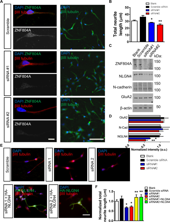
Figure 2
Small interfering RNA (siRNA)-mediated knockdown of zinc finger binding protein 804A (ZNF804A) in CTX0E16 neurons. (A) Representative images of ZNF804A staining and neurite length in early stage (DD7)...
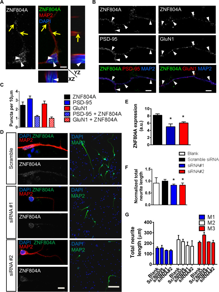
Figure 3
Localization and small interfering RNA (siRNA)-mediated knockdown of zinc finger binding protein 804A (ZNF804A) in young human induced pluripotent stem cell (hiPSC)-derived neurons. (A) In 35-day-old ...
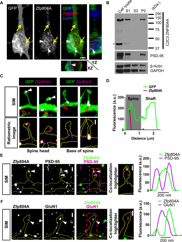
Figure 4
Zinc finger binding protein 804A (Zfp804A) is present at synapses in mature primary cortical neurons. (A) Representative confocal image, including orthogonal views in the XZ and YZ planes, of a 25-day...
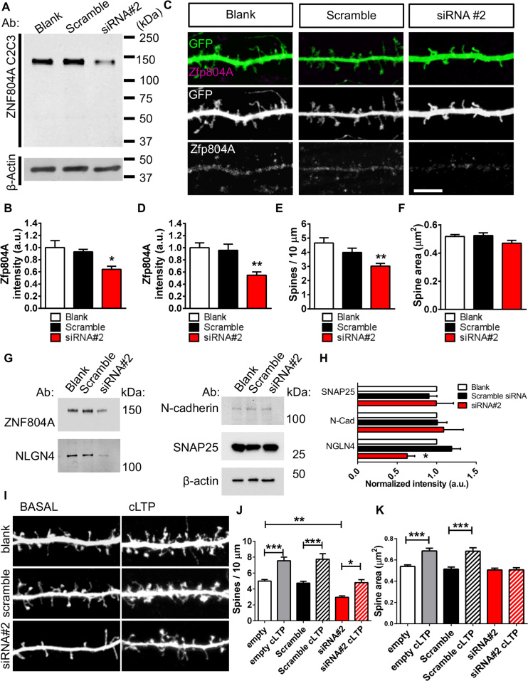
Figure 5
Small interfering RNA (siRNA)-mediated knockdown of zinc finger binding protein 804A (Zfp804A) in rat primary cortical neurons. (A) Representative Western blot of Zfp804A in 26-day in vitro (DIV26) pr...
Figure images are served from the NIH/NLM PubMed Central Open Access Subset or Europe PMC; copyright remains with the publishers and authors.
💬 Discussion
0 commentsNo comments yet. Be the first to start a discussion!
Leave a Comment