Abstract
The multifunctional nuclear protein positive cofactor 4 (PC4) is involved in various cellular processes including transcription, replication, and chromatin organization. Recently, PC4 has been identified as a suppressor of oxidative mutagenesis in Escherichia coli and Saccharomyces cerevisiae. To investigate a potential role of PC4 in mammalian DNA repair, we used a combination of live cell microscopy, microirradiation, and fluorescence recovery after photobleaching analysis. We found a clear accumulation of endogenous PC4 at DNA damage sites introduced by either chemical agents or laser microirradiation. Using fluorescent fusion proteins and specific mutants, we demonstrated that the rapid recruitment of PC4 to laser-induced DNA damage sites is independent of poly(ADP-ribosyl)ation and gammaH2AX but depends on its single strand binding capacity. Furthermore, PC4 showed a high turnover at DNA damages sites compared with the repair factors replication protein A and proliferating cell nuclear antigen. We propose that PC4 plays a role in the early response to DNA damage by recognizing single-stranded DNA and may thus initiate or facilitate the subsequent steps of DNA repair.
🔬 Techniques
🔭 Microscopes
🧬 Organisms
✨ Fluorophores
🧪 Sample Preparation
🔬 Cell Lines
🏭 Microscope Brands
🧪 Reagent Suppliers
🔎 Objectives
💻 Software Details
🏛️ Research Organizations (ROR)
Affiliated research institutions:
📋 Methods
Cell culture and transfection Human
HeLa, wild-type MEFs, PARP1 −/− MEFs ( Trucco et al., 1998 ), H2AX −/− MEFs ( Celeste et al., 2002 ), and mouse C2C12 cells were cultured in DME containing 50 μg/ml gentamicin supplemented with 10 and 20% FCS, respectively. PARP1 −/− and H2AX −/− MEFs were provided by V. Schreiber (Ecole Supérieure de Biotechnologie de Strasbourg, Strasbourg, France) and A. Nussenzweig (National Cancer Institute, National Institutes of Health, Bethesda, MD), respectively. Cells grown on μ-slides (Ibidi) or on gridded coverslips were cotransfected with jetPEI (PolyPlus Transfection) according to the manufacturer's instructions. For microirradiation experiments, cells were sensitized by incubation in medium containing 10 μg/ml BrdU for 24–48 h. HU, H 2 O 2 , and Aph were obtained from Sigma-Aldrich.
Expression plasmids
The generation of PC4 deletion and point mutants was previously described ( Kretzschmar et al., 1994 ; Werten et al., 1998 ). Corresponding GFP-PC4 fusion constructs were constructed by ligation of either restriction fragments (NdeI–ClaI for GFP-PC4; EcoRI–ClaI for the constructs GFP-PC4β2β3, GFP-PC4W89A, GFP-PC4 22–127, GFP-PC4 CTD β2β3, and GFP-PC4 CTDW89A; and XhoI–PstI for GFP-PC4 62–127) or PCR products (forward primer, 5′ GAAGATCTCCGGTTATTCTTCATATGCC 3′; reverse primer, 5′ TGGAATTCTCAATCATCTCTG 3′; BglII–EcoRI cloning for GFP-PC4 1–61) into matching restriction sites of pEGFP-C1 (Clontech Laboratories, Inc.). GFP-PC4 fusion constructs were verified by sequencing and tested by expression in HeLa cells followed by Western blot analysis. A red variant of PC4 was generated by replacing GFP with RFP ( Campbell et al., 2002 ) and termed RFP-PC4. The mRFP1 expression vector was supplied by R. Tsien (University of California, San Diego, La Jolla, CA). Mammalian expression constructs encoding translational fusions of human RPA34 and PCNA with either GFP or RFP were previously described ( Sporbert et al., 2005 ). In all cases, expression was under the control of the cytomegalovirus promoter and correct expression of fusion proteins was verified by Western blot analysis.
Show full methods section
Cell culture and transfection Human
HeLa, wild-type MEFs, PARP1 −/− MEFs ( Trucco et al., 1998 ), H2AX −/− MEFs ( Celeste et al., 2002 ), and mouse C2C12 cells were cultured in DME containing 50 μg/ml gentamicin supplemented with 10 and 20% FCS, respectively. PARP1 −/− and H2AX −/− MEFs were provided by V. Schreiber (Ecole Supérieure de Biotechnologie de Strasbourg, Strasbourg, France) and A. Nussenzweig (National Cancer Institute, National Institutes of Health, Bethesda, MD), respectively. Cells grown on μ-slides (Ibidi) or on gridded coverslips were cotransfected with jetPEI (PolyPlus Transfection) according to the manufacturer's instructions. For microirradiation experiments, cells were sensitized by incubation in medium containing 10 μg/ml BrdU for 24–48 h. HU, H 2 O 2 , and Aph were obtained from Sigma-Aldrich.
Expression plasmids
The generation of PC4 deletion and point mutants was previously described ( Kretzschmar et al., 1994 ; Werten et al., 1998 ). Corresponding GFP-PC4 fusion constructs were constructed by ligation of either restriction fragments (NdeI–ClaI for GFP-PC4; EcoRI–ClaI for the constructs GFP-PC4β2β3, GFP-PC4W89A, GFP-PC4 22–127, GFP-PC4 CTD β2β3, and GFP-PC4 CTDW89A; and XhoI–PstI for GFP-PC4 62–127) or PCR products (forward primer, 5′ GAAGATCTCCGGTTATTCTTCATATGCC 3′; reverse primer, 5′ TGGAATTCTCAATCATCTCTG 3′; BglII–EcoRI cloning for GFP-PC4 1–61) into matching restriction sites of pEGFP-C1 (Clontech Laboratories, Inc.). GFP-PC4 fusion constructs were verified by sequencing and tested by expression in HeLa cells followed by Western blot analysis. A red variant of PC4 was generated by replacing GFP with RFP ( Campbell et al., 2002 ) and termed RFP-PC4. The mRFP1 expression vector was supplied by R. Tsien (University of California, San Diego, La Jolla, CA). Mammalian expression constructs encoding translational fusions of human RPA34 and PCNA with either GFP or RFP were previously described ( Sporbert et al., 2005 ). In all cases, expression was under the control of the cytomegalovirus promoter and correct expression of fusion proteins was verified by Western blot analysis.
Immunofluorescence and detergent extraction
Cells were fixed in 3.7% formaldehyde for 10 min and permeabilized with 0.5% Triton X-100 or ice-cold methanol for 5 min. The following primary antibodies (diluted in PBS containing 4% BSA) were used: anti-γH2AX (Ser139) mouse monoclonal antibodies (Millipore), anti-PAR mouse monoclonal antibodies (Trevigen), anti-RPA34 mouse monoclonal antibodies (EMD), anti-PC4 rabbit polyclonal antibodies (SA2249; generated by standard techniques; Eurogentech), and anti-PCNA rat monoclonal antibodies ( Spada et al., 2007 ). Primary antibodies were detected using secondary antibodies (diluted 1:200 in PBS containing 4% BSA) conjugated to AlexaFluor 488 or 555 (Invitrogen). Cells were counterstained with DAPI and mounted in Vectashield (Vector Laboratories). For in situ extraction, cells were permeabilized for 30 s with 0.5% Triton X-100 in PBS before fixation. Live cell microscopy, microirradiation, and photobleaching experiments Live cell imaging, microirradiation, and photobleaching experiments were performed with a confocal laser scanning microscope (TCS SP2/AOBS or SP5/AOBS; Leica), each equipped with a UV-transmitting HCX PL 63x/1.4 oil objective. GFP and RFP were excited with a 488-nm Ar laser line and a 561-nm diode pumped solid state laser line (Leica), respectively. The microscopes were equipped with a heated environmental chamber set to 37°C. Confocal image series were typically recorded with a frame size of 256 × 256 pixels and a pixel size of 90 nm. Microirradiation was performed as previously described ( Mortusewicz et al., 2006 , 2007 ). In brief, a preselected spot ∼1 μm in diameter within the nucleus was microirradiated for 1 s with a 405-nm diode laser (Leica) set to 50–80 μW. The laser power was measured after passing through the objective lens with a laser power meter (Coherent). Before and after microirradiation, confocal image series of one mid z section were recorded at 2-s time intervals (typically 6 pre- and 150 postirradiation frames). For evaluation of the recruitment kinetics, fluorescence intensities at the irradiated region were corrected for background and for total nuclear loss of fluorescence over the time course and normalized to the preirradiation value. For FRAP analysis, a region of interest was selected and photobleached for 300 ms with all laser lines of the Ar laser and the 561-nm diode pumped solid state laser set to maximum power at 100% transmission. Before and after bleaching, confocal image series were recorded at 150-ms time intervals (typically 10 pre- and 200 postbleach frames). Mean fluorescence intensities of the bleached region were corrected for background and for total nuclear loss of fluorescence over the time course and normalized to the mean of the last four prebleach values. For the quantitative evaluation of microirradiation and photobleaching experiments, data of at least nine nuclei were averaged and the mean curve and the standard error of the mean calculated and displayed using Excel software (Microsoft). Images of fixed cells were taken with a widefield epifluorescence microscope (Axiophot 2; Carl Zeiss, Inc.) using a Plan Apochromat 63x/1.40 oil objective (Carl Zeiss, Inc.) and a cooled charge-coupled device camera (Visitron Systems). Online supplemental material Fig. S1 shows a schematic outline of fusion proteins used in this study and a comparison of the crystal structure of PC4 ( Brandsen et al., 1997 ) with RPA70 ( Bochkarev et al., 1997 ) and RPA34 ( Bochkarev et al., 1999 ). Fig. S2 shows that recruitment of PC4 to laser-induced DNA damage sites occurs in all S phase stages. Fig. S3 shows that the recruitment of PC4 is independent of its N-terminal CK2 phosphorylation sites, poly(ADP-ribosyl)ation, and phosphorylation of H2AX. Online supplemental material is available at http://www.jcb.org/cgi/content/full/jcb.200808097/DC1 .
Online supplemental material Fig. S1 shows a schematic outline of fusion proteins used in this study and a comparison of the crystal structure of PC4 ( Brandsen et al., 1997 ) with RPA70 ( Bochkarev et al., 1997 ) and RPA34 ( Bochkarev et al., 1999 ). Fig. S2 shows that recruitment of PC4 to laser-induced DNA damage sites occurs in all S phase stages. Fig. S3 shows that the recruitment of PC4 is independent of its N-terminal CK2 phosphorylation sites, poly(ADP-ribosyl)ation, and phosphorylation of H2AX. Online supplemental material is available at http://www.jcb.org/cgi/content/full/jcb.200808097/DC1 .
Supplementary Material [Supplemental Material Index]
📊 Figures
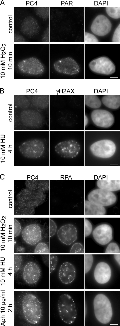
Figure 1.
PC4 accumulates at DNA damage sites. HeLa cells were treated with 10 mM HU, 10 mM H 2 O 2 , or 10 u03bcg/ml Aph for the indicated time points and in situ extracted with 0.5% Triton X-100 for 30 s befo...
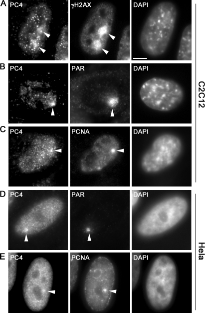
Figure 2.
PC4 accumulates at laser-induced DNA damage sites. Widefield fluorescence images of mouse C2C12 and human HeLa cells are shown. Fixation and immunostaining was performed u223c5 min after laser microir...
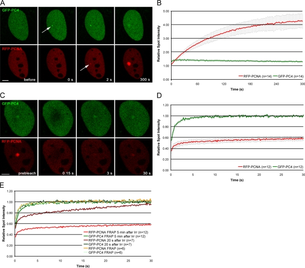
Figure 3.
Recruitment and mobility of PC4 and PCNA at DNA damage sites in living cells. (A) Live cell imaging of a microirradiated C2C12 cell coexpressing GFP-PC4 and RFP-PCNA. Accumulation of GFP-PC4 can be ob...
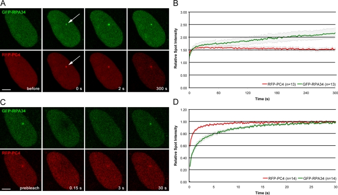
Figure 4.
Comparison of recruitment and binding capacities of PC4 with RPA34 in living cells. (A) Live cell imaging of a microirradiated C2C12 cell coexpressing GFP-RPA34 and RFP-PC4. Both GFP-RPA34 and RFP-PC4...
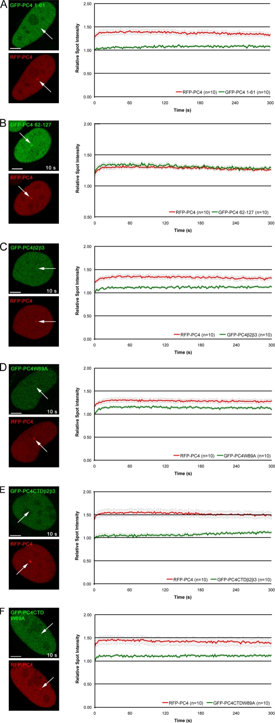
Figure 5.
The single strand binding capacity of PC4 is needed for efficient recruitment to DNA damage sites. Deletion proteins containing either the N-terminal domain (1u201361) or the CTD (62u2013127) of PC4 w...
Figure images are served from the NIH/NLM PubMed Central Open Access Subset or Europe PMC; copyright remains with the publishers and authors.
💬 Discussion
0 commentsNo comments yet. Be the first to start a discussion!
Leave a Comment