Abstract
Background: Angiogenesis is a dynamic process that involves expansion of a preexisting vascular network that can occur in a number of physiological and pathological settings. Despite its importance, the origin of the new angiogenic vasculature is poorly defined. In particular, the primary subtype of endothelial cells (capillary, venous, arterial) driving this process remains undefined. Methods: Endothelial cells were fate-mapped with the use of genetic markers specific to arterial and capillary cells. In addition, we identified a novel venous endothelial marker gene ( Gm5127 ) and used it to generate inducible venous endothelium-specific Cre and Dre driver mouse lines. Contributions of these various types of endothelial cells to angiogenesis were examined during normal postnatal development and in disease-specific setting. Results: Using a comprehensive set of endothelial subtype-specific inducible reporter mice, including tip, arterial, and venous endothelial reporter lines, we showed that venous endothelial cells are the primary endothelial subtype responsible for the expansion of an angiogenic vascular network. During physiological angiogenesis, venous endothelial cells proliferate, migrating against the blood flow and differentiating into tip, capillary, and arterial endothelial cells of the new vasculature. Using intravital 2-photon imaging, we observed venous endothelial cells migrating against the blood flow to form new blood vessels. Venous endothelial cell migration also plays a key role in pathological angiogenesis. This was observed both in formation of arteriovenous malformations in mice with inducible endothelium-specific Smad4 deletion mice and in pathological vessel growth seen in oxygen-induced retinopathy. Conclusions: Our studies establish that venous endothelial cells are the primary endothelial subtype responsible for normal expansion of vascular networks, formation of arteriovenous malformations, and pathological angiogenesis. These observations highlight the central role of the venous endothelium in normal development and disease pathogenesis.
🔬 Techniques
🔭 Microscopes
🧬 Organisms
💻 Software
✨ Fluorophores
🧪 Sample Preparation
🔬 Cell Lines
🏭 Microscope Brands
🧪 Reagent Suppliers
🔴 Lasers
💻 Software Details
💾 Data Repositories
🏛️ Research Organizations (ROR)
Affiliated research institutions:
📋 Methods
Endothelial cells were fate-mapped using genetic markers specific to arterial, capillary cells. In addition, we identified a novel venous endothelial marker gene ( Gm5127 ) used it to generate inducible venous endothelial-specific Cre and Dre driver mouse lines. Contributions of these various types of endothelial cells to angiogenesis were examined during normal postnatal development and in disease-specific setting.
METHODS Mice Cdh5 (PAC)CreER T2 (pan-endothelial Cre line) 10 , Bmx (PAC)CreER T2 (arterial-endothelial Cre line) 11 , Esm1 (PAC)CreER T2 (tip-endothelial Cre line) 4 and Rosa26 RC::RG (Dre reporter line) 12 animals have been previously described. Rosa26 mTmG and Rosa26 nTnG animals were obtained from Jackson Laboratory 13 . All experiments were performed using littermates on a C57BL/6 background unless otherwise indicated. For endothelial subtype lineage tracing experiments utilizing the pan-endothelial (Cdh5(PAC)CreER T2 ), tip-endothelial ( Esm1 (PAC)CreER T2 ), arterial-endothelial ( Bmx (PAC)CreER T2 ) and venous-endothelial ( Gm5127 (BAC)CreER T2 ) specific inducible Cre mice, the animals were intraperitoneally injected with 4-hydroxytamoxifen (4-OHT, Sigma-Aldrich) (15ug for pups and 150ug for adult mice) dissolved in 10% Ethanol (Sigma-Aldrich) with corn oil (Sigma-Aldrich) (v/v) by a 30-gauge needle (Sigma-Aldrich). Mice were then harvested, as previously described 14 , at specified time points. The P0 time point was defined by checking experimental breeding pairs every 12hr for new litters. The following day was counted as postnatal day 1(P1). All mouse protocols and experimental procedures were conducted with approval of the Yale University Institutional Animal Care and Use Committees. BAC clone modification for transgenic construct. The BAC clone (RP23–113I19) derived from RPCI-23 mouse genomic library was identified using the ENSEMBL database of the Wellcome Trust Sanger Institute ( http://www.ensembl.org ). The BAC clone was obtained from Source Bioscience (Berlin, Germany) and used for labda Red recombinase mediated recombination to introduce an inducible Cre recombinase cassette (CreER T2 ) 15 or inducible Dre recombinase cassette (DreER T2 ). The CreER T2 cassette construct was obtained from Addgene (pCAG-CreERT2) 7 and the DreER T2 cassette was subcloned from pCAG-NLS-HA-Dre construct (Addgene) 16 . Briefly, the replacement is achieved via homologous recombination between a CreER T2 /DreER T2 cassette construct and a locus on the BAC clone containing the start codon of transgene. The CreER T2 /DreER T2 cassette constructs for recombination were generated by PCR amplification incorporating the coding regions of CreER T2 /DreER T2 and FRT site-flanked Neo/Kan cassette. Those PCR amplificons were designed to harbor 50bp overhangs at either ends that mediate integration into the BAC at the intended position. The primes used for PCR reaction were: CreForward : TTTACTCCAGGAATCCTGCCCAACTTCACATTTTAAAAAATCGAGACCATGCCACCATGGCCAATTTACTGA, CreReverse : TGACTGTTTGACAAAACAACGGGGCCATTATATCTCACAGGCCTGAGAAAATCCGAAGTTCCTATACTTTC, DreForward : TTTACTCCAGGAATCCTGCCCAACTTCACATTTTAAAAAATCGAGACCATCGCCACCATGTCTGAGCTGA and DreReverse : TTTACTCCAGGAATCCTGCCCAACTTCACATTTTAAAAAATCGAGACCATTGTAATACGACTCACTATAG, respectively. The homologous recombination was performed in E. coli transformed with the BAC clone using the pRed/ET system 17 (Gene Bridges GmbH). Cells harboring pRed/ET were cultured aerobically in 1.4 mL LB medium at a temperature of 30°C. At an OD600 of approximately 0.3, expression of Red recombinase protein was induced by the addition of 50 μL 10% (w/v) L-arabinose (Sigma-Aldrich). At the same time, the temperature was increased to 37°C to ensure maximal expression and activity of recombination proteins. After 18hr plating on LB plate with kanamycin, colonies were picked up and genotyped with PCR reaction using an AccuPower PCR PreMix (Bioneer). After selection, the Kan cassette was removed by inducing the Flp recombinase (707-FLP, Gene Bridges GmbH). To remove an open reading frame for Cysltr1, which is located on the opposite strand of same clone, one more recombination step was performed. Then, modified DNA sequence were retrieved into low copy plasmid (pHW) using gap repair homologous recombination. Transgenesis and genotyping. BAC DNA was purified using NucleoBond BAC 100 (Takara). It was linearized by SgrDI digestion, then purified by phenol/chloroform extraction (Sigma-Aldrich) and the quality of the linearized DNA was checked by pulse-field gel electrophoresis (Bio-Rad). The linearized DNA was then injected at a concentration of 5 ng/uL into fertilized eggs of B6;SJLF2 hybrid mice (Jackson Laboratories) at the Yale Genome Editing Center. Transgenic animals were identified by performing PCR on genomic DNA purified from tail biopsies using the Phire Animal Tissue Direct PCR Kit (Thermo Scientific). The primers used for genotyping were: CreForward : GTTTCACTGGTTATGCGGCG and CreReverse : GGTGCTAACCAGCGTTTTCG. DreForward : TGGTGGATTCCTGCGAAACA and DreReverse : GCTACGAACAGGAAAGCCCT, respectively. Edu staining. For detection of proliferating cells in vivo, a stock of 50mg 5-ethynyl-2-deoxyuridine (EdU) (Sigma-Aldrich) dissolved in 1mL of DMSO. This stock solution was diluted with PBS to make a working solution (10mg/mL ). Edu solution were injected intraperitoneally (200mg/kg) 4hr before the animals were sacrificed. Retinas were isolated and fixed as previously described 14 , and the EdU-positive cells were detected according to the user manual of the Click-iT EdU Alexa Fluor- 647 or 488 Imaging Kit (Invitrogen). The EdU-stained tissues were visualized by Leica confocal laser scanning microscope SP8 (Leica, Wetzlar, Germany). Retina whole mount preparation. After 4-OHT administration, mice were euthanized at specific timepoint for analysis of the retinal vasculature as previously described 6 . Briefly, whole mount retinas were fixed by immersion in freshly made 4% paraformaldehyde (Sigma-Aldrich) in PBS buffer for overnight at 4°C, permeabilized with 1% Triton X-100 (Sigma-Aldrich) in TBS (TBS-T), and then exposed to 1% bovine serum albumin in PBS-T for antigen blocking before immunostaining. Vibratome section. Brain tissues were fixed in 4% paraformaldehyde (Sigma-Aldrich) in phosphate buffer for overnight at 4°C. Brains were cut using Leica VT1000 S vibratome (150μm thickness) and washed with 1% Triton X-100 (Sigma-Aldrich) in PBS 3 times for further immunostaining. Frozen Tissue Section. Embryonic tissues were fixed in freshly made 4% paraformaldehyde overnight at 4°C, rinsed with PBS at room temperature, incubated in 15% sucrose overnight at 4°C, and transferred to 30% sucrose at 4°C until the tissue sank. Fixed tissues were infiltrated with Tissue-Tek O.C.T. embedding medium for 30 minutes at room temperature, transferred to an embedding mold filled with OCT, frozen on dry ice, and stored at −70°C. Frozen sections (10μm-thick) were cut at −20°C, and slides were kept at −70°C until use for Immunohistochemistry. Immunohistochemistry. In brief, the tissues/sections were permeabilized in ice-cold 70% ethanol (Sigma-Aldrich) for 10min at −20°C and washed with 1% Triton X-100 (Sigma-Aldrich) in TBS (TBS-T) 3 times for further immunostaining. The specimens were first incubated in blocking buffer (1% FBS, 3% BSA, 0.5% Triton-X100, 0.01% sodium deoxycholate in TBS) for 30min at room temperature and subsequently incubated in blocking buffer containing antibodies for overnight at 4°C. Next day, specimens were washed 3 times with TBS-T (10 min/wash), incubated in secondary antibody at room temperature for 1hr. After they were washed three times with TBS-T in the dark (10 min/wash), the specimens were mounted on glass slides with anti-fading mounting medium (10% 0.1M Tris buffer (pH 9.0), 0.2mg/mL p-phenylenediamine hydrochloride (Sigma-Aldrich) and 4mM sodium azide (Sigma-Aldrich) in glycerol). Confocal microscopy was performed with Nikon ECLIPSE 80i fluorescence microscope, Leica spinning disk confocal microscope or a Leica SP8 confocal microscope. ImageJ (NIH) was used for the data analysis CLARITY tissue clearing. Dissected brain tissues were treated using CLARITY tissue clearing protocol as previously described. 18 Brains were dissected in PBS buffer and fixed in 4% PFA consequently. After 24hr fixation, the brains were sliced into 500μm section by Leica VT1000 S Vibratome, followed by 3 times washing step with PBS buffer for 10mins at room temperature. Next, brain sections were embedded with hydrogel monomers (acrylamide/bisacrylamide) (Sigma-Aldrich) 24hr at 4°C, followed by oil laid over the top of hydrogel solution and shaken at 37 °C for 4hr. The brain sections then washed with 8% sodium dodecyl sulfate (SDS) for 4 consecutive days at 37°C. After SDS washing step the brain sections became transparent, the brain sections then were washed for 2hr with a changing 1% PBS-T every 15minutes at room temperate. The brain sections were subsequently incubated with the primary antibody(1:200) in 1% PBS-T and 5% donkey serum overnight at 4°C. The sections were then washed with 1% PBS-T for one day 6 times at room temperature followed by a 4°C washing step with the same buffer. Next, brain sections incubated with a second antibody in 1% PBS-T and 5% donkey serum) for 24hr at 4°C and washed with PBS buffer for 2 consequence days at room temperate, with the washing buffer changed 6 times per day. The brain sections were finally re-cleaned with RIMS (88% Histodenz wt/vol in 0.02M phosphate buffer). The whole mount images were obtained by Leica confocal laser scanning microscope SP8 using navigator mode (Leica, Wetzlar, Germany). Cranial window installation and 2-photon imaging. Postnatal mice ( Cdh5 (PAC)CreER T2 Rosa26 +/nTnG ) were used for cranial window installation. At P1, pups were intraperitoneally injected with 15 μg of 4-OHT. On the following day (P2), a cranial window was installed in anesthetized mice for in vivo multiphoton imaging. Briefly, P2 mice were intraperitoneally injected with 150uL of glucose solution (0.2M glucose in saline) and anesthetized using 2% isoflurane anesthesia. A tail-pinch test was used to check the level of anesthesia before surgery. Then, the skin covering the skull was sterilized by wiping it with 70% ethanol. Under a surgical microscope (Stereo Microscope Fluorescent Leica M165-FC), approximately 25mm 2 of the skin covering the skull was removed using sterilized surgical scissors and the fascia of the skull was carefully removed using sterilized forceps. A sterile custom-made titanium bar was attached to the head using dental cement and saline solution was applied via pipette to the exposed tissue to prevent dehydration during the experimental process. Then, the handle of titanium bar was attached to a goniometer (GN05/M Goniometer, Thorlab) and mice were placed under a water immersion, long-working distance Olympus 20X objective lens (NA 0.95) of the 2-photon microscope. During imaging, mice were anesthetized with 0.9% isoflurane and kept on a heating pad (SmartStage, BioTherm) to maintain normal body temperature. Images were acquired using an Olympus BX61WI fluorescence microscope with a ×20, 0.95NA water immersion Olympus objective and dedicated single beam LaVision TriM scanning laser (LaVision Biotec) that controlled by Imspector software. The microscope was outfitted with a Chameleon Vision II Ti:Sapphire Laser (Coherent) with pulse precompensation. Emission wavelengths of 390–480 nm (blue), 500–550 nm (green, GFP), and 565–665 nm (orange-red) were collected with an array of 3 photomultiplier tubes (Hamamatsu).
Show full methods section
Endothelial cells were fate-mapped using genetic markers specific to arterial, capillary cells. In addition, we identified a novel venous endothelial marker gene ( Gm5127 ) used it to generate inducible venous endothelial-specific Cre and Dre driver mouse lines. Contributions of these various types of endothelial cells to angiogenesis were examined during normal postnatal development and in disease-specific setting.
METHODS Mice Cdh5 (PAC)CreER T2 (pan-endothelial Cre line) 10 , Bmx (PAC)CreER T2 (arterial-endothelial Cre line) 11 , Esm1 (PAC)CreER T2 (tip-endothelial Cre line) 4 and Rosa26 RC::RG (Dre reporter line) 12 animals have been previously described. Rosa26 mTmG and Rosa26 nTnG animals were obtained from Jackson Laboratory 13 . All experiments were performed using littermates on a C57BL/6 background unless otherwise indicated. For endothelial subtype lineage tracing experiments utilizing the pan-endothelial (Cdh5(PAC)CreER T2 ), tip-endothelial ( Esm1 (PAC)CreER T2 ), arterial-endothelial ( Bmx (PAC)CreER T2 ) and venous-endothelial ( Gm5127 (BAC)CreER T2 ) specific inducible Cre mice, the animals were intraperitoneally injected with 4-hydroxytamoxifen (4-OHT, Sigma-Aldrich) (15ug for pups and 150ug for adult mice) dissolved in 10% Ethanol (Sigma-Aldrich) with corn oil (Sigma-Aldrich) (v/v) by a 30-gauge needle (Sigma-Aldrich). Mice were then harvested, as previously described 14 , at specified time points. The P0 time point was defined by checking experimental breeding pairs every 12hr for new litters. The following day was counted as postnatal day 1(P1). All mouse protocols and experimental procedures were conducted with approval of the Yale University Institutional Animal Care and Use Committees. BAC clone modification for transgenic construct. The BAC clone (RP23–113I19) derived from RPCI-23 mouse genomic library was identified using the ENSEMBL database of the Wellcome Trust Sanger Institute ( http://www.ensembl.org ). The BAC clone was obtained from Source Bioscience (Berlin, Germany) and used for labda Red recombinase mediated recombination to introduce an inducible Cre recombinase cassette (CreER T2 ) 15 or inducible Dre recombinase cassette (DreER T2 ). The CreER T2 cassette construct was obtained from Addgene (pCAG-CreERT2) 7 and the DreER T2 cassette was subcloned from pCAG-NLS-HA-Dre construct (Addgene) 16 . Briefly, the replacement is achieved via homologous recombination between a CreER T2 /DreER T2 cassette construct and a locus on the BAC clone containing the start codon of transgene. The CreER T2 /DreER T2 cassette constructs for recombination were generated by PCR amplification incorporating the coding regions of CreER T2 /DreER T2 and FRT site-flanked Neo/Kan cassette. Those PCR amplificons were designed to harbor 50bp overhangs at either ends that mediate integration into the BAC at the intended position. The primes used for PCR reaction were: CreForward : TTTACTCCAGGAATCCTGCCCAACTTCACATTTTAAAAAATCGAGACCATGCCACCATGGCCAATTTACTGA, CreReverse : TGACTGTTTGACAAAACAACGGGGCCATTATATCTCACAGGCCTGAGAAAATCCGAAGTTCCTATACTTTC, DreForward : TTTACTCCAGGAATCCTGCCCAACTTCACATTTTAAAAAATCGAGACCATCGCCACCATGTCTGAGCTGA and DreReverse : TTTACTCCAGGAATCCTGCCCAACTTCACATTTTAAAAAATCGAGACCATTGTAATACGACTCACTATAG, respectively. The homologous recombination was performed in E. coli transformed with the BAC clone using the pRed/ET system 17 (Gene Bridges GmbH). Cells harboring pRed/ET were cultured aerobically in 1.4 mL LB medium at a temperature of 30°C. At an OD600 of approximately 0.3, expression of Red recombinase protein was induced by the addition of 50 μL 10% (w/v) L-arabinose (Sigma-Aldrich). At the same time, the temperature was increased to 37°C to ensure maximal expression and activity of recombination proteins. After 18hr plating on LB plate with kanamycin, colonies were picked up and genotyped with PCR reaction using an AccuPower PCR PreMix (Bioneer). After selection, the Kan cassette was removed by inducing the Flp recombinase (707-FLP, Gene Bridges GmbH). To remove an open reading frame for Cysltr1, which is located on the opposite strand of same clone, one more recombination step was performed. Then, modified DNA sequence were retrieved into low copy plasmid (pHW) using gap repair homologous recombination. Transgenesis and genotyping. BAC DNA was purified using NucleoBond BAC 100 (Takara). It was linearized by SgrDI digestion, then purified by phenol/chloroform extraction (Sigma-Aldrich) and the quality of the linearized DNA was checked by pulse-field gel electrophoresis (Bio-Rad). The linearized DNA was then injected at a concentration of 5 ng/uL into fertilized eggs of B6;SJLF2 hybrid mice (Jackson Laboratories) at the Yale Genome Editing Center. Transgenic animals were identified by performing PCR on genomic DNA purified from tail biopsies using the Phire Animal Tissue Direct PCR Kit (Thermo Scientific). The primers used for genotyping were: CreForward : GTTTCACTGGTTATGCGGCG and CreReverse : GGTGCTAACCAGCGTTTTCG. DreForward : TGGTGGATTCCTGCGAAACA and DreReverse : GCTACGAACAGGAAAGCCCT, respectively. Edu staining. For detection of proliferating cells in vivo, a stock of 50mg 5-ethynyl-2-deoxyuridine (EdU) (Sigma-Aldrich) dissolved in 1mL of DMSO. This stock solution was diluted with PBS to make a working solution (10mg/mL ). Edu solution were injected intraperitoneally (200mg/kg) 4hr before the animals were sacrificed. Retinas were isolated and fixed as previously described 14 , and the EdU-positive cells were detected according to the user manual of the Click-iT EdU Alexa Fluor- 647 or 488 Imaging Kit (Invitrogen). The EdU-stained tissues were visualized by Leica confocal laser scanning microscope SP8 (Leica, Wetzlar, Germany). Retina whole mount preparation. After 4-OHT administration, mice were euthanized at specific timepoint for analysis of the retinal vasculature as previously described 6 . Briefly, whole mount retinas were fixed by immersion in freshly made 4% paraformaldehyde (Sigma-Aldrich) in PBS buffer for overnight at 4°C, permeabilized with 1% Triton X-100 (Sigma-Aldrich) in TBS (TBS-T), and then exposed to 1% bovine serum albumin in PBS-T for antigen blocking before immunostaining. Vibratome section. Brain tissues were fixed in 4% paraformaldehyde (Sigma-Aldrich) in phosphate buffer for overnight at 4°C. Brains were cut using Leica VT1000 S vibratome (150μm thickness) and washed with 1% Triton X-100 (Sigma-Aldrich) in PBS 3 times for further immunostaining. Frozen Tissue Section. Embryonic tissues were fixed in freshly made 4% paraformaldehyde overnight at 4°C, rinsed with PBS at room temperature, incubated in 15% sucrose overnight at 4°C, and transferred to 30% sucrose at 4°C until the tissue sank. Fixed tissues were infiltrated with Tissue-Tek O.C.T. embedding medium for 30 minutes at room temperature, transferred to an embedding mold filled with OCT, frozen on dry ice, and stored at −70°C. Frozen sections (10μm-thick) were cut at −20°C, and slides were kept at −70°C until use for Immunohistochemistry. Immunohistochemistry. In brief, the tissues/sections were permeabilized in ice-cold 70% ethanol (Sigma-Aldrich) for 10min at −20°C and washed with 1% Triton X-100 (Sigma-Aldrich) in TBS (TBS-T) 3 times for further immunostaining. The specimens were first incubated in blocking buffer (1% FBS, 3% BSA, 0.5% Triton-X100, 0.01% sodium deoxycholate in TBS) for 30min at room temperature and subsequently incubated in blocking buffer containing antibodies for overnight at 4°C. Next day, specimens were washed 3 times with TBS-T (10 min/wash), incubated in secondary antibody at room temperature for 1hr. After they were washed three times with TBS-T in the dark (10 min/wash), the specimens were mounted on glass slides with anti-fading mounting medium (10% 0.1M Tris buffer (pH 9.0), 0.2mg/mL p-phenylenediamine hydrochloride (Sigma-Aldrich) and 4mM sodium azide (Sigma-Aldrich) in glycerol). Confocal microscopy was performed with Nikon ECLIPSE 80i fluorescence microscope, Leica spinning disk confocal microscope or a Leica SP8 confocal microscope. ImageJ (NIH) was used for the data analysis CLARITY tissue clearing. Dissected brain tissues were treated using CLARITY tissue clearing protocol as previously described. 18 Brains were dissected in PBS buffer and fixed in 4% PFA consequently. After 24hr fixation, the brains were sliced into 500μm section by Leica VT1000 S Vibratome, followed by 3 times washing step with PBS buffer for 10mins at room temperature. Next, brain sections were embedded with hydrogel monomers (acrylamide/bisacrylamide) (Sigma-Aldrich) 24hr at 4°C, followed by oil laid over the top of hydrogel solution and shaken at 37 °C for 4hr. The brain sections then washed with 8% sodium dodecyl sulfate (SDS) for 4 consecutive days at 37°C. After SDS washing step the brain sections became transparent, the brain sections then were washed for 2hr with a changing 1% PBS-T every 15minutes at room temperate. The brain sections were subsequently incubated with the primary antibody(1:200) in 1% PBS-T and 5% donkey serum overnight at 4°C. The sections were then washed with 1% PBS-T for one day 6 times at room temperature followed by a 4°C washing step with the same buffer. Next, brain sections incubated with a second antibody in 1% PBS-T and 5% donkey serum) for 24hr at 4°C and washed with PBS buffer for 2 consequence days at room temperate, with the washing buffer changed 6 times per day. The brain sections were finally re-cleaned with RIMS (88% Histodenz wt/vol in 0.02M phosphate buffer). The whole mount images were obtained by Leica confocal laser scanning microscope SP8 using navigator mode (Leica, Wetzlar, Germany). Cranial window installation and 2-photon imaging. Postnatal mice ( Cdh5 (PAC)CreER T2 Rosa26 +/nTnG ) were used for cranial window installation. At P1, pups were intraperitoneally injected with 15 μg of 4-OHT. On the following day (P2), a cranial window was installed in anesthetized mice for in vivo multiphoton imaging. Briefly, P2 mice were intraperitoneally injected with 150uL of glucose solution (0.2M glucose in saline) and anesthetized using 2% isoflurane anesthesia. A tail-pinch test was used to check the level of anesthesia before surgery. Then, the skin covering the skull was sterilized by wiping it with 70% ethanol. Under a surgical microscope (Stereo Microscope Fluorescent Leica M165-FC), approximately 25mm 2 of the skin covering the skull was removed using sterilized surgical scissors and the fascia of the skull was carefully removed using sterilized forceps. A sterile custom-made titanium bar was attached to the head using dental cement and saline solution was applied via pipette to the exposed tissue to prevent dehydration during the experimental process. Then, the handle of titanium bar was attached to a goniometer (GN05/M Goniometer, Thorlab) and mice were placed under a water immersion, long-working distance Olympus 20X objective lens (NA 0.95) of the 2-photon microscope. During imaging, mice were anesthetized with 0.9% isoflurane and kept on a heating pad (SmartStage, BioTherm) to maintain normal body temperature. Images were acquired using an Olympus BX61WI fluorescence microscope with a ×20, 0.95NA water immersion Olympus objective and dedicated single beam LaVision TriM scanning laser (LaVision Biotec) that controlled by Imspector software. The microscope was outfitted with a Chameleon Vision II Ti:Sapphire Laser (Coherent) with pulse precompensation. Emission wavelengths of 390–480 nm (blue), 500–550 nm (green, GFP), and 565–665 nm (orange-red) were collected with an array of 3 photomultiplier tubes (Hamamatsu).
Volocity software
(Improvision) was used for image acquisition and Adobe Premiere Pro software was used for the video edit and annotation.
Analysis of Publicly
Available scRNAseq databases. To check the expression of Gm5127 in various tissues and different developmental stages, we used publicly available processed data and corresponding metadata of BrainEC, Tabula Muris, MOCA and EC atlas to avoid any discrepancies between processing and annotation of the data. For BrainEC atlas, pre-processed and normalized data as well as the accompanying metadata were used from the Vanlandewijck et al. For Tabula Muris atlas, tSNE plot and processed Robjects were acquired from their website ( https://tabula-muris.ds.czbiohub.org/ and https://figshare.com/projects/Tabula_Muris_Transcriptomic_characterization_of_20_organs_and_tissues_from_Mus_musculus_at_single_cell_resolution/27733 ) For MOCA (Mouse Organogenesis Cell Atlas), processed data for 100,000 cell from filtered data set was extracted from their website ( https://oncoscape.v3.sttrcancer.org/atlas.gs.washington.edu.mouse.rna/downloads ). For EC atlas, normalized dataset was downloaded from EndoDB ( https://endotheliomics.shinyapps.io/ec_atlas/ ). The R (R-3.6.3) implementation of the ggplot2 and Seurat 3.0 software was used for t-SNE, UMAP and bar graph visualization. Antibodies. The following primary antibodies were used for immunostaining: NR2F2 (Invitrogen, Cat: PA5-35141); Cre (Cell Signaling, Cat: 15036); CD31 (RND, Cat: AF3628); GM130 (BDbiosciences, Cat: 610822); SOX17 (RND, Cat: AF1924) and Isolectin-B4 (Invitrogen); The secondary antibodies used were anti-mouse, rabbit or goat Alexa antibodies from the Invitrogen. Statistics. Data are presented as the mean ± SEM. A Student unpaired, two-tailed t test and one-way ANOVA followed by a multiple comparison procedure for pairwise comparisons were used for statistical analysis using GraphPad Prism 8.
Data Availability
The datasets generated during and/or analyzed during the current study are available from the corresponding author on reasonable request.
Code availability
Custom-written scripts used in this study are available from the corresponding authors upon reasonable request.
Supplementary Material Brain Ec migration moview Supplemental Figures I - XII Suppl figures Supplemental Movie I
📊 Figures
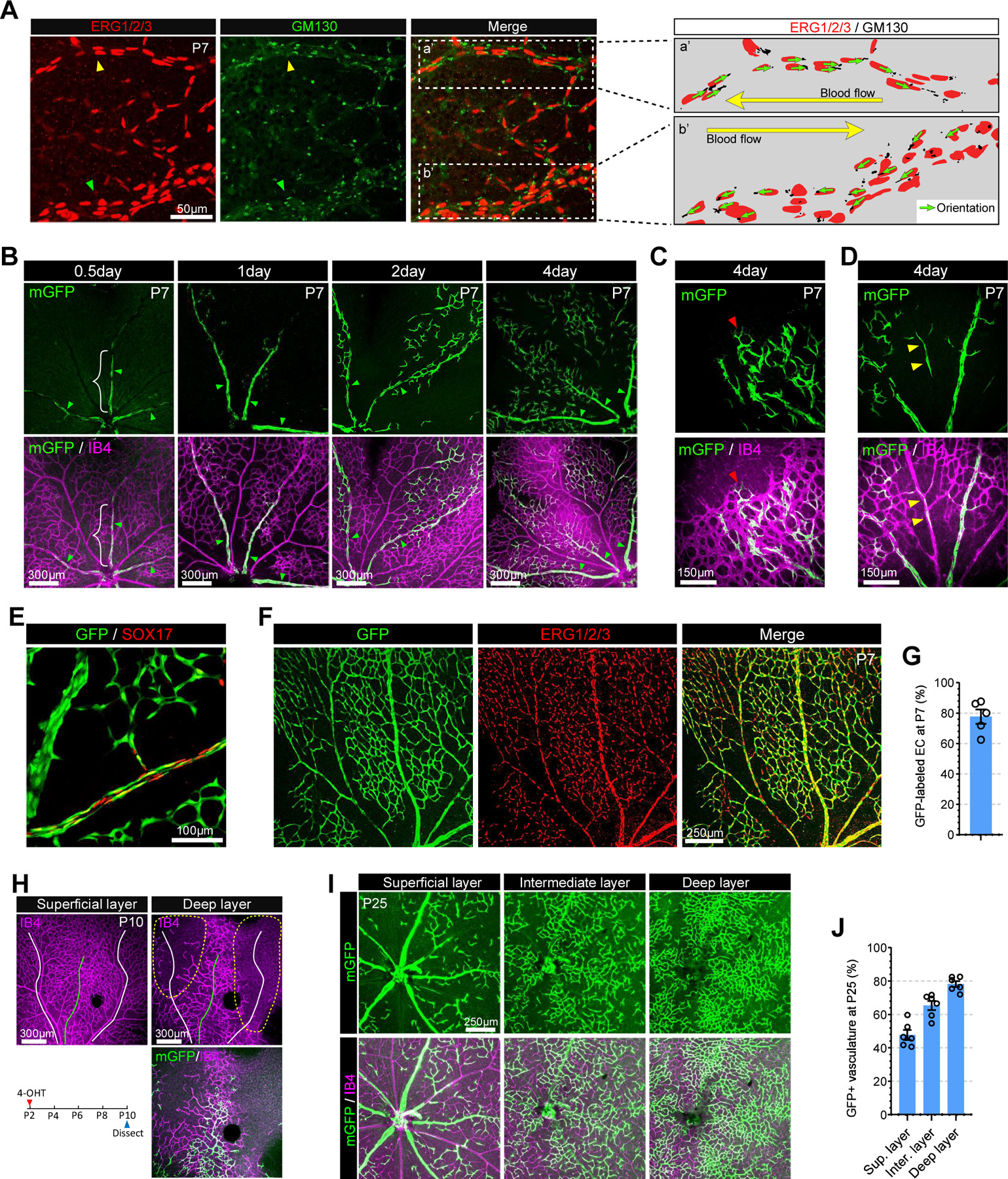
Figure 1.
VECs differentiate into tip, capillary and arterial ECs in the retinal vasculature.
A. Confocal images showing ERG1/2/3 (red, EC nucleus marker) and GM130 (green, golgi marker) immunostaining in P7 retinal vasculature. Right panel represents endothelial nucleus and golgi in the area ...
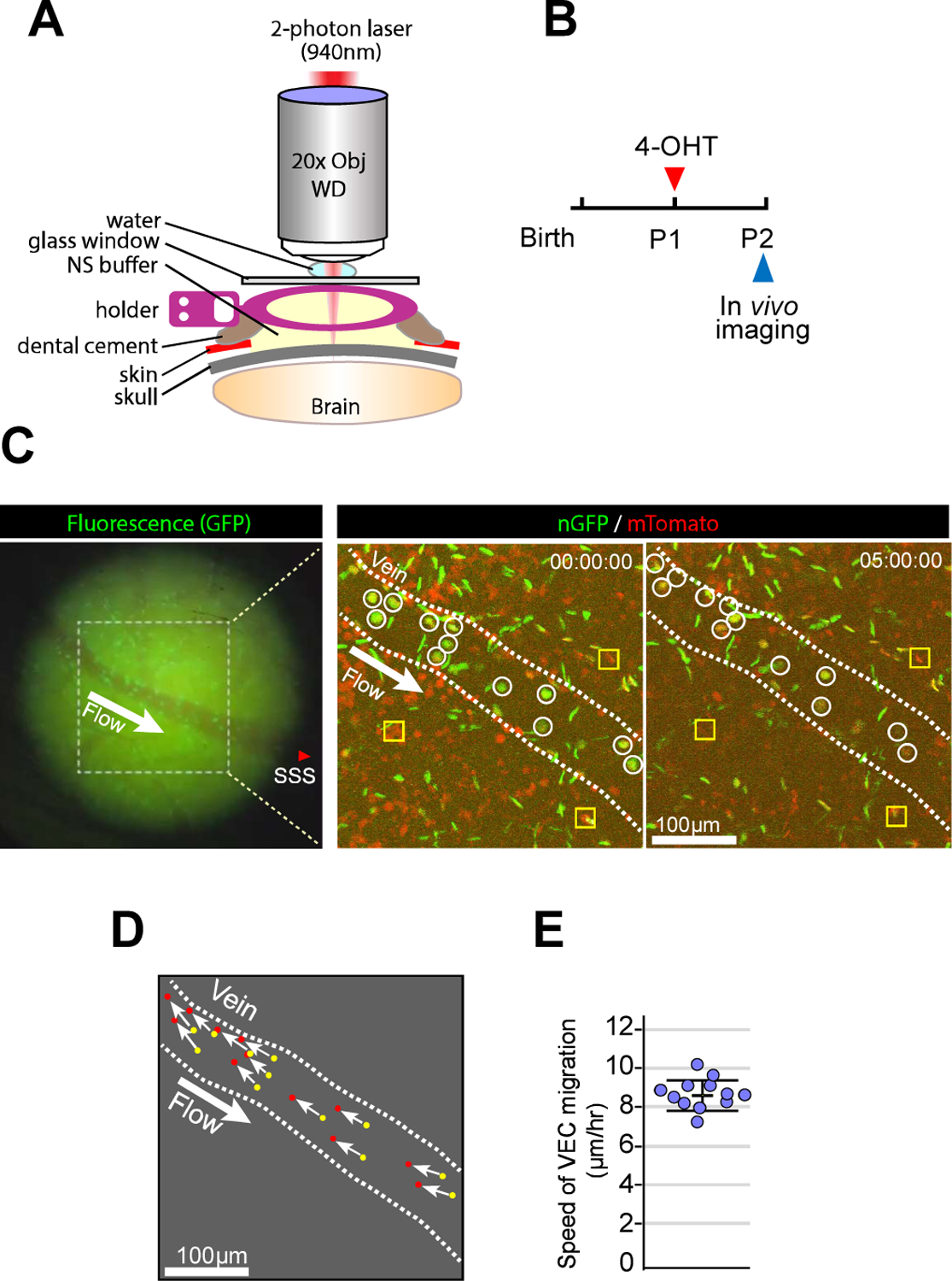
Figure 2.
VECs migrate against blood flow.
A. Schematic illustration of in vivo 2-photon imaging procedure. B. Timeline of the experimental procedure for 4-OHT administration and imaging using pan-EC nReporter mice. C. Fluorescence image from ...
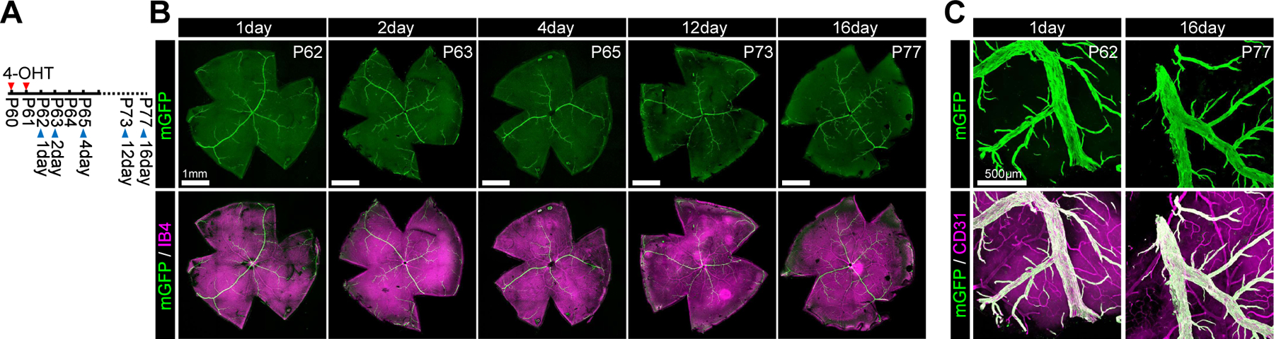
Figure 3.
VECs on quiescent stage shows lack of migration.
A. Timeline of the experimental procedure for 4-OHT administration and imaging. B. Confocal images of the whole mount retina immunohistochemistry showing mGFP-positive VECs (green) and vasculature (Is...
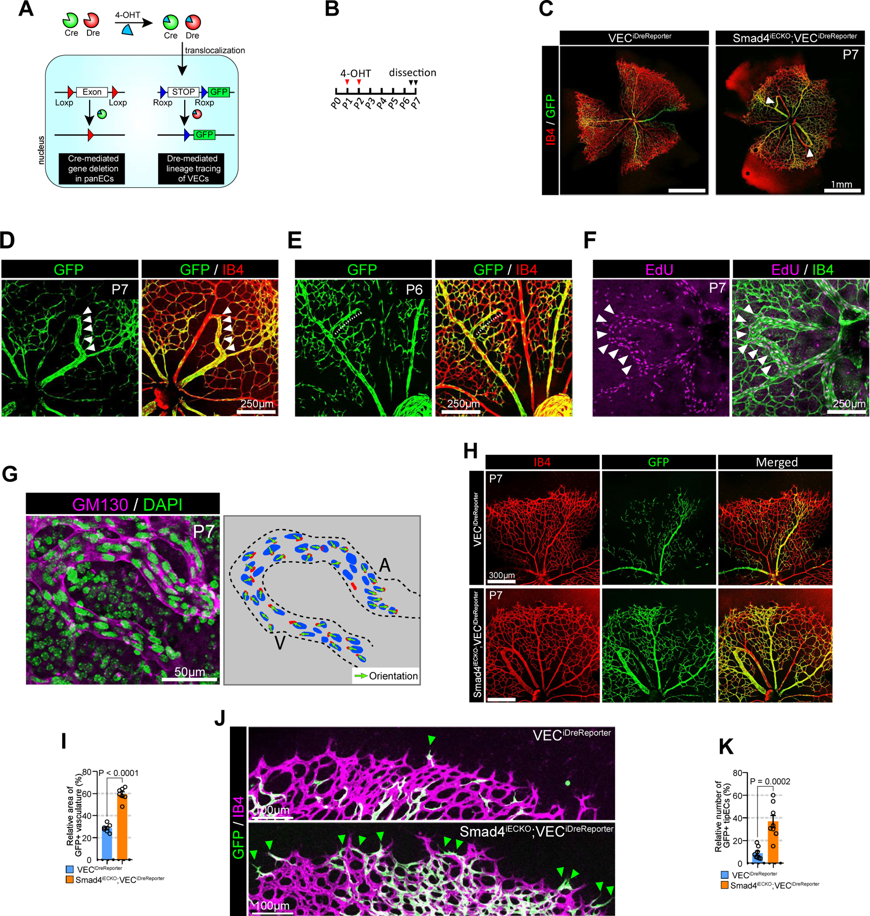
Figure 4.
Misdirected migration of VECs results in AV shunt formation.
A. Schematic diagram showing a strategy for generation of Smad4 iECKO ;VEC iDrereporter mice. Smad4 is deleted by inducible panEC specific Cre ( Cdh5 (PAC)CreER T2 ) while VECs is labeled with GFP usi...
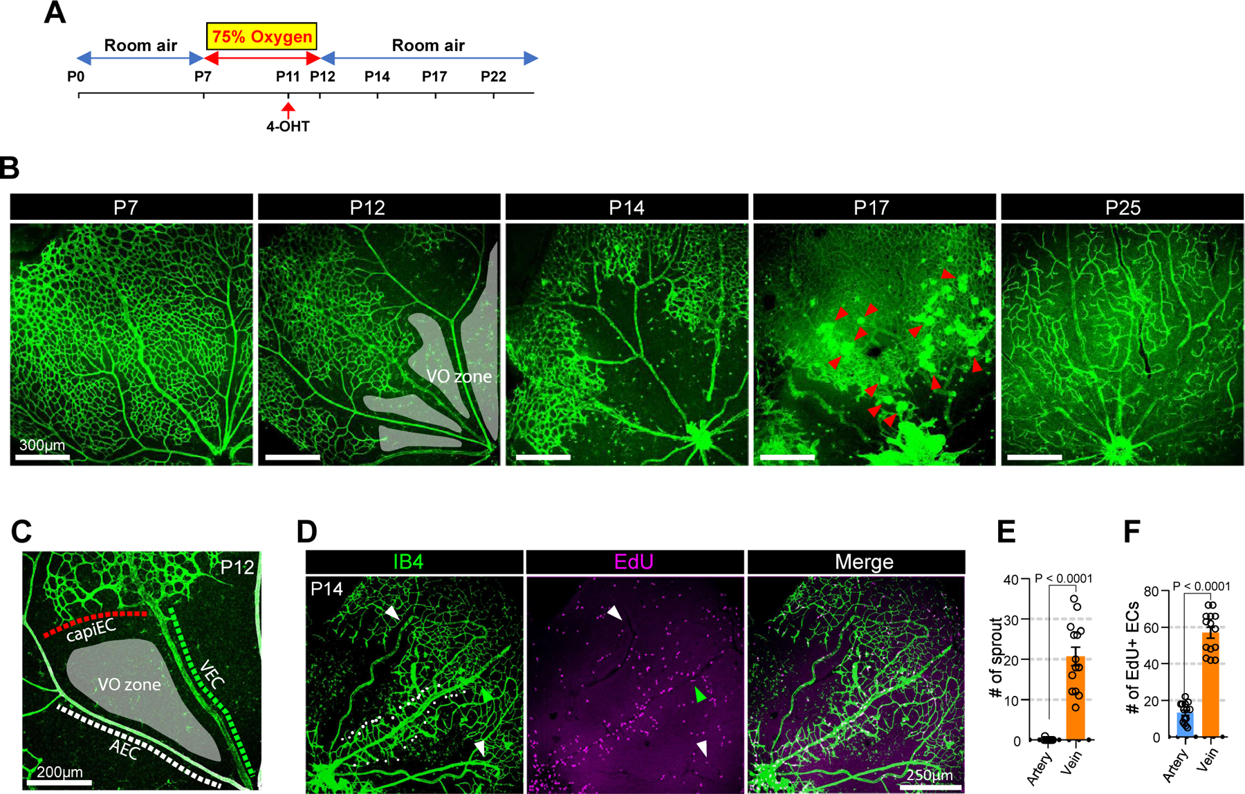
Figure 5.
Vein is the primary site for neoangiogenic spout initiation in OIR-induced neovascularization.
A. schematic diagram showing the experimental design of the OIR model. Postnatal pups along with their nursing female were exposed to 75% oxygen from P7 to P12 to induce vaso-obliteration and returned...
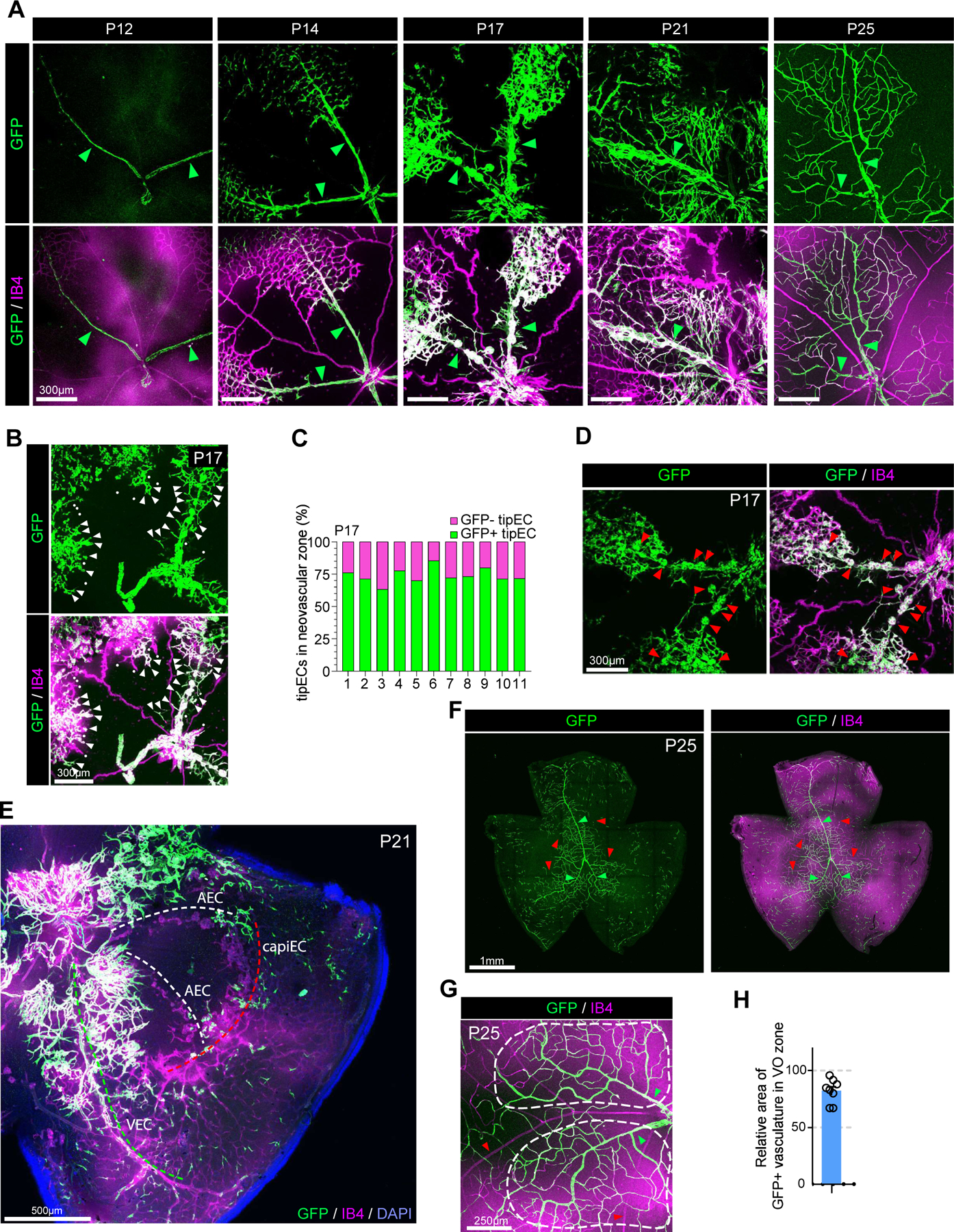
Figure 6.
VEC is the primary source of ECs in OIR-induced neovascularization.
A. Time course observation of the vasculature (Isolectin B4, purple) and GFP+ VECs (green) in VEC iDrereporter mice during OIR at indicated time points (P12, P14, P17, P21 and P25, respectively). Gree...
Figure images are served from the NIH/NLM PubMed Central Open Access Subset or Europe PMC; copyright remains with the publishers and authors.
💬 Discussion
0 commentsNo comments yet. Be the first to start a discussion!
Leave a Comment