Abstract
Although, drugs are required in the various skin compartments such as viable epidermis, dermis, or hair follicles, to efficiently treat skin diseases, drug delivery into and across the skin is still challenging. An improved understanding of skin barrier physiology is mandatory to optimize drug penetration and permeation. The various barriers of the skin have to be known in detail, which means methods are needed to measure their functionality and outside-in or inside-out passage of molecules through the various barriers. In this review, we summarize our current knowledge about mechanical barriers, i.e., stratum corneum and tight junctions, in interfollicular epidermis, hair follicles and glands. Furthermore, we discuss the barrier properties of the basement membrane and dermal blood vessels. Barrier alterations found in skin of patients with atopic dermatitis are described. Finally, we critically compare the up-to-date applicability of several physical, biochemical and microscopic methods such as transepidermal water loss, impedance spectroscopy, Raman spectroscopy, immunohistochemical stainings, optical coherence microscopy and multiphoton microscopy to distinctly address the different barriers and to measure permeation through these barriers in vitro and in vivo.
🔬 Techniques
🔭 Microscopes
✨ Fluorophores
🧪 Sample Preparation
🏭 Microscope Brands
🏛️ Research Organizations (ROR)
Affiliated research institutions:
📋 Methods
3.1. Physical/Chemical Methods The various methods described in this chapter are summarized in Table 1 . Figure 3 gives an overview of the areas of skin comprised by the measurements with the various physical/chemical methods and the distinct barriers localized in these areas. 3.1.1. Transepidermal Water Loss (TEWL) The measurement of the TEWL is a well-established method to determine the amount of water that permeates through the skin, which means through interfollicular epidermis, HFs and glands. The recent review by Alexander et al. [ 119 ] summarizes the principles of TEWL measurements. TEWL measurements were frequently applied to quantify disease-related skin dehydration occurring e.g., in atopic skin. In this context, increased TEWL is mechanistically linked to an impaired skin barrier. For instance, the molecular correlation between skin barrier impairment and TEWL has been shown in the skin of AD patients with mutated filaggrin [ 120 ]. Other reports suggest a correlation between the skin lipid composition and the regulation of the TEWL [ 15 ]. Recently, TEWL measurements were used to quantify the opening of the skin barrier by penetration enhancers and the clear correlation between increased TEWL and enhanced accumulation of the model drug cidofovir was shown [ 121 ]. However, TEWL is of course not a direct measurement of the drug. This method is very valuable to measure bulk changes of the skin barrier for inside-out movement of water as a surrogate for other molecules over time. It can easily be used in vivo when environmental influences are controlled. Yet, TEWL measurements cannot be connected to single molecular structures but reflect the sum of water loss via distinct skin components (interfollicular epidermis, HFs and sweat glands) and various skin-barrier related features (e.g., filaggrin expression, lipid composition or tight junctions). This lack of specificity may also prevent detailed insights into the molecular action of penetration enhancers or other barrier disrupting treatments. Figure 3 Localization of mechanical barriers in the skin (light red: stratum corneum (SC), Tight junctions (TJs) and basement membrane (BM)) and graphical representations of the various physical/chemical methods described in this review: ( A ) Fourier transform infrared (FTIR) spectroscopy ( B ) electrical impedance spectroscopy, ( C ) chemical analyses, ( D ) transepidermal water loss (TEWL), ( E ) transepithelial electrical resistances (TEER), ( F ) dye permeation analyses, and ( G ) Raman spectroscopy. Brackets denote the area measured by a specific method not discriminating between the different barriers within this area. ( B ) and ( C ) can be used to measure complete skin but can also be used—by additional preparation steps or more sophisticated methods—to discriminate between SC, viable epidermis and dermis. BV: blood vessel, HF: hair follicle, SbG: sebaceous gland, SwG: sweat gland. ( A ) from [ 122 ], ( C ) from [ 123 ], ( F ) from [ 74 ], ( G ) from [ 124 ]. 3.1.2. Transepithelial Electrical Resistance (TEER) and Electrical Impedance Spectroscopy (EIS) TEER measures the resistance at direct current. It is commonly used in monolayer cell cultures grown on transwell filters [ 60 , 125 ] applying “chopstick” electrodes or EndOhm chambers. In recent years it was also used for 3D cultures of reconstructed human epidermis/skin [ 74 , 84 , 126 , 127 ] or human skin [ 128 ]. While this is a good tool to measure overall barrier function to ions, which means comprising transcellular and paracellular barrier as well as barrier of the SC and the viable epidermis including TJs, it is not able to distinguish between these barriers. To this end, more sophisticated EIS methods are needed. EIS measures the resistance at alternating currents of different frequencies. Accessible are two-dimensional cell cultures and the measured impedance provide information on the paracellular passage of ions commonly referred to as barrier resistance, the transcellular capacity coupled current and the ventral distance between the cell and the cell binding substrate [ 129 , 130 ]. This technique enables the longitudinal recording of the epidermal barrier in real time. By using two-path impedance spectroscopy it is also possible to differentiate between paracellular and transcellular epithelial resistance [ 131 ]. Recently, EIS was used to measure the ion barrier of murine skin in vivo. Papain induced destruction of the skin barrier, as indicated by an impaired SC and disrupted TJs, resulted in a reduced resistance. Interestingly, the measured impedance correlated clearly with TEWL measurements [ 132 ]. However, the three-dimensional epidermis complicates the interpretation of the measured impedance and distinction between SC and TJ (and further barrier components) is not possible with the usual techniques. Additional mathematic modelling [ 133 ] and/or advanced techniques [ 134 ] are required to fully understand the obtained results. Drug or penetration enhancer-related changes of epidermal ion barrier are accessible by EIS. However, changes in EIS do not always correlate with the permeation rate of the drug/substance, as was e.g. shown by using different penetration enhancers to deliver theophylline, or determination of p-chloronitrobenzene flux after DMSO-induced skin damage [ 135 , 136 ]. 3.1.3.
Show full methods section
3.1. Physical/Chemical Methods The various methods described in this chapter are summarized in Table 1 . Figure 3 gives an overview of the areas of skin comprised by the measurements with the various physical/chemical methods and the distinct barriers localized in these areas. 3.1.1. Transepidermal Water Loss (TEWL) The measurement of the TEWL is a well-established method to determine the amount of water that permeates through the skin, which means through interfollicular epidermis, HFs and glands. The recent review by Alexander et al. [ 119 ] summarizes the principles of TEWL measurements. TEWL measurements were frequently applied to quantify disease-related skin dehydration occurring e.g., in atopic skin. In this context, increased TEWL is mechanistically linked to an impaired skin barrier. For instance, the molecular correlation between skin barrier impairment and TEWL has been shown in the skin of AD patients with mutated filaggrin [ 120 ]. Other reports suggest a correlation between the skin lipid composition and the regulation of the TEWL [ 15 ]. Recently, TEWL measurements were used to quantify the opening of the skin barrier by penetration enhancers and the clear correlation between increased TEWL and enhanced accumulation of the model drug cidofovir was shown [ 121 ]. However, TEWL is of course not a direct measurement of the drug. This method is very valuable to measure bulk changes of the skin barrier for inside-out movement of water as a surrogate for other molecules over time. It can easily be used in vivo when environmental influences are controlled. Yet, TEWL measurements cannot be connected to single molecular structures but reflect the sum of water loss via distinct skin components (interfollicular epidermis, HFs and sweat glands) and various skin-barrier related features (e.g., filaggrin expression, lipid composition or tight junctions). This lack of specificity may also prevent detailed insights into the molecular action of penetration enhancers or other barrier disrupting treatments. Figure 3 Localization of mechanical barriers in the skin (light red: stratum corneum (SC), Tight junctions (TJs) and basement membrane (BM)) and graphical representations of the various physical/chemical methods described in this review: ( A ) Fourier transform infrared (FTIR) spectroscopy ( B ) electrical impedance spectroscopy, ( C ) chemical analyses, ( D ) transepidermal water loss (TEWL), ( E ) transepithelial electrical resistances (TEER), ( F ) dye permeation analyses, and ( G ) Raman spectroscopy. Brackets denote the area measured by a specific method not discriminating between the different barriers within this area. ( B ) and ( C ) can be used to measure complete skin but can also be used—by additional preparation steps or more sophisticated methods—to discriminate between SC, viable epidermis and dermis. BV: blood vessel, HF: hair follicle, SbG: sebaceous gland, SwG: sweat gland. ( A ) from [ 122 ], ( C ) from [ 123 ], ( F ) from [ 74 ], ( G ) from [ 124 ]. 3.1.2. Transepithelial Electrical Resistance (TEER) and Electrical Impedance Spectroscopy (EIS) TEER measures the resistance at direct current. It is commonly used in monolayer cell cultures grown on transwell filters [ 60 , 125 ] applying “chopstick” electrodes or EndOhm chambers. In recent years it was also used for 3D cultures of reconstructed human epidermis/skin [ 74 , 84 , 126 , 127 ] or human skin [ 128 ]. While this is a good tool to measure overall barrier function to ions, which means comprising transcellular and paracellular barrier as well as barrier of the SC and the viable epidermis including TJs, it is not able to distinguish between these barriers. To this end, more sophisticated EIS methods are needed. EIS measures the resistance at alternating currents of different frequencies. Accessible are two-dimensional cell cultures and the measured impedance provide information on the paracellular passage of ions commonly referred to as barrier resistance, the transcellular capacity coupled current and the ventral distance between the cell and the cell binding substrate [ 129 , 130 ]. This technique enables the longitudinal recording of the epidermal barrier in real time. By using two-path impedance spectroscopy it is also possible to differentiate between paracellular and transcellular epithelial resistance [ 131 ]. Recently, EIS was used to measure the ion barrier of murine skin in vivo. Papain induced destruction of the skin barrier, as indicated by an impaired SC and disrupted TJs, resulted in a reduced resistance. Interestingly, the measured impedance correlated clearly with TEWL measurements [ 132 ]. However, the three-dimensional epidermis complicates the interpretation of the measured impedance and distinction between SC and TJ (and further barrier components) is not possible with the usual techniques. Additional mathematic modelling [ 133 ] and/or advanced techniques [ 134 ] are required to fully understand the obtained results. Drug or penetration enhancer-related changes of epidermal ion barrier are accessible by EIS. However, changes in EIS do not always correlate with the permeation rate of the drug/substance, as was e.g. shown by using different penetration enhancers to deliver theophylline, or determination of p-chloronitrobenzene flux after DMSO-induced skin damage [ 135 , 136 ]. 3.1.3.
Chemical and Radiochemical Analyses
Several marker molecules to test skin barrier function as well as drugs can be detected and quantified by chemical analyses such as high performance liquid chromatography (HPLC), ultra-performance liquid chromatography (UPLC), liquid chromatography tandem-mass spectrometry (LC-MS/MS), ultra-high performance liquid chromatography tandem mass spectrometry (UHPLC-MS/MS) or high performance thin layer chromatography [ 137 , 138 , 139 ]. In addition, also radioactively labelled surrogates for hydrophilic (caffeine) or lipophilic (testosterone) molecules are often used [ 140 ]. To this end, the molecular markers/drugs are applied in vitro (reconstructed human epidermis/skin, e.g., [ 141 , 142 ]), ex vivo (e.g., excised skin in horizontal or vertical diffusion cells—e.g., Franz cells—, Saarbrücken penetration model, Hamburg model of penetration, [ 138 , 143 , 144 ]) or in vivo [ 145 , 146 ] onto the skin. Often, molecular markers/drugs are detected after complete permeation of the skin by investigating blood levels or concentrations in the acceptor compartment of the diffusion (Franz) cell or in culture medium. Although measurements can be conducted at different time points, thus resulting in the ability to perform permeation kinetics, there is no information where drug diffusion was eventually decelerated within the skin. To get more information about this item, the skin samples can be separated into different compartments by tape-stripping (allowing detailed analysis of the various SC layers), or sectioning (e.g., SC, epidermis and dermis) or heating (separation epidermis/dermis). The segregated compartments can be investigated independently [ 137 , 145 , 147 , 148 ]. This also allows the calculation of penetration kinetics, especially when using tape strips [ 149 ]. Even though these approaches to separate different skin compartments are already a big step forward, they still have several limitations: In vivo testing of drug delivery is not possible because a skin biopsy is needed for evaluation after drug application. In addition, these tests can only be performed at one point in time per tissue sample. Furthermore, barrier structures within a compartment (e.g., TJs within the epidermis) cannot be specifically addressed. Finally, it is difficult to distinguish between interfollicular epidermis and hair follicles and glands. 3.1.4.
Dye Permeation Analyses
Topical application of usually fluorescent dyes, such as Lucifer yellow and fluorescein isothocyanates (FITCs) of different sizes are used to measure kinetics of dye permeation in 2D cell cultures but also in reconstructed human epidermis and in skin [ 60 , 74 , 125 , 128 , 150 ] to determine skin barrier function. To this end, specimens are taken from the acceptor/basal compartment and measured using fluorescent readers. This method can also be used for fluorescent-(labelled) drugs or drug delivery systems, e.g., 5-amino-levulinic acid [ 121 , 151 ]. This is a very useful technique to quantify penetration over time. Yet, it does not discriminate between the various barriers of the skin. Combining this technique with microscopical techniques can visualize local concentrations of the dye [ 33 , 152 ] (see also Section 3.2 ). 3.1.5.
Raman Spectroscopy
Vibrational spectroscopy techniques such as Raman spectroscopy are common analytical tools in skin research [ 153 , 154 ]. They can also provide insights into molecular changes which are potentially related to skin barrier functions such as skin hydration [ 155 ]. Beside the examination of the skin physiology, Raman spectroscopy appears to be also suited for the tracking of drugs and to determine drug penetration depth and quantitative information on drug accumulation in the skin [ 156 ]. In principle, the collected data give information on chemical compounds within the skin and they can provide a means to track characteristic drugs without the need of labelling. Every substance could provide characteristic spectra, especially large delocalized electron systems produce comparable strong Raman signals. This has e.g., enabled the detection of ibuprofen, lidocaine or caffeine skin permeation [ 154 , 157 , 158 ]. However, the high complexity of skin generates also very complex Raman spectra blurring drug related signals and prevent a straightforward interpretation of the obtained results [ 156 ]. To overcome these limitations, the combination of Raman spectroscopy with high resolution microscopy techniques such as confocal laser scanning microscopy or multiphoton microscopy is in the focus of recent research [ 159 ]. The outstanding advantage of such Raman microscopy is the high spatial resolution in the µm range in all three dimensions and the non-invasive, label free profiling of the chemical skin composition [ 160 , 161 ]. What is more, the acquired intensity signals connected to the concentration of the probed molecules allowing quantitative analysis of the skin [ 162 ]. However, long acquisition times, high levels of background noise and time-consuming image analysis limit the widespread use of Raman microscopy in dermatological research.
Coherent anti-Strokes Raman spectroscopy
(CARS) is a modified technical approach that significantly increases the imaging speed. The development of the combined multiphoton/CARS microscope could be considered as an important step towards a clinical application of Raman spectroscopy [ 163 ]; yet, further quenching of background signals and automatized image analysis is required to grant a close implementation into routine examination in dermatology. Further progress in the development of user-friendly devices and automatized image acquisition and analysis will open a broader usage of Raman spectroscopy in (trans)dermal drug delivery research [ 164 ]. 3.1.6. Fourier Transform Infrared (FTIR) Spectroscopy Another vibrational technique often used in combination with Raman spectroscopy is Fourier transform infrared (FTIR) spectroscopy. Compared to other spectroscopic methods, FTIR spectroscopy samples the absorbance of molecular vibrations resulting in characteristic fingerprints for many biomolecules. FTIR spectroscopy has numerous advances compared to classical dispersive spectroscopy [ 165 , 166 ]. First, the light yield is increased by up to factor 200 and therefore a much better signal-to-noise ratio can be achieved (the so called “Jacquinot” advantage). Second, the emission/absorption spectrum is not measured sequentially with regard to the wavelength but in parallel over the whole frequency range which, too, increases the signal-to-noise ratio (“Fellgett” or multiplex advantage). Third, the wavelength scale can be calibrated by a Helium-neon (HeNe) laser as a reference beam resulting in much better wavelength accuracy (“Connes” advantage). For real-time measurements of highly dynamic processes, fast scanning FTIR spectrometers with microsecond time-resolution are currently in development [ 167 ]. Attenuated total reflection (ATR) can be used as an extension to standard transmission FTIR spectroscopy enabling direct measurements of samples in a solid or liquid state without any preceding sample preparation and a penetration depth of 0.5 to 2 μm. For using ATR, the sample has to be brought in direct contact with special ATR crystals with a high refractive index (e.g., diamond). Concerning the skin barrier, the molecular conformation of the lipid matrix within the SC is of special interest. Boncheva et al. provide a method for estimating the molecular lipid structure by using ATR-FTIR spectroscopy for the measurement of the CH2 scissoring bandwidth. Besides comparing the molecular organization of SC lipids in human, porcine and reconstructed skin, they estimate the changes following the topical application of penetration enhancers such as oleic acid [ 13 ]. In general, the method allows conclusions whether the intercellular lipids in the SC are mainly arranged in the orthorhombic, hexagonal or liquid lateral packing. In addition, the secondary structure of keratin can be described. Finally also water content in the skin can be delineated [ 168 ].
3.2.
Microscopical Methods
The various methods described in this chapter are summarized in Table 1 . Figure 4 gives an overview of the skin compartments addressed by the various microscopical methods and the barriers/skin areas that can be visualized. 3.2.1.
Immunohistochemical Analysis
To investigate TJ-related barrier function, the simultaneous analysis of molecular tracers which can be primarily or secondarily detected by fluorescence microscopy and that were injected into the dermis (or applied to the basal compartment of 3D cell cultures), in combination with labelling of distinct TJ proteins by antibody-linked immunofluorescence, have frequently been used in skin samples of different species, applying normal fluorescence microscopy or confocal laser scanning microscopy (CLSM) [ 64 , 65 , 74 , 97 , 109 , 169 , 170 , 171 ]. This method which is called “biotinylation assay” when using EZ-Link TM -Sulfo-NHS-LC-Biotin (Biotin-SH) gives insight into barrier function of TJs in interfollicular epidermis of healthy and diseased/knock-out skin [ 64 , 65 , 74 , 169 , 170 ], HFs [ 97 , 171 ] and glands [ 109 ]. This method warrants high molecular resolution of the localization of the stop of tracers of different sizes (up to now 556 Da (“Biotin-SH”), 1500 Da, 5000 Da, 31 kDa) in correlation with the TJ barrier. By using antibodies directed to SC components this could also be used to determine barriers within the SC from outside-in when tracers are applied topically or from inside-out in situations with impaired TJ barrier However, even though this method is very valuable in basic research, it has several disadvantages to investigate drug delivery and thus plays, up to now, only a marginal role in drug formulation design. First of all, it can only be performed ex vivo or in vitro with processed tissue samples with processing for immunohistochemical stainings always including the possibility of changing the position of the drug investigated. In addition, it is not possible to perform successive measurements but only one point in time per sample can be investigated. Furthermore, quantification is, at least at the moment, time consuming [ 74 ]. Finally, the drug or the delivery system must be fluorescent or detectable by antibodies. Tagging the drug by a fluorescence dye might change the penetration behavior of the drug by changing its size and polarity and therefore the results may only give limited information on the penetration behavior of the drug itself. Thus, this technique might only be utilizable for very large drugs where the addition of the tag only results in minor changes. For 3D imaging, one could combine 3D tissue antibody staining [ 170 ] with CLSM/multiphoton microscopy (MPM), optimally with drugs that can be detected by fluorescence lifetime imaging (see Section 3.2.4 and Section 3.2.5 ). Figure 4 Localization of mechanical barriers in the skin (light red: stratum corneum (SC), Tight junctions (TJs) and basement membrane (BM)) and graphical presentation of the various microscopical methods described in this review: ( A1 – A6 ) Multiphoton microscopy with fluorescence lifetime imaging and second harmonic generation signal detection ( B ) transmission electron microscopy ( C ) confocal laser scanning microscopy of immunofluorescence stainings ( D ) Immunohistochemical analysis of a biotinylation assay ( E ) optical coherence tomography (OCT). The bracket denotes the area measured by OCT not discriminating between the different barriers within this area. Dotted black lines denote specific barriers/skin areas addressed by a method. Arrow in E denotes a hair follicle. BV: blood vessel, HF: hair follicle, SbG: sebaceous gland, SwG: sweat gland, ( B ) from [ 172 ], ( C ) from [ 170 ], ( E ) from [ 173 ]. 3.2.2.
Transmission Electron Microscopy
Another approach to investigate detailed localization of the barrier function of the skin to tracers is to use electron dense tracers like lanthanum combined with ultrastructural demonstration of TJ and SC structures in electron microscopy [ 174 ]. However, this method includes even more processing and is more time consuming than the light microscopical approach mentioned above. Nonetheless, transmission electron microscopy was used for monitoring dermal penetration of e.g., nanoparticles with different outcomes [ 175 , 176 ]. 3.2.3.
Optical Coherence Tomography
(OCT) and Optoacoustic Imaging OCT has been introduced in dermatology more than 20 years ago and proven as a useful tool for non-invasive morphological skin analysis [ 177 , 178 ]. It provides a large field of view (6 × 6 mm) and an optical penetration depth of up to 2 mm with a lateral and axial resolution in the 10 µm range [ 179 ]. Accordingly, OCT enables the discrimination between the SC and the living epidermis, papillary ridges and the subjacent dermis. Further advances resulted in the development of high definition (HD)-OCT. In contrast to OCT, HD-OCT provides an increased lateral and axial resolution in the 3 µm range whereas penetration depth and the field of view is decreased to approximately 1 mm and 2 mm, respectively. OCT and HD-OCT are valuable tools for diagnostic purposes and they are suitable to detect disease-related changes of the skin [ 179 ]. Currently, only few groups applied OCT to track the permeation of exogenously added agents through the skin. In this context, OCT was used to follow nanoparticle delivery through HFs [ 173 , 180 ] and epidermis [ 181 ]. OCT was also used to track glucose diffusion across the skin of rhesus monkeys [ 182 ]. Though, the majority of reports focused on micro needled-based dermal delivery systems in which OCT was used to visualize the position of the needles within the skin [ 183 , 184 ]. Further studies used OCT to document the effect of drugs on skin morphology which may only indirectly prove dermal drug delivery [ 185 , 186 , 187 ]. Optoacoustic or photoacoustic imaging is non-invasive and fast. It depends on the light illumination of the tissue trough e.g., pulsed lasers and the detection of light induced pressure waves through broadband ultrasound detectors. Upon multispectral light excitation, naturally occurring light adsorbers such as melanin or hemoglobin generate characteristic spectra which enable their discrimination in the tissue [ 188 ]. Real-time imaging with handheld devices envisions the broad clinical application of optoacoustic instruments in the near future [ 189 ]. Optoacoustic imaging provides a three-dimensional view into the skin with an imaging depth ranging from several centimeters to micrometers and a spatial resolution ranging from hundreds of micrometers to hundreds of nanometers, respectively. This is in contrast to conventional light microscopic approaches, where immense light scattering prevent access to deeper tissues. Therefore, optoacoustic imaging allows to differentiate between single skin layers (SC, viable epidermis and dermis) and enables visualization of the blood capillary loops and the vascular plexus (impact of the vascular barrier on drug delivery). Addressing cancer-induced morphologic (e.g., angiogenesis) or metabolic changes (e.g., tissue glycation) within the skin, several studies already document the applicability of optoacoustic imaging for diagnostic purposes [ 190 , 191 , 192 ] or potentially cancer-related changes of tissue glycation [ 193 ]. Although not required for imaging, light adsorbing substances such as fluorophores can be identified by optoacoustics enabling the in vivo tracking of drug permeation through the epidermis towards the blood circulation. Parallel assessment of disease related features such as epidermis thickness or vasodilatations [ 159 ] offer a potential tool that combines morphological skin layer evaluation, disease severity and drug permeation. To our knowledge there is no current study investigating (trans)-dermal drug delivery by optoacoustic imaging. However, especially from cancer-related research, several examples showed the use of optoacoustic imaging for the detection of fluorescently labelled drugs or drug carriers, like Cy7 conjugated iron oxide nanoparticles (Cy7-SPION) or PEGylated indocyanine green (ICG) liposomes, within experimental tumors such as breast cancer [ 194 , 195 , 196 ]. Interestingly, recent developments suggest the applicability of optoacoustic induced power waves to transiently open the SC and thus to facilitate drug permeation. Further developments may provide devices that allow diagnostic imaging, promote drug penetration and simultaneous tracking of the drugs [ 197 ]. 3.2.4. Confocal Laser Scanning Microscopy (CLSM) CLSM and especially confocal fluorescence microscopy can be a valuable tool to investigate the skin barrier and the delivery of drugs in vitro, ex vivo and in vivo. It can be very beneficial for the investigation of immunofluorescence stainings in 3D samples [ 170 ] (see also Section 3.2.1 ). Compared to classical bright field and fluorescence microscopy, CLSM uses a focused laser beam to excite the sample point-wise on a raster in x and y direction. The emission is then spatially filtered by a confocal pinhole to block out-of-focus light, sacrificing image intensity in exchange for improved image contrast compared to bright field microscopy. Image acquisition at multiple steps in z direction and subsequent three-dimensional reconstruction allows for investigation of skin structures in all three space dimensions. Besides imaging of endogenous fluorophores like reduced nicotinamide adenine dinucleotide (NADH), collagen or melanin, to illuminate the spatial structure of the skin, dermal drug delivery can be probed by fluorescence labelling of the drug or—in special cases, if the drug itself has fluorescent properties—without labelling [ 198 ]. For example, Alvarez-Román et al. investigated the skin penetration and distribution of polymeric nanoparticles in porcine skin ex vivo by CLSM and observed an accumulation in HFs and skin furrows [ 199 ]. Another application of CLSM is reflectance confocal microscopy (RCM), which uses backscattered light instead of fluorescence emission for imaging. In vivo RCM is frequently used as a diagnostic tool in dermatological routine [ 200 ], but has also many potential use cases in skin barrier research due to almost real-time imaging capabilities and relatively modest acquisition costs. 3.2.5. Multiphoton Microscopy (MPM) After its first operability demonstration 30 years ago in a laboratory setting [ 201 ], MPM overcomes the limit of explorative practice and developed as a powerful instrument in the area of skin basic research and increasingly clinical applications [ 202 , 203 ]. Three photonic component parts define MPM’s applicability for the characterization of human skin. Based on the nonlinear excitation of naturally occurring endogenous fluorophores such as NADH, flavin adenine dinucleotide, tryptophan, porphyrins and melanin [ 204 ], and protein structures (e.g., elastin, collagen) [ 205 , 206 ], the morphological pattern and layering of the skin can be evaluated on a subcellular level. Beyond the reflection in the outermost barrier of the epidermis—the SC—the first living cell in the viable epidermis can be assessed in detail with the incidental finding of a precise discrimination of the SC/SG transition. The principle of two-photon excitation overcomes the limitations of fluorescence imaging, e.g., operation of CLSM in fluorescence mode, for innocuous in vivo utilization. Here, with a negligible linear energy transfer at the boundary layer, the excitation almost exclusively takes place within the target volume of examination without damaging the surrounding tissue [ 207 ]. The additional utilization of the nonlinear optical process second harmonic generation (SHG), observed in noncentrosymmetric molecular structures such as collagen [ 163 , 208 ], allows, next to its application in the area of dermal fibril characterization up to a penetration depth of approximately 200 µm [ 209 , 210 , 211 ], the exact discrimination of the dermo–epidermal junction and consecutively the epidermal thickness. Third, MPM additionally offers the applicability of fluorescence lifetime imaging microscopy (FLIM) [ 163 , 212 ]. Since the lifetime of a fluorophore—the time it spends in the excited state before returning to the ground state by photon emission—is characteristic for a fluorophore in its biochemical environment [ 213 , 214 ], FLIM facilitates a distinct in vivo identification of fluorescent molecules that share an overlap in their morphological or spectroscopic pattern within human skin. By time-correlated single photon counting [ 213 ] or phasor analysis [ 215 ] selective visualization of endogenous fluorophores or topical drug uptake was successful at high spatial resolutions [ 216 ]. Therefore, MPM FLIM enables for non-invasive high-resolution examination of human skin in vitro, ex vivo and in clinical in vivo applications. On this basis, clinical studies focused on both high-resolution morphological patterns and pathophysiological variances on a cellular level e.g., in the process of skin wound healing [ 217 , 218 ]. Next to MPM FLIM application in advanced skin cancer diagnosis [ 203 , 219 ], also the skin barrier function itself became the focus of attention: Volz as well as Frombach used high-resolution Cluster-FLIM in in vitro- and ex vivo-approaches to explicitly inspect the dynamics of both drug penetration and the penetrated skin area itself [ 163 , 216 , 220 , 221 , 222 ]. Transferring skin barrier analysis to ‘patient’s bedside’, Ogawa-Fuse examined skin hydration utilizing a combination of already mentioned techniques such as CARS/Raman and MPM FLIM [ 220 ]. Moreover, the same technical combination facilitated the measurement of pharmaceutical and cosmetic drug penetration and delivery through the skin, e.g., minocycline and tazarotene [ 163 , 216 , 221 , 222 ]. The combination of mentioned non-invasive spectroscopic and microscopic techniques such as CARS/Raman spectroscopy, MPM and associated techniques provide a huge potential for the investigation of the skin barrier and the related impact on dermal or even transdermal drug delivery. Sophisticated image analysis through artificial intelligence may open up the broad application of high-resolution skin microscopy in skin research and clinics [ 164 ].
📊 Figures
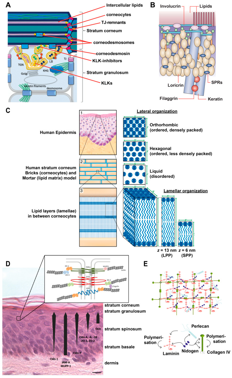
Figure 1
Overview of epidermal molecular structures important for skin barrier function. ( A ) Schematic drawing of the uppermost stratum granulosum and stratum corneum . KHG: keratohyalin granula KLK: kallikr...
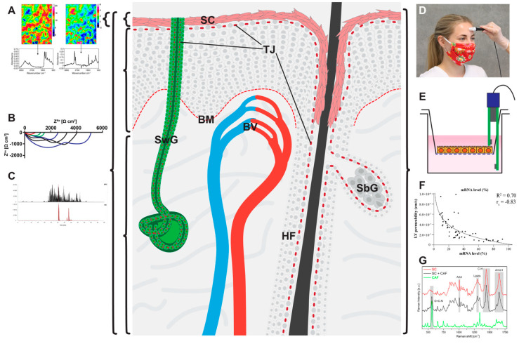
Figure 3
Localization of mechanical barriers in the skin (light red: stratum corneum (SC), Tight junctions (TJs) and basement membrane (BM)) and graphical representations of the various physical/chemical metho...
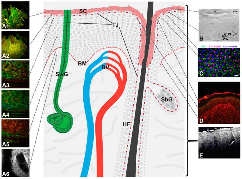
Figure 4
Localization of mechanical barriers in the skin (light red: stratum corneum (SC), Tight junctions (TJs) and basement membrane (BM)) and graphical presentation of the various microscopical methods desc...
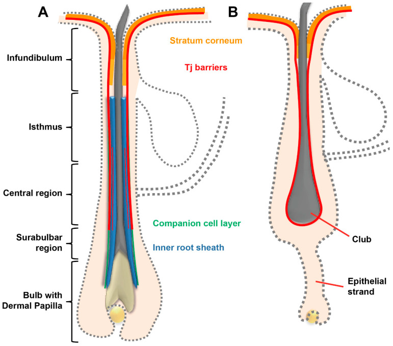
Figure 2
Schematic drawing of a hair follicle in anagen ( A ) and catagen ( B ) phase denoting stratum corneum (SC) and tight junction (TJ) barriers. More intense color of SC denotes SC similar to epidermal SC...
Figure images are served from the NIH/NLM PubMed Central Open Access Subset or Europe PMC; copyright remains with the publishers and authors.
💬 Discussion
0 commentsNo comments yet. Be the first to start a discussion!
Leave a Comment