Abstract
Abstract Target protection proteins confer resistance to the host organism by directly binding to the antibiotic target. One class of such proteins are the antibiotic resistance (ARE) ATP-binding cassette (ABC) proteins of the F-subtype (ARE-ABCFs), which are widely distributed throughout Gram-positive bacteria and bind the ribosome to alleviate translational inhibition from antibiotics that target the large ribosomal subunit. Here, we present single-particle cryo-EM structures of ARE-ABCF-ribosome complexes from three Gram-positive pathogens: Enterococcus faecalis LsaA, Staphylococcus haemolyticus VgaA LC and Listeria monocytogenes VgaL. Supported by extensive mutagenesis analysis, these structures enable a general model for antibiotic resistance mediated by these ARE-ABCFs to be proposed. In this model, ABCF binding to the antibiotic-stalled ribosome mediates antibiotic release via mechanistically diverse long-range conformational relays that converge on a few conserved ribosomal RNA nucleotides located at the peptidyltransferase center. These insights are important for the future development of antibiotics that overcome such target protection resistance mechanisms.
🔬 Techniques
🧬 Organisms
💻 Software
✨ Fluorophores
🧪 Sample Preparation
🏭 Microscope Brands
🧪 Reagent Suppliers
📷 Detectors
💻 Software Details
💾 Data Repositories
🏛️ Research Organizations (ROR)
Affiliated research institutions:
📋 Methods
Strains and plasmids
All strains and plasmids used in this work are listed in Table S5 . Primers are listed in Table S6 . E. faecalis OG1RF and TX5332, a LsaA disruption mutant of OG1RF 30 , were kindly provided by Dr. Barbara E. Murray (Health Science Center, University of Texas). All cloning was performed by Protein Expertise Platform at Umeå University. E. faecalis LsaA ORF was PCR amplified from pTEX5333 plasmid and cloned into pCIE vector 69 for cCF10-induced expression. The LsaA ORF was supplemented with C-terminal His 6 -TEV-FLAG 3 -tag (HTF tag) and the ribosome-binding site was optimized for high expression yield. Point mutations E 142 Q and E 452 Q were introduced to LsaA resulting in pCIE_LsaA-EQ 2 -HTF. S. haemolyticus vga(A) LC gene was PCR-amplified from a S. haemolyticus isolate held in the O’Neill strain collection at the University of Leeds, using oligonucleotide primers vgaA LC -F (5′-GGTGGT GGTAC CAGGATGAGGAAATATGAAAA-3′) and vgaA LC -R (5′-GGTGGT GAATTC GGTAATTTATTTATCTAAATTTCTT-3′) (engineered restriction sites shown underlined). The protein encoded by this gene is identical to that previously reported 50 (accession number DQ823382 ). The fragment was digested with Kpn I and Eco RI and ligated into the tetracycline-inducible expression vector pRMC2 (ref. 70 ). Constructs encoding the VgaA LC protein fused with a C-terminal FLAG 3 tag were obtained by synthesis (Genewiz), with E 105 Q, E 410 Q and EQ 2 mutants subsequently created by site-directed mutagenesis. Generation of other point mutants of untagged Vga(A) LC was performed by NBS Biologicals, again using chemical synthesis to generate the original vga(A) LC template, followed by site-directed mutagenesis. L. monocytogenes VgaL (Lmo0919). In order to construct L. monocytogenes EGDe::∆ lmo0919 , regions corresponding to the upstream and downstream flanking regions of lmo0919 , present on the EGDe genome were amplified with primer pairs VKT35 (5′-GGGGGGATCCATCACTAGCCGAATCCAAAC-3′), VKT36 (5′-gggggaattcaaaaaataacctcctgaatattttcagag-3′) and VHKT37 (5′-GGGGGAATTCAAAAAATAACCTCCTGAATATTTTCAGAG-3′), VHKT38 (5′-GGGGCCATGGCGTGCTGTACGGTATGC-3′), respectively. Fragments were then cloned in tandem into the pMAD vector using Bam HI, Eco RI and Nco RI restriction sites. The resulting vector, VHp689, was then sequenced to ensure wild-type sequences of clones. Gene deletion was then performed as per Arnaud et al. 71 . lmo0919 was amplified from EGDe genomic DNA using primers VHKT12 (5′-CCCCCCATGGCATCTACAATCGAAATAAATC-3′) and VHKT39 (5′-GGGGCTGCAGTTAACTAAATTGCTGTCTTTTTG-3′), and cloned into pIMK3 using Nco I and Pst I restriction sites, resulting in plasmid VHp690. Overlap extension PCR was used in order to introduce a HTF tag at the C-terminus of lmo0919 (ref. 72 ). The lmo0919 locus and HTF tag were amplified with primer pairs VHKT12, VHKT15 (5′-ATGATGATGGCCGCCACTAAATTGCTGTCTTTTTG-3′) and VHKT14 (5′-AGACAGCAATTTAGTGGCGGCCATCATCATCATC-3′), VHKT13 (5′-GGGGCTGCAGTTAGCCTTTGTCATCGTC-3′) using EGDe genomic DNA and VHp100 template DNA, respectively, producing fragments with overlapping ends. VHKT12 and VHKT13 were then used to fuse the fragments and the resulting PCR product was cloned into pIMK3 using Nco I and Pst I sites resulting in VHp692. To introduce two EQ mutations (E104Q and E408Q) simultaneously into the VHp692 plasmid, primers VHT266 (5′-TCTTGATCAACCAACCAACTATTTGGATATCTACGCAATGGAA-3′) and VHT267 (5′-TTGTTGGTTGGTCTGCTAGGAGAACACTTGGATTTTGGCGCA-3′) containing both mutations were used to extend out from lmo0919 HTF to amplify the VHp692 backbone. Primers VHT264 (5′-AGCAGACCAACCAACAAGCAATCTTGATGTCG-3′) and VHT265 (5′-TGGTTGGTTGATCAAGAATCAAGAAATTGGCGT-3′) also containing lmo0919 EQ2 mutations were used to amplify a fragment with overlapping sequence to the backbone fragment. Both PCR products were then assembled using NEBuilder® HiFi DNA Assembly Master Mix (NEB), resulting in VHp693. B. subtilis To construct the VHB109 [ trpC2 Δ vmlR thrC::P hy-spnak - lsaA kmR ] strain untagged LsaA under the control of an IPTG-inducible P hy-spank promotor, a PCR product encoding lsa(A) was PCR-amplified from pTEX5333 using the primers VHT127 (5′-CGACGAAGGAGAGAGCGATAATGTCGAAAATTGAACTAAAACAACTATC-3′) and VHT128 (5′-CACCGAATTAGCTTGCATGCTTATGATTTCAAGACAATTTTTTTATCTGTTA-3′). The second PCR fragment encoding a kanamycin-resistance marker, a polylinker downstream of the Phy-spank promoter and the lac repressor ORF—all inserted in the middle of the thrC gene—was PCR-amplified from pHT009 plasmid using primers VHT123 (5′-CATTATCGCTCTCTCCTTCGTCGACTAAGCTAATTG-3′) and VHT125 (5′-TAAGCATGCAAGCTAATTCGGTGGAAACGAGG-3′). The two fragments were ligated using the NEBuilder HiFi DNA Assembly master mix (New England BioLabs, Ipswich, MA) yielding the pHT009-lsaA plasmid (VHp369) which was used to transform the VHB5 [ trpC2 Δ vmlR ] strain. Selection for kanamycin resistance yielded the desired VHB109 strain. To construct the VHB168 [ trpC2 Δ vmlR thrC::P hy-spnak -lsaAK244A kmR ] strain, VHp369 plasmid was subjected to site-directed mutagenesis using primer VHP303 (5′-GCATCACCTTCACGGTTCATCGACCATTCCGCT-3′) and VHP304 (5′-GTACGGCAACGCTAAGGAAAAAGGGAGCGGGGCGA-3′), according to the directions of Phusion Site-Directed Mutagenesis Kit (Thermo Fisher Scientific), yielding VHp526 (pHT009- lsaAK244A ) plasmid which was used to transform the VHB5 [ trpC2 Δ vmlR ] strain. Selection for kanamycin resistance yielded the desired VHB168 strain. To construct the VHB169 [ trpC2 Δ vmlR thrC ::P hy-spnak - lsaAF257A kmR ] strain, VHp369 plasmid was subjected to site-directed mutagenesis using primer VHP305 (5′-CAATCGCCCCGCTCCCTTTTTCCTTAGCGT-3′) and VHP306 (5′-CGGATACAGGAGCCATTGGTGCCCGGGCA-3′), according to the directions of Phusion Site-Directed Mutagenesis Kit (Thermo Fisher Scientific), yielding, yielding VHp527 (pHT009- lsaAF257A ) plasmid which was used to transform the VHB5 [ trpC2 Δ vmlR ] strain. Selection for kanamycin resistance yielded the desired VHB169 strain.
Show full methods section
Strains and plasmids
All strains and plasmids used in this work are listed in Table S5 . Primers are listed in Table S6 . E. faecalis OG1RF and TX5332, a LsaA disruption mutant of OG1RF 30 , were kindly provided by Dr. Barbara E. Murray (Health Science Center, University of Texas). All cloning was performed by Protein Expertise Platform at Umeå University. E. faecalis LsaA ORF was PCR amplified from pTEX5333 plasmid and cloned into pCIE vector 69 for cCF10-induced expression. The LsaA ORF was supplemented with C-terminal His 6 -TEV-FLAG 3 -tag (HTF tag) and the ribosome-binding site was optimized for high expression yield. Point mutations E 142 Q and E 452 Q were introduced to LsaA resulting in pCIE_LsaA-EQ 2 -HTF. S. haemolyticus vga(A) LC gene was PCR-amplified from a S. haemolyticus isolate held in the O’Neill strain collection at the University of Leeds, using oligonucleotide primers vgaA LC -F (5′-GGTGGT GGTAC CAGGATGAGGAAATATGAAAA-3′) and vgaA LC -R (5′-GGTGGT GAATTC GGTAATTTATTTATCTAAATTTCTT-3′) (engineered restriction sites shown underlined). The protein encoded by this gene is identical to that previously reported 50 (accession number DQ823382 ). The fragment was digested with Kpn I and Eco RI and ligated into the tetracycline-inducible expression vector pRMC2 (ref. 70 ). Constructs encoding the VgaA LC protein fused with a C-terminal FLAG 3 tag were obtained by synthesis (Genewiz), with E 105 Q, E 410 Q and EQ 2 mutants subsequently created by site-directed mutagenesis. Generation of other point mutants of untagged Vga(A) LC was performed by NBS Biologicals, again using chemical synthesis to generate the original vga(A) LC template, followed by site-directed mutagenesis. L. monocytogenes VgaL (Lmo0919). In order to construct L. monocytogenes EGDe::∆ lmo0919 , regions corresponding to the upstream and downstream flanking regions of lmo0919 , present on the EGDe genome were amplified with primer pairs VKT35 (5′-GGGGGGATCCATCACTAGCCGAATCCAAAC-3′), VKT36 (5′-gggggaattcaaaaaataacctcctgaatattttcagag-3′) and VHKT37 (5′-GGGGGAATTCAAAAAATAACCTCCTGAATATTTTCAGAG-3′), VHKT38 (5′-GGGGCCATGGCGTGCTGTACGGTATGC-3′), respectively. Fragments were then cloned in tandem into the pMAD vector using Bam HI, Eco RI and Nco RI restriction sites. The resulting vector, VHp689, was then sequenced to ensure wild-type sequences of clones. Gene deletion was then performed as per Arnaud et al. 71 . lmo0919 was amplified from EGDe genomic DNA using primers VHKT12 (5′-CCCCCCATGGCATCTACAATCGAAATAAATC-3′) and VHKT39 (5′-GGGGCTGCAGTTAACTAAATTGCTGTCTTTTTG-3′), and cloned into pIMK3 using Nco I and Pst I restriction sites, resulting in plasmid VHp690. Overlap extension PCR was used in order to introduce a HTF tag at the C-terminus of lmo0919 (ref. 72 ). The lmo0919 locus and HTF tag were amplified with primer pairs VHKT12, VHKT15 (5′-ATGATGATGGCCGCCACTAAATTGCTGTCTTTTTG-3′) and VHKT14 (5′-AGACAGCAATTTAGTGGCGGCCATCATCATCATC-3′), VHKT13 (5′-GGGGCTGCAGTTAGCCTTTGTCATCGTC-3′) using EGDe genomic DNA and VHp100 template DNA, respectively, producing fragments with overlapping ends. VHKT12 and VHKT13 were then used to fuse the fragments and the resulting PCR product was cloned into pIMK3 using Nco I and Pst I sites resulting in VHp692. To introduce two EQ mutations (E104Q and E408Q) simultaneously into the VHp692 plasmid, primers VHT266 (5′-TCTTGATCAACCAACCAACTATTTGGATATCTACGCAATGGAA-3′) and VHT267 (5′-TTGTTGGTTGGTCTGCTAGGAGAACACTTGGATTTTGGCGCA-3′) containing both mutations were used to extend out from lmo0919 HTF to amplify the VHp692 backbone. Primers VHT264 (5′-AGCAGACCAACCAACAAGCAATCTTGATGTCG-3′) and VHT265 (5′-TGGTTGGTTGATCAAGAATCAAGAAATTGGCGT-3′) also containing lmo0919 EQ2 mutations were used to amplify a fragment with overlapping sequence to the backbone fragment. Both PCR products were then assembled using NEBuilder® HiFi DNA Assembly Master Mix (NEB), resulting in VHp693. B. subtilis To construct the VHB109 [ trpC2 Δ vmlR thrC::P hy-spnak - lsaA kmR ] strain untagged LsaA under the control of an IPTG-inducible P hy-spank promotor, a PCR product encoding lsa(A) was PCR-amplified from pTEX5333 using the primers VHT127 (5′-CGACGAAGGAGAGAGCGATAATGTCGAAAATTGAACTAAAACAACTATC-3′) and VHT128 (5′-CACCGAATTAGCTTGCATGCTTATGATTTCAAGACAATTTTTTTATCTGTTA-3′). The second PCR fragment encoding a kanamycin-resistance marker, a polylinker downstream of the Phy-spank promoter and the lac repressor ORF—all inserted in the middle of the thrC gene—was PCR-amplified from pHT009 plasmid using primers VHT123 (5′-CATTATCGCTCTCTCCTTCGTCGACTAAGCTAATTG-3′) and VHT125 (5′-TAAGCATGCAAGCTAATTCGGTGGAAACGAGG-3′). The two fragments were ligated using the NEBuilder HiFi DNA Assembly master mix (New England BioLabs, Ipswich, MA) yielding the pHT009-lsaA plasmid (VHp369) which was used to transform the VHB5 [ trpC2 Δ vmlR ] strain. Selection for kanamycin resistance yielded the desired VHB109 strain. To construct the VHB168 [ trpC2 Δ vmlR thrC::P hy-spnak -lsaAK244A kmR ] strain, VHp369 plasmid was subjected to site-directed mutagenesis using primer VHP303 (5′-GCATCACCTTCACGGTTCATCGACCATTCCGCT-3′) and VHP304 (5′-GTACGGCAACGCTAAGGAAAAAGGGAGCGGGGCGA-3′), according to the directions of Phusion Site-Directed Mutagenesis Kit (Thermo Fisher Scientific), yielding VHp526 (pHT009- lsaAK244A ) plasmid which was used to transform the VHB5 [ trpC2 Δ vmlR ] strain. Selection for kanamycin resistance yielded the desired VHB168 strain. To construct the VHB169 [ trpC2 Δ vmlR thrC ::P hy-spnak - lsaAF257A kmR ] strain, VHp369 plasmid was subjected to site-directed mutagenesis using primer VHP305 (5′-CAATCGCCCCGCTCCCTTTTTCCTTAGCGT-3′) and VHP306 (5′-CGGATACAGGAGCCATTGGTGCCCGGGCA-3′), according to the directions of Phusion Site-Directed Mutagenesis Kit (Thermo Fisher Scientific), yielding, yielding VHp527 (pHT009- lsaAF257A ) plasmid which was used to transform the VHB5 [ trpC2 Δ vmlR ] strain. Selection for kanamycin resistance yielded the desired VHB169 strain.
Bacterial transformation E. faecalis
Electrocompetent cells were prepared as per Bhardwaj et al. 73 . In short, an overnight culture grown in the presence of appropriate antibiotics was diluted to OD 600 of 0.05 in 50 mL of BHI media (supplemented with 2 mg/mL kanamycin in case of TX5332), grown to OD 600 of 0.6–0.7 at 37 °C with moderate shaking (160 r.p.m.). Cells were collected by centrifugation at 3200 × g at 4 °C for 10 min. Cells were resuspended in 0.5 mL of sterile lysozyme buffer (10 mM Tris-HCl pH 8; 50 mM NaCl, 10 mM EDTA, 35 µg/mL lysozyme), transferred to 1.5 mL Eppendorf tube and incubated at 37 °C for 30 min. Cells were pelleted at 8700 × g at 4 °C for 10 min and washed three times with 1.5 mL of ice-cold electroporation buffer (0.5 M sucrose, 10% glycerol(w/v)). After last wash the cells were resuspended in 500 µL of ice-cold electroporation buffer and aliquoted and stored at –80 °C. For electroporation 35 µL of electrocompetent cells were supplemented with 1 µg of plasmid DNA, transferred to ice-cold 1 mm electroporation cuvette and electroporated at 1.8 keV. Immediately after electroporation 1 mL of ice-cold BHI was added to the cells, the content of the cuvette was transferred to 1.5 mL Eppendorf tubes and the cells were recovered at 37 °C for 2.5 h and plated onto BHI plates containing appropriate antibiotics (10 µg/mL chloramphenicol and 2 mg/mL kanamycin). S. aureus Preparation and transformation of S. aureus electrocompetent cells followed the method of Schenk and Laddaga 74 , though used TSBY (Tryptone soya broth [Oxoid] containing 2.5% yeast extract) in place of B2 medium. Briefly, bacteria were grown with vigorous aeration in TSBY to an OD 600 of 0.6, harvested by centrifugation, and washed three times in an equal volume of sterile, deionized water. Subsequent wash steps used decreasing volumes of 10% glycerol; first 1/5 the original culture volume, then 1/10, finally resuspending in ~1/32 volume and storing the resultant electrocompetent cells at −80 °C. For electroporation, 60 µL of electrocompetent cells were mixed with ≧1 µg of plasmid DNA in a 1 mm electroporation cuvette at room temperature and pulsed at 2.3 kV, 100 Ω, 25 μFD. Immediately after electroporation, 390 µL room temperature TSBY was added to the cells and incubated with aeration at 37 °C for 1–2 h, before plating onto tryptone soya agar with appropriate antibiotic selection. Using this method, sequence-verified constructs established in E. coli were first transferred into the restriction deficient S. aureus RN4220 strain 75 , before recovery and introduction into S. aureus SH1000 (refs. 76 , 77 ). L. monocytogenes L. monocytogenes EGD-e was transformed with pIMK3 integrative plasmids via conjugation. E. coli S17.1 harbouring pIMK3 and its derivatives was grown at 37 °C overnight in LB media supplemented with 50 µg/mL kanamycin; 1 mL of culture was washed three times with sterile BHI media to remove antibiotics. Two hundred microliters of washed E. coli culture was mixed with an equal volume of L. monocytogenes overnight culture grown at 37 °C in BHI media. Two hundred microliters of mixed bacterial suspension was then dropped onto a conjugation filter (Millipore #HAEP047S0) placed onto a BHI agar plate containing 0.2 µg/mL penicillin-G. After overnight incubation at 37 °C, bacterial growth from the filter was resuspended in 1 mL of BHI and 100–300 µL plated onto BHI agar plates supplemented with 50 µg/mL kanamycin (to select for pIMK3), 50 µg/mL nalidixic acid and 10 µg/mL colistin sulfate (Sigma-Aldrich C4461-100MG). Resulting colonies were checked for correct integration via PCR and subsequent sequencing using primers VHKT42 and VHKT43.
Antibiotic susceptibility testing
Minimum inhibitory concentrations (MIC) were determined based on guidelines from the European Committee on Antimicrobial Susceptibility Testing (EUCAST) ( http://www.eucast.org/ast_of_bacteria/mic_determination ). E. faecalis Bacteria were grown in BHI media supplemented with 2 mg/mL kanamycin (to prevent lsa revertants), 0.1 mg/mL spectinomycin (to maintain the pCIE spec plasmid), 100 ng/mL of cCF10 peptide (to induce expression of LsaA protein) as well as increasing concentrations of antibiotics was inoculated with 5 × 10 5 CFU/mL (OD 600 of approximately 0.0005) of E. faecalis Δ lsaA ( lsa::Kan ) strain TX5332 transformed either with empty pCIE spec plasmid or with pCIE spec encoding LsaA. After 16–20 h at 37 °C without shaking, the presence or absence of bacterial growth was scored by eye. S. aureus Bacteria were grown in cation-adjusted Mueller-Hinton Broth (MHB) at 37 °C with vigorous aeration, supplemented with 10 mg/L chloramphenicol to maintain the pRMC2 plasmid. Upon reaching an absorbance of OD 625 of 0.6, anhydrotetracycline (ATC) (Sigma-Aldrich, UK) was added at a final concentration of 100 ng/mL to induce expression from pRMC2, and incubated for a further 3 h. Cultures were then diluted to 5 × 10 5 CFU/mL using MHB supplemented with ATC (100 ng/mL) and used in MIC determinations essentially as described above (though cultures were shaken). L. monocytogenes Bacteria were grown in BHI media supplemented with 50 µg/mL kanamycin (to prevent loss of the integrated pIMK3 plasmid), 1 mM of IPTG (to induce expression of VgaL protein) as well as increasing concentrations of antibiotics was inoculated with 5 × 10 5 CFU/mL (OD 600 of approximately 0.0003) of L. monocytogenes EGD-e wild-type strain or EGD-e::Δ lmo0919 strain transformed either with empty pIMK3 plasmid or with pIMK3 encoding VgaL variants. After 16–20 h at 37 °C without shaking, the presence or absence of bacterial growth was scored by eye. B. subtilis (for LsaA mutants) B. subtilis strains were pre-grown on LB plates supplemented with 1 mM IPTG overnight at 30 °C. Fresh individual colonies were used to inoculate filtered LB medium in the presence of 1 mM IPTG, and OD 600 adjusted to 0.01. The cultures were seeded on a 100-well honeycomb plate (Oy Growth Curves AB Ltd, Helsinki, Finland), and plates incubated in a Bioscreen C (Labsystems, Helsinki, Finland) at 37 °C with continuous medium shaking. After 90 min (OD 600 ≈ 0.1), antibiotics were added and growth was followed for an additional 6 h.
Preparation of bacterial lysates
Preparation of bacterial biomass
E. faecalis : E. faecalis TX5332 transformed with pCIE plasmids (either empty vector and expressing either wild type or EQ 2 variants of C-terminally HTF-tagged LsaA) were grown overnight from single colony in BHI supplemented with 2 mg/mL kanamycin and 10 µg/mL of chloramphenicol. Next day overnight cultures were diluted to starting OD 600 of 0.05 in 160 mL BHI supplemented with 0.5 mg/mL kanamycin and 10 µg/mL of chloramphenicol. Cells were grown with intensive shaking at 37 °C till OD 600 of 0.6 and were induced with 300 ng/mL of cCF10 peptide for 30 min prior harvesting by centrifugation at 10,000 × g for 15 min at 4 °C. S. aureus : S. aureus SH1000 transformed with pRMC2 plasmids (empty vector, wild type and EQ 2 VgaA LC -FLAG 3 ) were grown in LB supplemented with 25 µg/mL of chloramphenicol. Saturated cultures were diluted to an OD 600 of 0.1 in 400 mL LB supplemented with 20 µg/mL of chloramphenicol and grown at 37 °C with vigorous aeration to an OD 600 of 0.6. Protein expression was induced with 100 ng/mL of anhydrotetracycline for 30 min prior to harvesting by centrifugation at 10 000 × g for 15 min at 4 °C. L. monocytogenes : L. monocytogenes EGD-e was transformed with pIMK3 plasmids (empty vector, wild type and EQ 2 VgaL-HTF) were grown overnight from single colony in LB supplemented with 50 µg/mL of kanamycin. Next day overnight cultures were diluted till starting OD 600 of 0.005 in 200 mL BHI supplemented with 50 µg/mL of kanamycin. Cells were grown at 37 °C with shaking at 160 r.p.m. till OD 600 of 0.6 and were induced with 1 mM IPTG for 60 min prior harvesting by centrifugation at 10,000 × g for 15 min at 4 °C.
Preparation of clarified lysates
Cell pellets were resuspended in 1.5 mL of cell lysis buffer (95 mM KCl, 5 mM NH 4 Cl, 20 mM HEPES pH 7.5, 1 mM DTT, 5 mM Mg(OAc) 2 , 0.5 mM CaCl 2 , 8 mM putrescine, 1 mM spermidine, 1 tablet of cOmplete™ EDTA-free Protease Inhibitor Cocktail (Roche) per 10 mL of buffer and in the absence or presence of either 0.5 or 0.75 mM ATP), resuspended cells were opened by a FastPrep homogeniser (MP Biomedicals) with 0.1 mm zirconium beads (Techtum) in four cycles by 20 s with 1 min chill on ice. Cell debris was removed after centrifugation at 14,800 × g for 15 min at 4 °C. Total protein concentration in supernatant was measured by Bradford assay (Bio-Rad), supernatant was aliquoted and frozen in liquid nitrogen.
Polysome fractionation and immunoblotting
Sucrose density gradient centrifugation After melting the frozen lysates on ice, 2 A 260 units of each extract was aliquoted into three tubes and supplemented with or without 0.5–0.75 mM ATP and was loaded onto 5–25 or 7–35% (w/v) sucrose density gradients in HEPES:Polymix buffer 78 , 5 mM Mg(OAc) 2 and supplemented or not with 0.5–0.75 mM ATP. Gradients were resolved at 245,000 × g for 2.5 h at 4 °C in an SW41 rotor (Beckman) and analysed and fractionated using Biocomp Gradient Station (BioComp Instruments) with A 280 as a readout.
Immunoblotting
LsaA and VgaA LC : Schleicher & Schuell Minifold II Slot Blot System SRC072/0 44-27570 manifold was used for transferring samples from sucrose gradient fractions to PVDF membranes (Immobilon PSQ, Merck Millipore). Shortly, 15–100 μL of each sucrose gradient fraction was added to 200 μL of slot-blotting buffer (20 mM HEPES:KOH pH 7.5, 95 mM KCl, 5 mM NH 4 Cl, 5 mM Mg(OAc) 2 ) in slots and blotted onto PVDF membrane that had been activated with methanol for 1 min, wetted in MilliQ water and equilibrated with Slot-blotting Buffer (1c PM 5 mM Mg 2+ without putrescine and spermidine) for 10 min. After blotting of the samples each slot was washed twice with 200 μL of Slot-blotting Buffer. The membrane was removed from the blotter, transferred to hybridization bottle, equilibrated for 10 min in PBS-T (1× PBS supplemented with 0.05% Tween-20) and blocked in PBS-T supplemented with 5% w/v nonfat dry milk for 1 h. Antibody incubations were performed for 1 h in 1% nonfat dry milk in PBS-T with five 5-min washes in fresh PBS-T between and after antibody incubations. HTF-tagged LsaA and FLAG 3 -tagged VgaA LC proteins were detected using anti-Flag M2 primary (Sigma-Aldrich, F1804; 1:10,000 dilution) antibodies combined with anti-mouse-HRP secondary (Rockland; 610-103-040; 1:10,000 dilution) antibodies. An ECL detection was performed on ImageQuant LAS 4000 (GE Healthcare) imaging system using Pierce® ECL western blotting substrate (Thermo Scientific). The blotting and all incubations were performed at room temperature in a hybridization oven. VgaL (Lmo0919) : Western blotting of lysates on sucrose gradient fractionation was performed as previously described 78 . In all, 1.5 mL of 99.5% ethanol was added to each 0.5 mL sucrose fraction and precipitated at −20 °C overnight. Samples were then pelleted via centrifugation for 30 min at 14,800 × g , air dried and resuspended in 2× SDS loading buffer (100 mM Tris-HCl pH 6.8, 4% SDS w/v, 0.02% bromophenol blue, 20% glycerol (w/v), 4% β-mercaptoethanol). Samples were resolved on a 12% SDS-PAGE gel and transferred to a nitrocellulose membrane (pore size 0.2 μM, BioTrace TM NT) using the Bio-Rad Trans-Blot Turbo Transfer apparatus (30 min, 1 A, 25 V). The membrane was then blocked for 1 h at room temperature in PBS-T (1× PBS, 0.05% Tween-20) with 5% (w/v) nonfat dry milk. VgaL-HTF was then detected using anti-Flag M2 primary antibodies as described above for Lsa and VgaA LC . VgaL-HTF was detected using anti-Flag M2 primary (Sigma-Aldrich, F1804; 1:10,000 dilution) antibodies combined with anti-mouse-HRP secondary (Rockland; 610-103-040; 1:10,000 dilution) antibodies. Affinity purification on anti-FLAG M2 affinity gel One hundred microlitres of well mixed anti-FLAG M2 Affinity Gel aliquots were loaded on columns (Micro Bio-Spin Columns, Bio-Rad) and washed two times with 1 mL of cell lysis buffer by gravity flow. All incubations, washings and elutions were done at 4 °C. The total protein concentration of each lysate was adjusted to 2 mg/mL with cell lysis buffer and 1 mL of each lysate was loaded on columns and incubated for 2 h with end-over-end mixing for binding. The columns were washed five times by 1 mL of cell lysis buffer by gravity flow. For elution of FLAG-tagged proteins and their complexes 100–300 µL of 0.1 mg/mL FLAG 3 peptide (Sigma) was added to samples, the solutions were incubated at 4 °C for 20 min with end-over-end mixing. Elutions were collected by centrifugation at 2000 × g for 2 min at 4 °C. Twenty microlitre-aliquots of collected samples (flow-through, washes and elutions) were mixed with 5 µL of 5× SDS loading buffer and heated up at 95 °C for 15 min. The beads remaining in the column were washed twice with 1 mL of cell lysis buffer and resuspended in 100 µL of 1× SDS loading buffer. Denatured samples were resolved on 12–15% SDS-PAGE. SDS-gels were stained by Blue-Silver Coomassie Staining 79 and washed with water for 6 h or overnight before imaging with LAS 4000 (GE Healthcare). tRNA microarrays To fully deacylate tRNAs, eluates and input lysate samples from two biological replicates were mixed with 80 µL 250 mM Tris-HCl, pH 9.0, 10 µL 0.2 M EDTA, 10 µL 1% SDS, and incubated for 45 min, and neutralized with 200 µL 1 M NaOAc, pH 5.5, before mixing 1:1 with acidic phenol:chloroform alcohol 5:1. The supernatant was precipitated with ethanol and dissolved in ddH 2 O. tRNA microarrays were performed as described 80 . Briefly, using the unique invariant single-stranded 3′-NCCA-ends of intact tRNA a Cy3-labelled or Atto647-labelled RNA/DNA hybrid oligonucleotide was ligated to the tRNA extracted from the ARE-immunoprecipitated samples and total E. faecalis tRNA (from the lysate), respectively. Labelled tRNA was purified by phenol:chloroform extraction and loaded on a microarray containing 24 replicates of full-length tDNA probes recognizing E. faecalis tRNA isoacceptors. Fluorescence signals were normalized to four in vitro-transcribed human tRNAs, spiked into each sample. Microarrays were statistically analysed with in-house scripts written in Python 3.7.0. Data are available at the Gene Expression Omnibus under accession GSE 162168. Grid preparation, cryo-electron microscopy and single-particle reconstruction Preparation of cryo-EM grids and data collection Elutions from LsaA and VgaL pull-downs were loaded on grids within 2 h after obtaining them without freezing, samples were kept on ice. The VgaA LC sample was frozen in liquid nitrogen after pull-down, defrosted and loaded later. After glow-discharging of grids, 3.5 μL of sample was loaded on grids in Vitrobot (FEI) in conditions of 100% humidity at 4 °C, blotted for 5 s and vitrified by plunge-freezing in liquid ethane. Samples were imaged on a Titan Krios (FEI) operated at 300 kV at a nominal magnification of ×130k (LsaA) or ×165k (VgaA LC and VgaL, 1.09 Å/pixel and 0.86 Å/pixel, respectively, later estimated to be 1.041 and 0.82 Å/pixel, respectively, by comparing refined maps to structures with known magnification) with a Gatan K2 Summit camera at an exposure rate of 5.80 electrons/pixel/s with a 4 s exposure and 40 frames (LsaA), or 20 frames (VgaA LC and VgaL) using the EPU software. Quantifoil 1.2/1.3 Cu 200 grids were used for LsaA and VgaA LC and Quantifoil 2/2 Cu 200 grids were used for VgaL.
Single-particle reconstruction
Motion correction was performed with MotionCor2 with 5 × 5 patches 81 . Relion 3.0 or 3.1 was used for further processing unless otherwise stated and resolutions are reported according to the so-called ‘gold standard’ criteria 82 – 84 . CTFFIND4 (LsaA dataset) or Gctf v1.06 (VgaA LC and VgaL datasets) was used for CTF estimation 85 , 86 . Particles were picked with Gautomatch ( https://www2.mrc-lmb.cam.ac.uk/research/locally-developed-software/zhang-software/#gauto , developed by K. Zhang) without supplying a reference, and in the case of LsaA, re-picked using RELION autopicker after templates were generated by 2D classification. Particles were initially extracted at three times the original pixel size and subjected to 2D classification. Classes that resembled ribosomes were used for 3D refinement, with a 60 Å low-pass filter applied to initial references. For 3D refinement of LsaA-70S, the initial reference was EMDB-0176, a B. subtilis 70S ribosome with no factor bound in the E-site 45 ; for VgaA LC -70S and VgaL-70S 3D refinements the RELION initial model job type was used to create a reference from particles selected after 2D classification. 3D classification was performed without angular sampling, and classes of interest were re-extracted at 1.041 Å/pixel (LsaA) or 0.82 Å/pixel (VgaA LC and VgaL) for further refinement. In the case of LsaA, after initial 3D classification, a soft mask around the A-site was used for partial signal subtraction followed by focussed classification. The classes with the strongest and weakest A-site density were selected for signal restoration and refinement. In the case of the VgaA LC dataset, initial 3D classification yielded a class with apparent sub-stoichiometric density in the E-site corresponding to VgaA LC . Micrographs with poor values from CTF estimation were discarded, particles were re-extracted, subjected to an additional 2D classification and 3D refinement, followed by Bayesian polishing and CTF refinement. An additional 3D classification yielded a class with strong E-site density corresponding to the factor. Refer to Supplementary Figs. 4 – 6 for details. For multibody refinements, soft masks around the small subunit body, small subunit head, and large subunit/ARD were applied. In the case of the VgaA LC dataset, particles were first re-extracted in a smaller box (360 × 360 pixels) and subjected to 3D refinement prior to multibody refinement. ResMap was used to estimate local resolution 87 . Maps were locally filtered using SPHIRE 88 .
Molecular modelling
For the E. faecalis and L. monocytogenes ribosomes, homology models were generated with SWISS-MODEL 89 , mostly from PDB 6HA1/6HA8 (ref. 45 ). PDBs 4YBB 90 5MDV 91 were used as additional templates and references where necessary, 4V9O 92 was used for bS21, 7K00 (ref. 93 ) for bL31, 5ML7 (ref. 94 ) and 3U4M 95 were used for the L1 stalk region, 5AFI 96 and 5UYQ 97 were used for tRNAs, and 6QNQ was used to help tentatively place metal ions 98 . PDB 5LI0 (ref. 57 ) was used as a starting model for the S. aureus ribosome. Where appropriate, individual components of multibody refinements were fitted into density from the corresponding locally filtered map to help modelling. Models were adjusted with Coot 99 and refined using locally filtered maps in Phenix version dev-2947-000 (ref. 100 ). Figures were created with PyMOL 2.0 (Schrödinger, LLC), UCSF Chimera 101 , UCSF ChimeraX 102 , RELION 82 , and Igor Pro (WaveMetrics, Inc.). Structures were aligned in PyMOL using the 23S rRNA unless otherwise noted. Subunit rotation was visualized in PyMOL using the modevectors script, which was initially developed by Sean Law and modified by others, and the rotation angle measured using the draw_axis script, made by Pablo Guardado Calvo. Figures were assembled with Adobe Illustrator (Adobe Inc.). Reporting summary Further information on research design is available in the Nature Research Reporting Summary linked to this article.
Supplementary information Supplementary Information Peer Review File Reporting Summary
📊 Figures
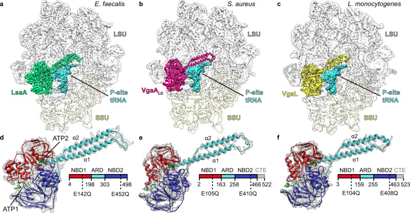
Fig. 1
Cryo-EM structures of ARE-ABCFu2013ribosome complexes.
a u2013 c Cryo-EM maps with isolated densities for a E. faecalis LsaA (green), b S. aureus VgaA LC (magenta), c L. monocytogenes VgaL (yellow) as well as P-site tRNA (cyan), small subunit (SSU, yellow...
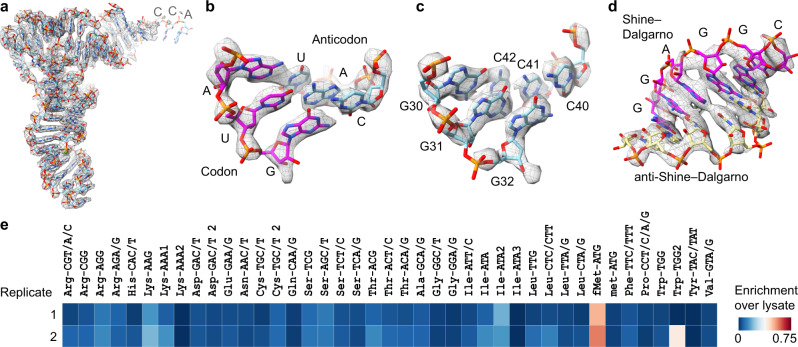
Fig. 2
The LsaA-70S complex contains an initiator tRNA and SD-helix.
a u2013 d Isolated density (grey mesh) with molecular models (sticks) for a initiator tRNA fMet (cyan), b interaction between AUG start codon of the mRNA (magenta) and anticodon of initiator tRNA fMet...
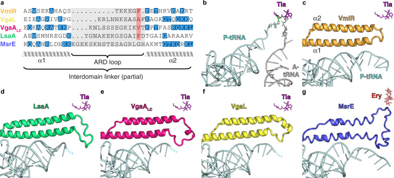
Fig. 3
Comparison of the ARD loops of different ARE-ABCFs.
a The sequence length of the ARD loops differs significantly for VmlR, VgaL, VgaA LC , LsaA and MsrE. Although the lack of sequence homology precludes accurate sequence alignment of the ARD loops, the...
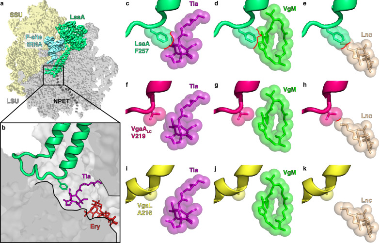
Fig. 4
Interaction of LsaA, VgaA LC and VgaL at the peptidyl transferase centre.
a u2013 b LsaA and distorted P-site tRNA superimposed on a transverse section of the large subunit (LSU, grey) to reveal a the ARD of LsaA extending into the nascent polypeptide exit tunnel (NPET) and...
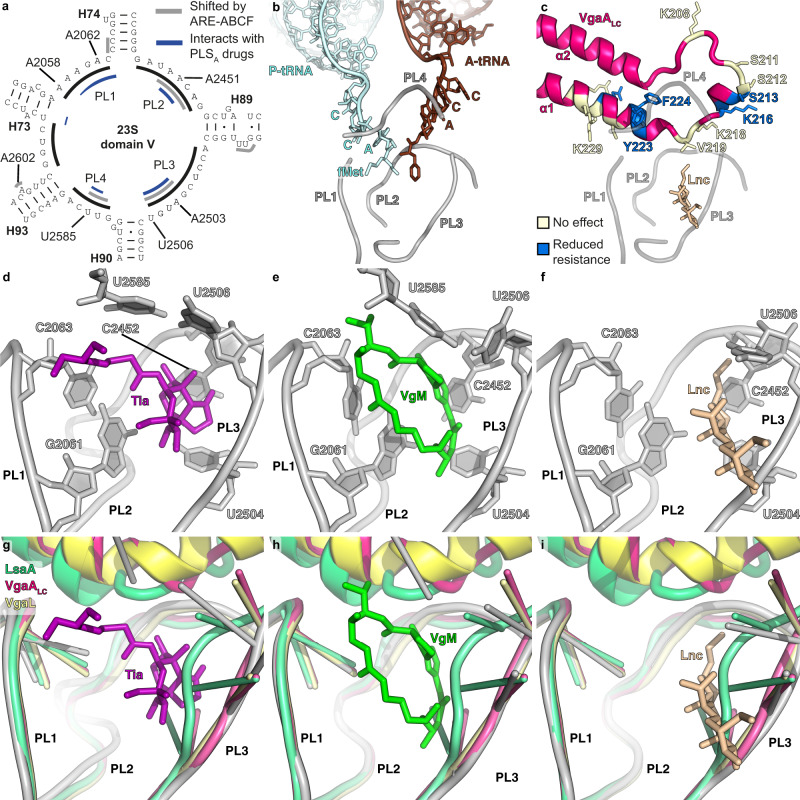
Fig. 5
ARE-ABCF binding induces conformational changes at the PTC.
a Secondary structure of peptidyl transferase ring within domain V of the 23S rRNA, highlighting residues within PTC loops 1u20134 (PL1u20134) that comprise the binding site of PLS A antibiotics (blue...
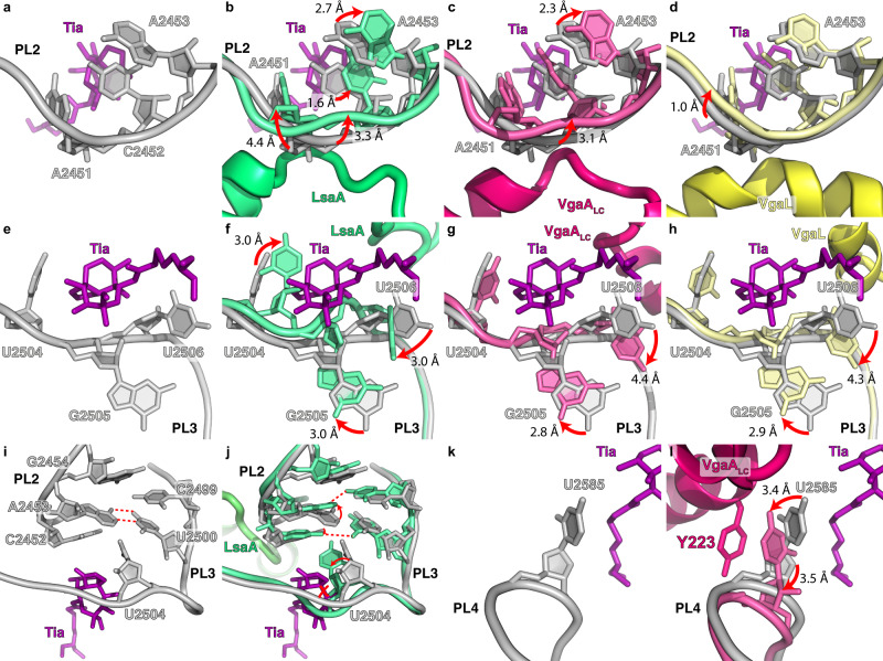
Fig. 6
Changes in the PTC induced by ARE-ABCF binding.
a u2013 d Effects of ARE binding on PL2 with respect to the tiamulin-binding site (PDB 1XBP) 2 . a The tiamulin-binding site only. b u2013 d Same as a but with the LsaA- ( b ), VgaA LC - ( c ), or Vga...
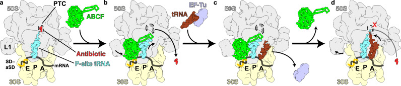
Fig. 7
Model for ribosome protection by ARE-ABCFs VmlR, LsaA, VgaA LC and VgaL.
a PLS A -stalled ribosomes containing an initiator tRNA in the P-site are recognized by the ARE-ABCFs such as VmlR, LsaA, VgaA LC and VgaL, which bind to the E-site of the ribosome with a closed ATP-b...
Figure images are served from the NIH/NLM PubMed Central Open Access Subset or Europe PMC; copyright remains with the publishers and authors.
💬 Discussion
0 commentsNo comments yet. Be the first to start a discussion!
Leave a Comment