Abstract
AbstractMany bacterial pathogens of animals and plants use a conserved type III secretion system (T3SS) to inject virulence effector proteins directly into eukaryotic cells to subvert host functions. Contact with host membranes is critical for T3SS activation, yet little is known about T3SS architecture in this state or the conformational changes that drive effector translocation. Here we use cryo-electron tomography and sub-tomogram averaging to derive the intact structure of the primordial Chlamydia trachomatis T3SS in the presence and absence of host membrane contact. Comparison of the averaged structures demonstrates a marked compaction of the basal body (4 nm) occurs when the needle tip contacts the host cell membrane. This compaction is coupled to a stabilization of the cytosolic sorting platform–ATPase. Our findings reveal the first structure of a bacterial T3SS from a major human pathogen engaged with a eukaryotic host, and reveal striking ‘pump-action’ conformational changes that underpin effector injection.
🔬 Techniques
✨ Fluorophores
🧪 Sample Preparation
🔬 Cell Lines
🏭 Microscope Brands
🧪 Reagent Suppliers
💻 Software Details
💾 Data Repositories
🏛️ Research Organizations (ROR)
Affiliated research institutions:
📋 Methods
Reagents, cell culture and Chlamydia propagation All cell culture reagents were purchased from Invitrogen. HeLa and U2OS cells were cultured in Dulbecco's modified eagle medium (high glucose with Glutamax) containing 10% fetal calf serum and penicillin–streptomycin. C. trachomatis LGV2 serovars were propagated in HeLa cells and stored at −80 °C in sucrose–phosphate–glutamate buffer 47 .
Sample preparation
For preparation of Chlamydia -infected cells for cryo-electron tomography, HeLa or U2OS cells were seeded into well plates and infected with C. trachomatis LGV2 (MOI 5) 18 . The following day adherent cells were seeded onto 200 mesh gold grids (R3.5/1; Quantifoil Micro Tools, Jena, Germany) at a density of one or two cells per grid square. At ∼48 h post infection, EM grids with or without host cells were introduced to the tissue culture dish and incubated with the newly released elementary body progeny (15 min–1 h at 37 °C). Grids were removed and rinsed in Hank's buffered salt solution. Four microlitres of BSA-coated gold (Sigma) was added to the grid before it was plunge frozen into liquid ethane (Vitrobot Mark IV, FEI). Salmonella enterica serovar Typhimurium strain ΔaraBAD1167::hilA+ Δtar-flhD2039 ΔminCDE::tetRA was grown in Luria-Bertani broth (LB) overnight at 37 °C with aeration. The following day, 50 μl of the overnight was subcultured into 5 ml LB and supplemented with 0.1% L -arabinose and 10 μg ml −1 tetracycline. After 5 h, rods and cellular debris were isolated by centrifugation at 2,000 g for 10 minutes at 4 °C. Minicells were isolated from the supernatant by centrifugation at 15,300 g for 10 min at 4 °C. The minicell pellet was resuspended in BSA-coated colloidal gold (Sigma) and 4 μl of the suspension was added to Quantifoil grids before being plunge frozen into liquid ethane.
Show full methods section
Reagents, cell culture and Chlamydia propagation All cell culture reagents were purchased from Invitrogen. HeLa and U2OS cells were cultured in Dulbecco's modified eagle medium (high glucose with Glutamax) containing 10% fetal calf serum and penicillin–streptomycin. C. trachomatis LGV2 serovars were propagated in HeLa cells and stored at −80 °C in sucrose–phosphate–glutamate buffer 47 .
Sample preparation
For preparation of Chlamydia -infected cells for cryo-electron tomography, HeLa or U2OS cells were seeded into well plates and infected with C. trachomatis LGV2 (MOI 5) 18 . The following day adherent cells were seeded onto 200 mesh gold grids (R3.5/1; Quantifoil Micro Tools, Jena, Germany) at a density of one or two cells per grid square. At ∼48 h post infection, EM grids with or without host cells were introduced to the tissue culture dish and incubated with the newly released elementary body progeny (15 min–1 h at 37 °C). Grids were removed and rinsed in Hank's buffered salt solution. Four microlitres of BSA-coated gold (Sigma) was added to the grid before it was plunge frozen into liquid ethane (Vitrobot Mark IV, FEI). Salmonella enterica serovar Typhimurium strain ΔaraBAD1167::hilA+ Δtar-flhD2039 ΔminCDE::tetRA was grown in Luria-Bertani broth (LB) overnight at 37 °C with aeration. The following day, 50 μl of the overnight was subcultured into 5 ml LB and supplemented with 0.1% L -arabinose and 10 μg ml −1 tetracycline. After 5 h, rods and cellular debris were isolated by centrifugation at 2,000 g for 10 minutes at 4 °C. Minicells were isolated from the supernatant by centrifugation at 15,300 g for 10 min at 4 °C. The minicell pellet was resuspended in BSA-coated colloidal gold (Sigma) and 4 μl of the suspension was added to Quantifoil grids before being plunge frozen into liquid ethane.
Cryo-electron tomography
For cryo-electron tomography, single-axis tilt series were collected with SerialEM 48 on a 300-kV Tecnai Polara electron microscope equipped with a K2 Summit direct electron detector and Quantum energy filter (Gatan). An energy window of 20 eV was used for recording zero-loss images and samples were maintained at liquid nitrogen temperatures. Images were recorded over a range of −60° to +45° with 3° increment at a 10-μm defocus and 5.4 Å pixel size. Tilt images were collected as eight sub-frames in electron-counting mode with a dose rate of six to seven electrons per pixel per second yielding a total dose of 1.5 electrons per Å 2 for each tilt image and a cumulative dose of 54 electrons per Å 2 for each tilt series. Sub-frames were aligned in Digital Micrograph before being incorporated into the final image stack in SerialEM. Tilt images were aligned in IMOD 49 using 10-nm gold fiducials and CTF correction was applied before tomogram reconstruction by weighted back-projection 50 . Nonlinear anisotropic filtering was applied to tomograms for sub-tomogram identification and selection 51 .
Image processing
Sub-tomogram positions were manually selected in IMOD and extracted from raw tomograms using the dtcrop function in Dynamo 52 . Initial alignment was performed manually with the Dynamo gallery. Further sub-tomogram alignment and averaging was conducted in Dynamo with a modification of splitting particles into two independent data sets for the final resolution measurement. First, asymmetric averages were generated by aligning the outer and the inner membranes separately. Six-fold rotational symmetry was detected in the sorting platform by rotational correlation ( Supplementary Fig. 6 ) and imposed for further refinement. Twelve-fold symmetry was applied to the Chlamydia and Salmonella basal body to increase the signal-to-noise ratio of the final structures, in accordance with symmetry determined by single-particle analysis 3 . Note that applying axial symmetry does not change interpretation of the data. After convergence, the final structures from each data set were merged at the midpoint of the volume and low-pass filtered to the detected resolution. Classification by multi-reference alignment was performed by first generating 5 or 10 initial references by adding 10% Gaussian noise to a global average (made from all ∼1,200 sub-tomograms) yielding a signal-to-noise ratio of 0.9. Starting from an initial global alignment, sub-tomograms were iteratively aligned to each reference using a bottle-like alignment mask that encompassed the basal body, inner membrane and sorting platform–ATPase. The maximum angular range used for searching was 6° and the maximum shift allowed was 5 voxels. After each round of alignment, sub-tomograms contribute once to the reference to which it had the highest correlation coefficient. Class averages were produced after convergence when the sub-tomograms no longer changed class memberships. Classification by principal component analysis and k-means did not cleanly separate sub-tomograms into groups with and without the sorting platform. Segmentation, surface renderings and measurements Automatic segmentation of membranes was performed with TomoSegMem 53 and refined manually in Amira (FEI Visualization Sciences Group, Massachusetts, USA). Segmentation of actin filaments was conducted in IMOD. The EM Package for Amira was used to map individual T3SS models into their corresponding positions in cryo-electron tomograms 54 . Multiple sequence alignments were produced by ClustalW Omega 55 and graphically illustrated with BOXSHADE. Distances between the bacterial outer membrane and host membrane were measured in individual tomographic slices where a T3SS needle contact was clearly resolved. Measurements were collected in IMOD by drawing a line along the T3SS needle from the centre of the outer membrane to the centre of the plasma membrane and calculating the number of pixels the line spanned. For non-T3SS-mediated contacts, measurements were collected from tomographic slices that depicted a constant spacing between the non-T3SS containing bacterial hemisphere and the host plasma membrane. A line perpendicular to the apposed membranes was drawn in IMOD and converted to distance in pixels.
Supplementary Material Supplementary Information Supplementary Figures 1-7 Supplementary Movie 1 Transition of the Chlamydia type III secretion system from the host-free to host-contact state. Basal body compaction and sorting platform-ATPase complex stabilization visualized as a linear morph between the host-free and host-active sub-tomogram averages. The video was created using the iMorph plugin for Fiji.
📊 Figures
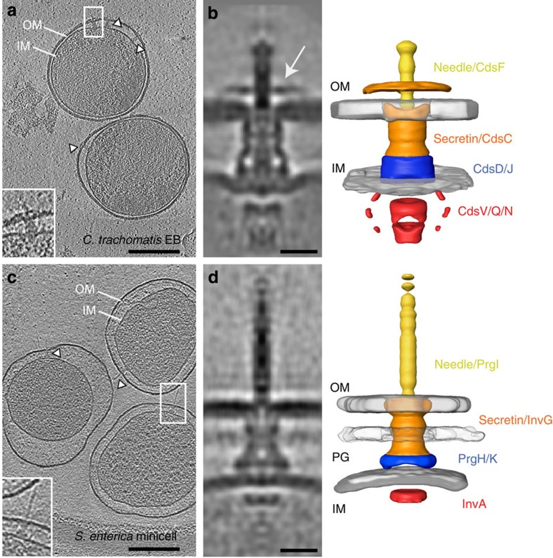
Figure 1
In situ sub-tomogram averages of host-free Chlamydia and Salmonella T3SS.
Central slices (10 nm thick) from denoised cryo-electron tomograms of plunge-frozen C. trachomatis LGV2 elementary bodies ( a ) and S. enterica minicells ( c ). Outer (OM) and inner membranes (IM), pe...
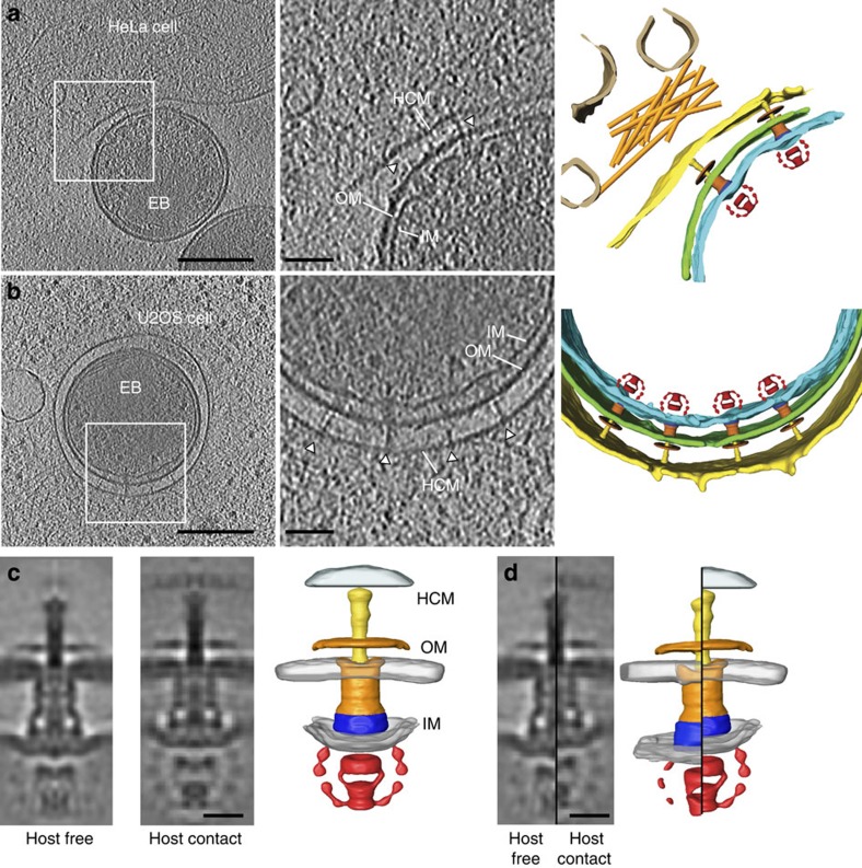
Figure 2
In situ structure of host-contact Chlamydia T3SS.
( a , b ) Central slices (10 nm thick) through denoised cryo-electron tomograms of an elementary body bound to a HeLa cell plasma membrane via a type III secretion system ( a ) and within a vacuole of...
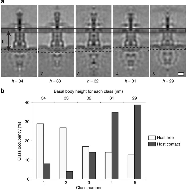
Figure 3
Structural variability of Chlamydia T3SS sub-tomograms by multi-reference alignment.
( a ) Central slices through five class averages of Chlamydia T3SSs generated by non-biased multi-reference alignment in Dynamo. The class averages are sorted by basal body height ( h ), which varies ...
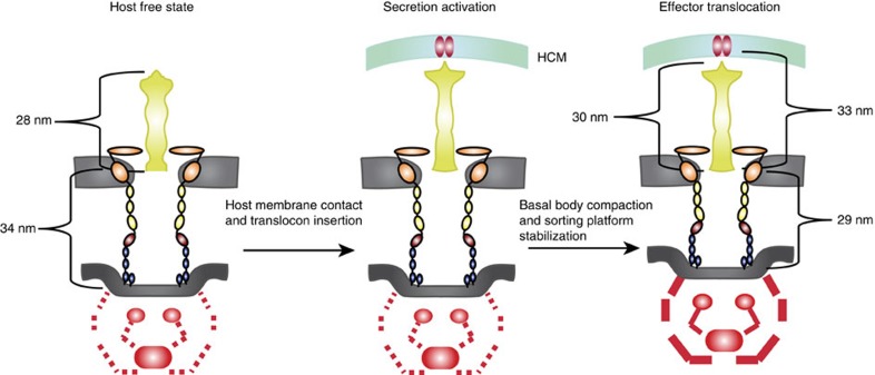
Figure 4
Model for activation of type III secretion.
(Left) The Chlamydia type III secretion system basal body is composed of inner membrane rings (CdsD and CdsJ; blue) and together with the outer membrane protein secretin (CdsC) form a multimeric chann...
Figure images are served from the NIH/NLM PubMed Central Open Access Subset or Europe PMC; copyright remains with the publishers and authors.
💬 Discussion
0 commentsNo comments yet. Be the first to start a discussion!
Leave a Comment