Abstract
Increasing evidences show that the actin cytoskeleton is a key parameter of the nuclear remodeling process in response to the modifications of cellular morphology. However, detailed information on the interaction between the actin cytoskeleton and the nuclear lamina was still lacking. We addressed this question by constraining endothelial cells on rectangular fibronectin-coated micropatterns and then using Structured Illumination Microscopy (SIM) to observe the interactions between actin stress fibers, nuclear lamina and LINC complexes at a super-resolution scale. Our results show that tension in apical actin stress fibers leads to deep nuclear indentations that significantly deform the nuclear lamina. Interestingly, indented nuclear zones are characterized by a local enrichment of LINC complexes, which anchor apical actin fibers to the nuclear lamina. Moreover, our findings indicate that nuclear indentations induce the formation of segregated domains of condensed chromatin. However, nuclear indentations and condensed chromatin domains are not irreversible processes and both can relax in absence of tension in apical actin stress fibers.
🔬 Techniques
💻 Software
✨ Fluorophores
🧪 Sample Preparation
🔬 Cell Lines
🏭 Microscope Brands
🧪 Reagent Suppliers
💻 Software Details
💾 Data Repositories
🏛️ Research Organizations (ROR)
Affiliated research institutions:
📋 Methods
Microcontact printing Circular and rectangular (aspect ratio of 1:10) microfeatures of 1600 μm 2 were drawn with the Clewin software and generated to a silicon master by deep reactive-ion etching (FH Vorarlberg University of Applied Sciences, Microtechnology, Dornbirn, Austria). The silicon surface was passivated under vacuum with vapors of fluorosilane (tridecafluoro-1,1,2,2-tetrahydrooctyl-1-trichlorosilane) for 30 min, and then molded with polydimethylsiloxane, PDMS, (Sylgard 184 Silicone Elastomer Kit; Dow Corning, Midland, MI). After 4 h of curing at 60°C, the PDMS layer was peeled off and stamps of 1 cm 2 were cut manually. The structured surface of the PDMS stamps were first oxidized in an UV/O 3 cleaner for 8 min and then inked for 1 h at room temperature with a 25 μg.ml −1 fibronectin (FN) solution from human plasma. FN-coated stamps were dried under filtered nitrogen and gently deposited on a flat PDMS-coated glass coverslip for 15 seconds, as described previously 42 43 . Uncoated regions were blocked by incubating micropatterned coverslips for 5 min in a 1% solution of Pluronic F-127 44 .
Cell culture
Primary human umbilical vein endothelial cells, HUVECs (Cells Applications, San Diego, CA) were grown in complete endothelial cell growth medium (Cells Applications, San Diego, CA) supplemented with 1% antibiotics and antimycotics and maintained at 37°C in a humidified atmosphere with 5% CO 2 . Cells between passages 2 and 8 were cultured on microprinted PDMS coverslips at a concentration of about 15,000 cells ml −1 . Immunohistochemistry and labeling After 24 h in culture on micropatterned substrates, HUVECs were fixed and permeabilized with 4% paraformaldehyde, 0.05% Triton X-100 in Phosphate buffered saline (PBS) for 15 min at 37°C and then washed three times in PBS. Fixed cells were incubated for 30 min at room temperature in a blocking solution (FBS 5%, BSA 1%) and labeled for F-actin (Alexa Fluor 488 Phalloidin 1:200, Molecular Probes), nuclear lamina (anti-lamin A/C antibody produced in mouse 1:200, Sigma-Aldrich SAB4200236) and LINC-complex (anti Syne-2 antibody produced in rabbit 1:100, Sigma-Aldrich HPA003435) for 45 min at 37°C in PBS. After three successive washes in PBS, cells were incubated for 45 min at 37°C with an anti-mouse antibody produced in goat and labeled with tetramethylrhodamine (Molecular Probes, Invitrogen, T2762), an anti-mouse antibody produced in rabbit and labeled with fluorescein isothiocyanate (Sigma-Aldrich, F9137) or an anti-rabbit antibody produced in goat an labeled with tetramethylrhodamine (Sigma-Aldrich, T6778). All secondary antibodies were used in a 1:200 concentration in PBS. Slides were mounted in Slow Fade Gold Antifade (Molecular Probes). Living cell relaxation To observe nuclear DNA in living cells, HUVECs were transduced with the BacMam 2.0 CellLight Histone 2B-GFP according to the manufacturer's protocol (Molecular Probes). Routine culture flasks were incubated with approximately 20 BacMam particles/cell for at least 24 hours. Labeled cells were then detached and grown on rectangular FN micropatterns (1:10 aspect ratio) for 24 hours and finally transferred to an inverted confocal microscope equipped with a temperature and CO 2 level controller. The GFP fluorescence signal was recorded during the relaxation of the elongated cells, which was induced by the addition of 1 ml of accutase after a gentle PBS wash.
Show full methods section
Microcontact printing Circular and rectangular (aspect ratio of 1:10) microfeatures of 1600 μm 2 were drawn with the Clewin software and generated to a silicon master by deep reactive-ion etching (FH Vorarlberg University of Applied Sciences, Microtechnology, Dornbirn, Austria). The silicon surface was passivated under vacuum with vapors of fluorosilane (tridecafluoro-1,1,2,2-tetrahydrooctyl-1-trichlorosilane) for 30 min, and then molded with polydimethylsiloxane, PDMS, (Sylgard 184 Silicone Elastomer Kit; Dow Corning, Midland, MI). After 4 h of curing at 60°C, the PDMS layer was peeled off and stamps of 1 cm 2 were cut manually. The structured surface of the PDMS stamps were first oxidized in an UV/O 3 cleaner for 8 min and then inked for 1 h at room temperature with a 25 μg.ml −1 fibronectin (FN) solution from human plasma. FN-coated stamps were dried under filtered nitrogen and gently deposited on a flat PDMS-coated glass coverslip for 15 seconds, as described previously 42 43 . Uncoated regions were blocked by incubating micropatterned coverslips for 5 min in a 1% solution of Pluronic F-127 44 .
Cell culture
Primary human umbilical vein endothelial cells, HUVECs (Cells Applications, San Diego, CA) were grown in complete endothelial cell growth medium (Cells Applications, San Diego, CA) supplemented with 1% antibiotics and antimycotics and maintained at 37°C in a humidified atmosphere with 5% CO 2 . Cells between passages 2 and 8 were cultured on microprinted PDMS coverslips at a concentration of about 15,000 cells ml −1 . Immunohistochemistry and labeling After 24 h in culture on micropatterned substrates, HUVECs were fixed and permeabilized with 4% paraformaldehyde, 0.05% Triton X-100 in Phosphate buffered saline (PBS) for 15 min at 37°C and then washed three times in PBS. Fixed cells were incubated for 30 min at room temperature in a blocking solution (FBS 5%, BSA 1%) and labeled for F-actin (Alexa Fluor 488 Phalloidin 1:200, Molecular Probes), nuclear lamina (anti-lamin A/C antibody produced in mouse 1:200, Sigma-Aldrich SAB4200236) and LINC-complex (anti Syne-2 antibody produced in rabbit 1:100, Sigma-Aldrich HPA003435) for 45 min at 37°C in PBS. After three successive washes in PBS, cells were incubated for 45 min at 37°C with an anti-mouse antibody produced in goat and labeled with tetramethylrhodamine (Molecular Probes, Invitrogen, T2762), an anti-mouse antibody produced in rabbit and labeled with fluorescein isothiocyanate (Sigma-Aldrich, F9137) or an anti-rabbit antibody produced in goat an labeled with tetramethylrhodamine (Sigma-Aldrich, T6778). All secondary antibodies were used in a 1:200 concentration in PBS. Slides were mounted in Slow Fade Gold Antifade (Molecular Probes). Living cell relaxation To observe nuclear DNA in living cells, HUVECs were transduced with the BacMam 2.0 CellLight Histone 2B-GFP according to the manufacturer's protocol (Molecular Probes). Routine culture flasks were incubated with approximately 20 BacMam particles/cell for at least 24 hours. Labeled cells were then detached and grown on rectangular FN micropatterns (1:10 aspect ratio) for 24 hours and finally transferred to an inverted confocal microscope equipped with a temperature and CO 2 level controller. The GFP fluorescence signal was recorded during the relaxation of the elongated cells, which was induced by the addition of 1 ml of accutase after a gentle PBS wash.
Structured illumination microscopy and confocal imaging
Immunostained preparations were observed in Structured Illumination Microscopy (SIM) performed on an Eclipse Ti inverted microscope equipped with a Nikon Plan Apo ×100 TIRF objective (NA 1.49, oil immersion) and an Andor DU-897X-5254 camera. Z-step size was set to 0.120 um, which is well within the Nyquist criterion. For each focal plane, 15 images (5 phases, 3 angles) were captured with the NIS-Elements software. SIM image processing, reconstruction and analysis were carried out using the N-SIM module of the NIS-Element Advanced Research software. Confocal imaging was performed with an inverted Nikon Eclipse Ti-E motorized microscope (Nikon C1 scanhead; Nikon, Japan) equipped with Plan Apo ×60 and ×100 (NA 1.45, oil immersion) objectives, two lasers (Ar ion 488 nm; HeNe, 543 nm) and a modulable diode (408 nm). Confocal images were acquired with NIS Elements Advanced Research 3.0 software (Nikon) by using small Z-depth increments between focal sections (0.150 μm).
Image analysis
Confocal and SIM images were acquired and analyzed with the NIS-Elements Advanced Research 3.0 software. Three-dimensional normal views were obtained in maximum intensity projection mode. Unless otherwise stated, orthogonal views were obtained in maximum intensity projection mode and cropped to represent a one micron-length of the nucleus. LINC complexes quantifications were obtained after the thresholding and the binarization of SIM slides or Extended Depth of Focus images for actin co-localization or quantification on the whole nuclear projected area, respectively. Nuclear compression experiments We used circular glass coverslips of 22 mm in diameter, 170 μm thick and 0.136 grams to apply a homogeneous pressure on the nucleus. After 20 hours in culture on FN-coated circular micropatterns, a glass coverslip was placed over HUVECs during 6 hours. The top side of glass coverslips was coated with a 1% solution of pluronic F-127 to avoid cellular adhesion and exerted a normal pressure proportional to its weight.
Data presentation and statistics
Results are presented as the means ± Standard Deviation (SD). Student's two-tailed t -test was performed in Origin 8.0 where two groups were compared. Values of p < 0.05 were considered statistically significant.
Supplementary Material Supplementary Information Supplementary Information Supplementary Information Movie S1 Supplementary Information Movie S2 Supplementary Information Movie S3 Supplementary Information Movie S4 Supplementary Information Movie S5
📊 Figures
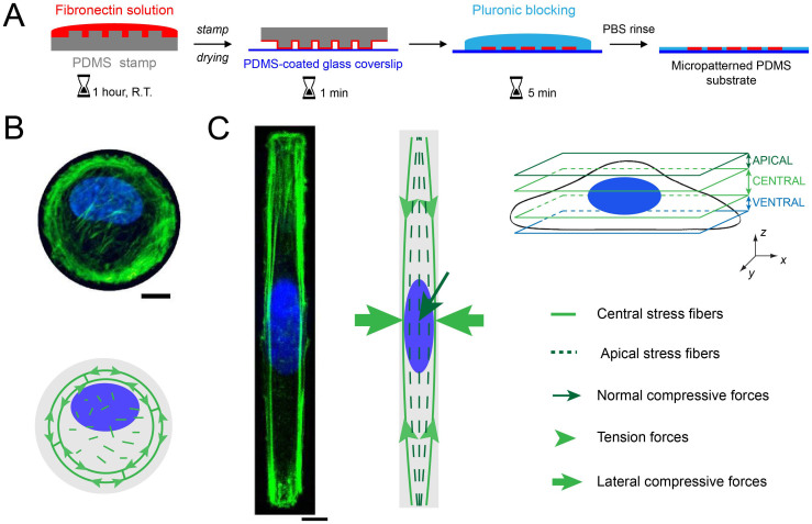
Figure 1
The control of the cell morphology by microcontact printing induces a modification of the nuclear shape.
(A) Successive steps of the microcontact printing method used to create circular and rectangular micropatterns of fibronectin (in red) on a glass coverslip coated with a layer of polydimethylsiloxane ...
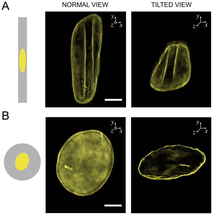
Figure 2
The top of the nucleus in elongated cells presents linear deformations of the nuclear lamina.
Normal views (left column) and tilted views (right column) obtained from 3D-SIM imaging of the nuclear lamina (in yellow) of (A) a rectangular-shaped and (B) a circular-shaped endothelial cells. Scale...
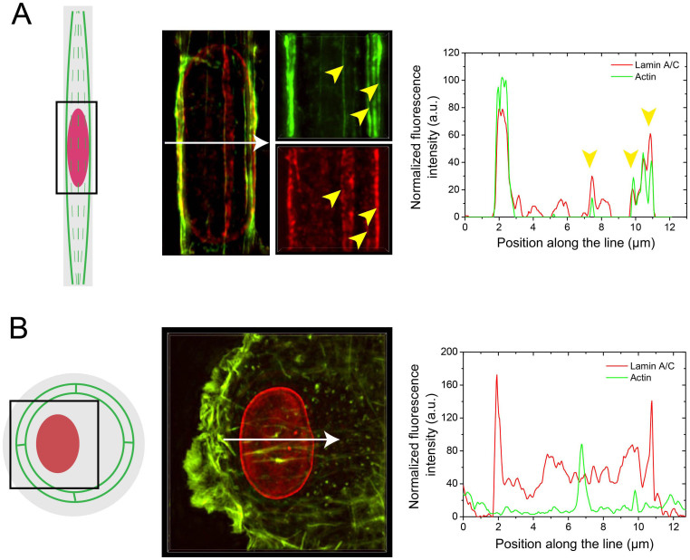
Figure 3
Nuclear lamina deformations correspond to the localization of apical stress fibers.
(A) Maximum intensity projections obtained from 3D-SIM microscopy of the actin microfilaments (in green) and the nuclear lamina (in red) of a single endothelial cell spread on a FN-coated rectangular ...
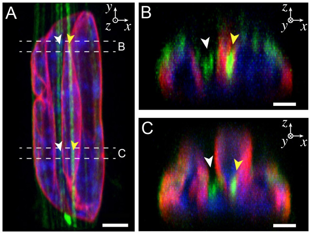
Figure 4
Apical actin fibers are located at the bottom of indentation sites that form deep valleys in the nucleus.
(A) Normal confocal view (XY) of an elongated nucleus indented by straight apical actin fibers. Lamins A/C are in red, DNA in blue and actin in green. Two cross-sectional views (XZ) obtained from the ...
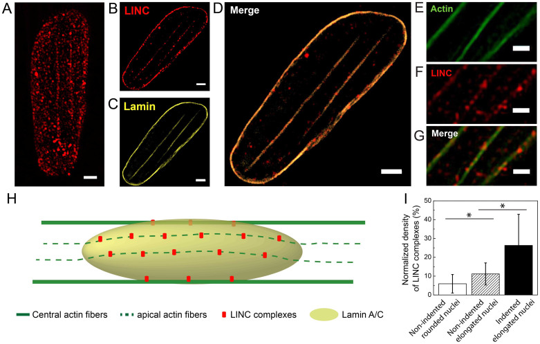
Figure 5
The density of LINC complexes is enriched at the sites of nuclear lamina indentations.
(A) SIM images of LINC complexes labeled with Syne-2 antibody (red) in a deformed nucleus. A focal plane in SIM images located in the central zone was selected to show (B) LINC complexes fluorescence ...
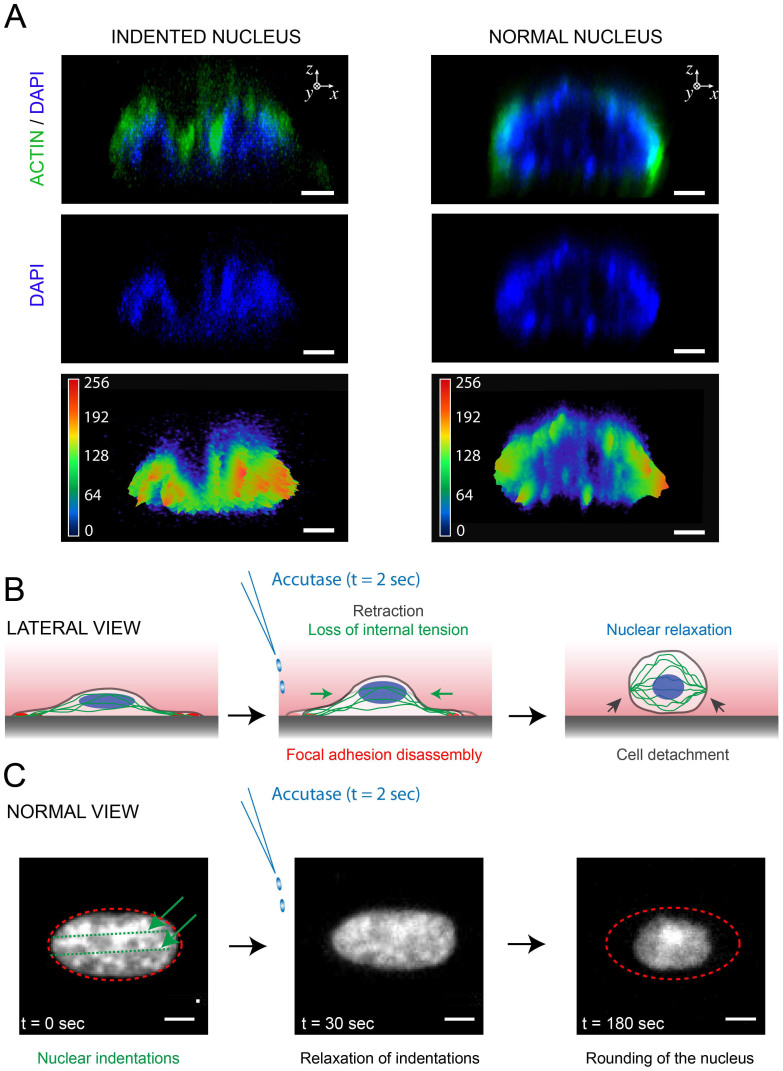
Figure 6
Indentation of the nuclear lamina induces chromatin segregation, which is a reversible process.
(A) 3D confocal cross-section views (XZ) of indented (left column) and normal (right column) nuclei show the formation of rich chromatin domains within an indented nucleus. DNA is in blue and actin fi...
Figure images are served from the NIH/NLM PubMed Central Open Access Subset or Europe PMC; copyright remains with the publishers and authors.
💬 Discussion
0 commentsNo comments yet. Be the first to start a discussion!
Leave a Comment