Abstract
AbstractLight is extensively used to study cells in real time (live cell imaging), separate cells using fluorescence activated cell sorting (FACS) and control cellular functions with light sensitive proteins (Optogenetics). However, photo-sensitive molecules inside cells and in standard cell culture media generate toxic by-products that interfere with cellular functions and cell viability when exposed to light. Here we show that primary cells from the rat central nervous system respond differently to photo-toxicity, in that astrocytes and microglia undergo morphological changes, while in developing neurons and oligodendrocyte progenitor cells (OPCs) it induces cellular death. To prevent photo-toxicity and to allow for long-term photo-stimulation without causing cellular damage, we formulated new photo-inert media called MEMO and NEUMO, and an antioxidant rich and serum free supplement called SOS. These new media reduced the detrimental effects caused by light and allowed cells to endure up to twenty times more light exposure without adverse effects, thus bypassing the optical constraints previously limiting experiments.
🔬 Techniques
🔭 Microscopes
💻 Software
✨ Fluorophores
🧪 Sample Preparation
🔬 Cell Lines
🏭 Microscope Brands
🧪 Reagent Suppliers
💻 Software Details
🏛️ Research Organizations (ROR)
Affiliated research institutions:
📋 Methods
Media formulation Modified Eagle’s Medium for Optogenetics
(MEMO) and NEUronal Media for Optogenetics (NEUMO) were developed by screening available formulations for DMEM and Neurobasal for photo-reactive components using cell viability assays and literature searches. Supplements for Optogenetic Survival (SOS) is formulated and based upon the components of the serum free supplements B27 36 , NS21 34 and N2 35 with removal of triiodo-L-thyronine (T3) and thyroxine (T4) and with additional proprietary modifications. MEMO, NEUMO and SOS were made in house, or sourced from Cell Guidance Systems; UK, as part of their LiveLight package and used according to manufacturer’s instructions. Cell-culture Unless otherwise stated all compounds were sourced from Sigma-Aldrich; UK. All cells were cultured at 37 °C in a humidified atmosphere containing 5% CO 2 . Primary mixed glial cultures and OPCs were prepared as described previously 54 . Briefly, brains from P2 postnatal rats were dissected, meninges removed, cortices chopped and digested with papain (Worthington Biochemical Corporation, USA) for 1 hour, followed with ovomucoid (0.5 mg/ml BSA, 1 mg/ml trypsin inhibitor, 40 µg/ml DNase I Type IV, 1% Penicillin and Streptomycin (Pen/Strep) in DMEM), trituration, and seeded as 3 cortical hemispheres per poly-D-lysine (PDL) coated T-75 flasks or glass coverslips. Mixed glia were grown for 10–12 days in vitro (d.i.v.) in DMEM with 10% fetal calf serum (FCS) (Life Technologies; UK) and 1% Pen/Strep. OPCs and microglia were separated from astrocytes by mechanical dissociation (175 r.p.m. on ~19 mm diameter (Ø) orbital shaker) for 16 hours at 37 °C. Microglia were enriched using adherence to non-treated dishes (Corning; UK) (15 ml/10 cm plate) for 15 min, and OPCs enriched by centrifuging (250 g max for 8 minutes) the remaining suspension. OPCs were resuspended in DMEM with modified SATO (100 µg/ml BSA, 60 ng/ml progesterone, 16.1 µg/ml putrescine, 5 ng/ml sodium selenite, 5 µg/ml insulin, 5 µg/ml N-acetyl-L-cysteine, 50 µg/ml holo-transferrin and 1% Pen/Strep) and growth factors (PDGF-aa at 10 ng/ml and FGF-b at 10 ng/ml; Peprotech; UK). OPCs were seeded at a density of 22 × 10 3 cells/cm 2 onto PDL coated glass coverslips or flasks with daily addition of growth factors yielding 86 ± 1.8% (n = 18) NG2 + cells after 3 days of proliferation. Microglia were dissociated from non-treated dishes with TrypLE Express (Life Technologies; UK), and seeded onto 22 mm Ø PDL coated glass coverslips (450,000 cells/coverslip) in 10% FCS, 1% Pen/Strep in DMEM yielding 87.3 ± 7.5% (n = 5) IB4 + cells. For primary neurons, cortices from E18 rat embryos were chopped and digested with papain for 15 min followed with ovomucoid in DMEM or Ca 2+ and Mg 2+ free EBSS, and then triturated using fire polished glass pipettes. Cells were clarified over a 4% BSA step in ovomucoid and seeded onto PDL coated glass coverslips in 6 well plates at 1 × 10 6 cells per plate in neuronal medium (Neurobasal, 2% B27 and 1% Glutamax; Life Technologies; UK) with 1% Pen/Strep and supplemented with 5% FCS. The next day media was changed to neuronal medium without FCS yielding 80 ± 2.8% β-III-tubulin + neurons and 5.75 ± 1.9% GFAP + astrocytes (n = 3). None of the cells we examined were modified to express light sensitive proteins such as ChR2, or recombinant proteins (such as GFP). OPC proliferation, migration, and differentiation assays OPCs were plated onto PDL coated coverslips in DMEM + SATO with growth factors (PDGF-aa and FGF-b) left overnight and placed into either DMEM + SATO or MEMO + SATO, with daily mitogen addition for 3 days. To measure proliferation, fixed cells were immunostained for NG2 and Ki67 co-localization. Alternatively, 100 nM EdU was applied 24 hours prior to fixation and detected according to manufacturer’s instructions (Life Technologies; UK). Migration assays were performed using an agarose drop assay 39 . OPCs were proliferated, dissociated and re-seeded as 1.5 µl drops of 1 × 10 6 cells/µl in 1% w/v low melting point agarose, onto PDL and matrigel (BD Biosciences; UK) coated coverslips. Plated drops were cooled at 4° C for 15 min, then 50 µl of 10% FCS in DMEM added, and the well flooded with MEMO + SATO or DMEM + SATO media, with growth factors, and fixed 4 days later. Migration distance (distance from the drop edge to the corona of migrated cells) was calculated from five photo-micrographs of each drop taken with a Zeiss AxioVision digital microscope. Data shown are of at least three replicates. For differentiation experiments, OPCs on PDL coated coverslips in DMEM + SATO media with T3 (400 ng/ml), were placed in either DMEM + SATO + T3 or MEMO + SATO + T3 and cells allowed to differentiate for 5 days. Terminal differentiation was detected with MBP immunostaining as described below. iPSC differentiation Human induced pluripotent stem cells (hiPSCs) following proliferation with FGF-2 (20 µg/ml) and heparin (2 µg/ml), were treated with a combination of retinoic acid (100 nM) and the sonic-hedgehog analog, puromorphamine (PM, 1 µM) for 15 days. Cells were then cultured as an embryonic body (EB) suspension in PM for another 11 days. After completion of this stage, cells were kept in DMEM:F12 composition media containing glia stimulating growth factors (60 ng/ml T3; 10 ng/ml PDGF-aa; 10 ng/ml IGF1; 10 ng/ml NT3; 1 µM cAMP; and 100 ng/ml d-biotin) for 50–100 d.i.v. Terminal plating of gliogenic EBs was on poly-ornithine/laminin-coated glass coverslips. Terminally plated iPSC-derived gliogenic EBs were grown in a DMEM:F12 formulation media with either N2 + B27 or SOS for five weeks, then fixed and stained for GFAP and β-III-tubulin as below.
Show full methods section
Media formulation Modified Eagle’s Medium for Optogenetics
(MEMO) and NEUronal Media for Optogenetics (NEUMO) were developed by screening available formulations for DMEM and Neurobasal for photo-reactive components using cell viability assays and literature searches. Supplements for Optogenetic Survival (SOS) is formulated and based upon the components of the serum free supplements B27 36 , NS21 34 and N2 35 with removal of triiodo-L-thyronine (T3) and thyroxine (T4) and with additional proprietary modifications. MEMO, NEUMO and SOS were made in house, or sourced from Cell Guidance Systems; UK, as part of their LiveLight package and used according to manufacturer’s instructions. Cell-culture Unless otherwise stated all compounds were sourced from Sigma-Aldrich; UK. All cells were cultured at 37 °C in a humidified atmosphere containing 5% CO 2 . Primary mixed glial cultures and OPCs were prepared as described previously 54 . Briefly, brains from P2 postnatal rats were dissected, meninges removed, cortices chopped and digested with papain (Worthington Biochemical Corporation, USA) for 1 hour, followed with ovomucoid (0.5 mg/ml BSA, 1 mg/ml trypsin inhibitor, 40 µg/ml DNase I Type IV, 1% Penicillin and Streptomycin (Pen/Strep) in DMEM), trituration, and seeded as 3 cortical hemispheres per poly-D-lysine (PDL) coated T-75 flasks or glass coverslips. Mixed glia were grown for 10–12 days in vitro (d.i.v.) in DMEM with 10% fetal calf serum (FCS) (Life Technologies; UK) and 1% Pen/Strep. OPCs and microglia were separated from astrocytes by mechanical dissociation (175 r.p.m. on ~19 mm diameter (Ø) orbital shaker) for 16 hours at 37 °C. Microglia were enriched using adherence to non-treated dishes (Corning; UK) (15 ml/10 cm plate) for 15 min, and OPCs enriched by centrifuging (250 g max for 8 minutes) the remaining suspension. OPCs were resuspended in DMEM with modified SATO (100 µg/ml BSA, 60 ng/ml progesterone, 16.1 µg/ml putrescine, 5 ng/ml sodium selenite, 5 µg/ml insulin, 5 µg/ml N-acetyl-L-cysteine, 50 µg/ml holo-transferrin and 1% Pen/Strep) and growth factors (PDGF-aa at 10 ng/ml and FGF-b at 10 ng/ml; Peprotech; UK). OPCs were seeded at a density of 22 × 10 3 cells/cm 2 onto PDL coated glass coverslips or flasks with daily addition of growth factors yielding 86 ± 1.8% (n = 18) NG2 + cells after 3 days of proliferation. Microglia were dissociated from non-treated dishes with TrypLE Express (Life Technologies; UK), and seeded onto 22 mm Ø PDL coated glass coverslips (450,000 cells/coverslip) in 10% FCS, 1% Pen/Strep in DMEM yielding 87.3 ± 7.5% (n = 5) IB4 + cells. For primary neurons, cortices from E18 rat embryos were chopped and digested with papain for 15 min followed with ovomucoid in DMEM or Ca 2+ and Mg 2+ free EBSS, and then triturated using fire polished glass pipettes. Cells were clarified over a 4% BSA step in ovomucoid and seeded onto PDL coated glass coverslips in 6 well plates at 1 × 10 6 cells per plate in neuronal medium (Neurobasal, 2% B27 and 1% Glutamax; Life Technologies; UK) with 1% Pen/Strep and supplemented with 5% FCS. The next day media was changed to neuronal medium without FCS yielding 80 ± 2.8% β-III-tubulin + neurons and 5.75 ± 1.9% GFAP + astrocytes (n = 3). None of the cells we examined were modified to express light sensitive proteins such as ChR2, or recombinant proteins (such as GFP). OPC proliferation, migration, and differentiation assays OPCs were plated onto PDL coated coverslips in DMEM + SATO with growth factors (PDGF-aa and FGF-b) left overnight and placed into either DMEM + SATO or MEMO + SATO, with daily mitogen addition for 3 days. To measure proliferation, fixed cells were immunostained for NG2 and Ki67 co-localization. Alternatively, 100 nM EdU was applied 24 hours prior to fixation and detected according to manufacturer’s instructions (Life Technologies; UK). Migration assays were performed using an agarose drop assay 39 . OPCs were proliferated, dissociated and re-seeded as 1.5 µl drops of 1 × 10 6 cells/µl in 1% w/v low melting point agarose, onto PDL and matrigel (BD Biosciences; UK) coated coverslips. Plated drops were cooled at 4° C for 15 min, then 50 µl of 10% FCS in DMEM added, and the well flooded with MEMO + SATO or DMEM + SATO media, with growth factors, and fixed 4 days later. Migration distance (distance from the drop edge to the corona of migrated cells) was calculated from five photo-micrographs of each drop taken with a Zeiss AxioVision digital microscope. Data shown are of at least three replicates. For differentiation experiments, OPCs on PDL coated coverslips in DMEM + SATO media with T3 (400 ng/ml), were placed in either DMEM + SATO + T3 or MEMO + SATO + T3 and cells allowed to differentiate for 5 days. Terminal differentiation was detected with MBP immunostaining as described below. iPSC differentiation Human induced pluripotent stem cells (hiPSCs) following proliferation with FGF-2 (20 µg/ml) and heparin (2 µg/ml), were treated with a combination of retinoic acid (100 nM) and the sonic-hedgehog analog, puromorphamine (PM, 1 µM) for 15 days. Cells were then cultured as an embryonic body (EB) suspension in PM for another 11 days. After completion of this stage, cells were kept in DMEM:F12 composition media containing glia stimulating growth factors (60 ng/ml T3; 10 ng/ml PDGF-aa; 10 ng/ml IGF1; 10 ng/ml NT3; 1 µM cAMP; and 100 ng/ml d-biotin) for 50–100 d.i.v. Terminal plating of gliogenic EBs was on poly-ornithine/laminin-coated glass coverslips. Terminally plated iPSC-derived gliogenic EBs were grown in a DMEM:F12 formulation media with either N2 + B27 or SOS for five weeks, then fixed and stained for GFAP and β-III-tubulin as below.
Light toxicity assays
Cells were irradiated with blue light (470 ± 10 nm) emitted from an LED plate; controlling power ( W : 0.1–1.5 mW/mm 2 at cell surface), flash duration (τ: 1–10 ms) and frequency ( f : 0.1–90 Hz) from an electronic stimulator housed outside the incubator (Fig. 1a ). Light power was measured using a LaserCheck photo-diode (Coherent; UK). Incubator temperature was adjusted to maintain 37 °C in the cell culture media using thermocouple measurements and placing the 6 well plate onto a metal heat dissipater. For light toxicity media comparison experiments, cells were transferred into DMEM or MEMO based media with mitogens for OPCs, or Neurobasal or NEUMO based media for neurons and astrocytes, for 4 hours prior to light treatment and with either SATO, 2% B27 or SOS. For the 2160 kJ/m 2 OPC stimulation experiments, cells were plated on PDL and matrigel coated coverslips and allowed to proliferate for three days, before transferring coverslips into MEMO + SOS media with growth factors, and allowed to equilibrate for 4 hours before light treatment. Non-viable cells were detected by adding 10 µg/ml propidium iodide (PI) (Life-Technologies; UK) to cells for 20 min prior to fixation.
Immunocytochemistry and fluorescence microscopy
All washes and incubation steps were performed in PBS with 0.01% w/v sodium azide (PBS-NaN 3 ) unless otherwise stated. Cell cultures were fixed for 15 min at RT in 4% w/v paraformaldehyde (PFA) in PBS. Fixed cells were blocked and permeabilized with 0.1% Triton X-100 with 10% v/v goat serum (Vector Laboratories; UK) in PBS for 1 hour at RT. Primary and secondary antibody sources and dilutions are shown in Table 1 . Primary antibody incubations were overnight at 4° C, followed with PBS wash, secondary antibody incubations at RT for 1 hour followed with PBS wash, stained for 10 min at RT with DAPI or Hoechst-33342 (Life Technologies; UK) followed with PBS wash prior to mounting in Mowiol with 2.5% w/v DABCO. Omission of primary antibodies was used to verify specificity as control in all experiments. Photo-micrographs were taken with using Leica SP5 confocal or Leica DM inverted microscopes (Leica; UK). Table 1 . Primary Antibodies Target Source Species Dilution NG2 Millipore; UK Rabbit 1:200 NG2 Millipore; UK Mouse 1:100 MBP Serotec; UK Rat 1:100 IB4-Alexa-488* Life-Technologies; UK N/A 1:100 Ki67 Thermo-Scientific; UK Rabbit 1:200 β -III-Tubulin Sigma-Aldrich; UK Mouse 1:100 GFAP Dako: UK Rabbit 1:500 GFAP Sigma-Aldrich; UK Mouse 1:500 Secondary Antibodies Target Source Fluorophore- λ ex Dilution Goat-Anti-Rabbit Life-Technologies; UK Alexa-488 1:1000 Goat-Anti-Mouse Life-Technologies; UK Alexa-488 1:1000 Goat-Anti-Rat Life-Technologies; UK Alexa-488 1:1000 Goat-Anti-Rabbit Life-Technologies; UK Alexa-555 1:1000 Goat-Anti-Mouse Life-Technologies; UK Alexa-555 1:1000 Goat-Anti-Rabbit Life-Technologies; UK Alexa-647 1:500 Goat-Anti-Mouse Life-Technologies; UK Alexa-647 1:500 *Isolecithin-B4 (IB4) is a lectin conjugated to the Alexa-488 fluorophore, N/A; not applicable, λ ex ; excitation wavelength in nm.
Image analysis
Binarised images of MBP and IB4 were used to calculate fraction areas with ImageJ v1.48, and normalized to controls for each biological replicate. For Sholl analysis, GFAP images were binarised and branches manually traced using the NeuronJ plugin in ImageJ followed with Sholl Analysis v3.4.1 plugin in ImageJ 55 . Sholl parameters were 10 µm starting radius with 2.5 µm steps. Viability was calculated as cell numbers with DAPI and cell specific marker (NG2 or β-III-tubulin) minus those with PI, and values normalized to control conditions.
Calcium Imaging
Coverslips with OPCs were placed in either DMEM + SATO or MEMO + SOS with mitogens for ~24 hrs, prior to loading with 4 µM Fura2-AM (Life Technologies; UK) for 1 hour at 37 °C. Coverslips were placed on an Olympus IX71 microscope superfused with buffered Ringer’s solution containing the following (in mM) 124 NaCl, 2.5 KCl, 2 MgCl 2 , 1 NaH 2 PO 4 , 26 NaHCO 3 , 10 Glucose and 2.5 CaCl 2 and bubbled with 95% O 2 , 5% CO 2 . Fluorescent images from 340 nm and 380 nm excitations were collected, on average from all experiments (n = 10), for 83.9 ± 1.54 minutes at an average of ~1 frame per second (0.78 ± 0.02), with an exposure time of 200 ms. The emission of Fura2-AM is measured at 510 nm after excitation at 340 nm and 380 nm and the ratio of these emission intensities correlates with the calcium concentration within the cell.
Statistical Analysis
Numbers of experiments are indicated on bargraphs, data shown as mean ± standard error of the mean (s.e.m.), and assumed to follow normal distribution. P values from Student’s two tailed unequal variance t-tests < 0.05 were considered significant.
📊 Figures
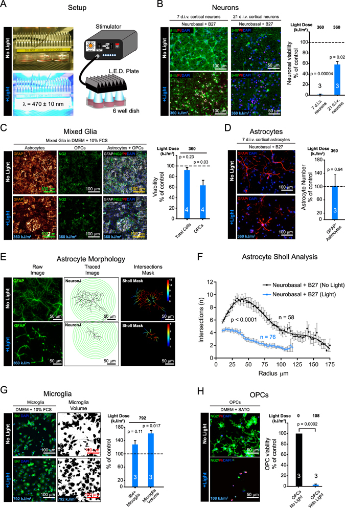
Figure 1
Light induces cytotoxicity in vitro . ( a ) Blue light (470u2009u00b1u200910u2009nm) LED setup with control unit regulating delivery of power, pulse duration and frequency of light to cells in 6 well ...
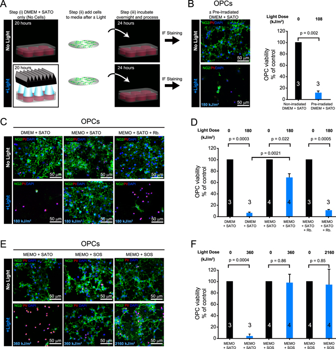
Figure 2
In vitro photo-toxicity can be resolved with a new culture medium. ( a ) Setup of experiment to test media for photo-reactive components. Step (i) media is treatedu2009u00b1u2009light stimulation ( W ...
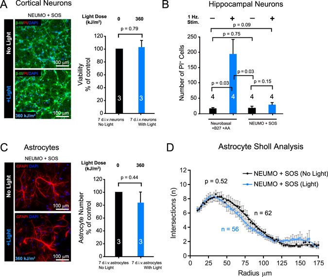
Figure 3
Light induced cytotoxicity can be prevented with new culture medium. ( a ) Viability of 7 d.i.v. u03b2-III + cortical neuronsu2009u00b1u2009light at indicated dose in NEUMO (in replacement of Neurobas...
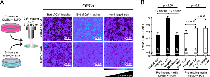
Figure 4
Preventing photo-toxicity during calcium imaging. ( a ) Schematic of OPCs taken from DMEMu2009+u2009SATO based media and MEMOu2009+u2009SOS based media for ratiometric Fura2-AM Ca 2+ -imaging (u03bb e...
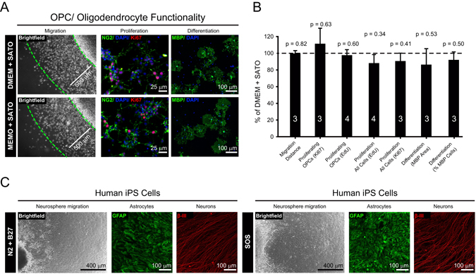
Figure 5
The new media preserves normal cell function. ( a ) Comparison of the effect of DMEMu2009+u2009SATO and MEMOu2009+u2009SATO based media upon OPC migration over 4 days from agarose drop (Brightfield im...
Figure images are served from the NIH/NLM PubMed Central Open Access Subset or Europe PMC; copyright remains with the publishers and authors.
💬 Discussion
0 commentsNo comments yet. Be the first to start a discussion!
Leave a Comment