Abstract
BACKGROUND: Bone metastasis is one of the most common complications of advanced breast cancer. During dissemination to bone, breast cancer cells locate in a putative 'metastatic niche', a microenvironment that regulates the colonisation, maintenance of tumour cell dormancy and subsequent tumour growth. The precise location and composition of the bone metastatic niche is not clearly defined. We have used in vivo models of early breast cancer dissemination to provide novel evidence that demonstrates overlap between endosteal, perivascular, HSC and the metastatic niche in bone. METHODS: Estrogen Receptor (ER) +ve and -ve breast cancer cells were labelled with membrane dyes Vybrant-DiD and Vybrant-CM-DiI and injected via different routes in BALBc/nude mice of different ages. Two-photon microscopy was used to detect and quantitate tumour cells and map their location within the bone microenvironment as well as their distance to the nearest bone surface compared to the nearest other tumour cell. To investigate whether the metastatic niche overlapped with the HSC niche, animals were pre-treated with the CXCR4 antagonist AMD3100 to mobilise hematopoietic (HSCs) prior to injection of breast cancer cells. RESULTS: Breast cancer cells displayed a characteristic pattern of homing in the long bones, with the majority of tumour cells seeded in the trabecular regions, regardless of the route of injection, cell-line characteristics (ER status) or animal age. Breast cancer cells located in close proximity to the nearest bone surface and the average distance between individual tumour cells was higher than their distance to bone. Mobilisation of HSCs from the niche to the circulation prior to injection of cell lines resulted in increased numbers of tumour cells disseminated in trabecular regions. CONCLUSION: Our data provide evidence that homing of breast cancer cells is independent of their ER status and that the breast cancer bone metastasis niche is located within the trabecular region of bone, an area rich in osteoblasts and microvessels. The increased number of breast cancer cells homing to bone after mobilisation of HSCs suggests that the HSC and the bone metastasis niche overlap.
🔬 Techniques
🔭 Microscopes
✨ Fluorophores
🧪 Sample Preparation
🔬 Cell Lines
🏭 Microscope Brands
🧪 Reagent Suppliers
🔴 Lasers
📷 Detectors
💻 Software Details
🏛️ Research Organizations (ROR)
Affiliated research institutions:
📋 Methods
Tumour cell lines MDA-MB-231-GFP-IV [29] , T47D and MCF7 breast cancer cell lines (ATCC) were cultured in RPMI 1640 supplemented with 10% FBS (Life Technologies, Paisley, UK /Invitrogen) at 37 °C 5%CO 2, MDA-MB-231-NW1-Luc2 cells were cultured in DMEM (Life Technologies, Paisley, UK) + Pyruvate medium enriched with 100 U/mL penicillin/streptomycin and 10% FBS (Sigma Aldrich Co Ltd, Poole, UK). Prior to the injections, tumour cells were labelled either with the lipophilic membrane dye Vybrant-CM-DiI or Vybrant-DiD (Life Technologies Ltd, Paisley, UK) according to the manufacturer's instructions.
Animal models Six-week old and twelve-week old female
BALB/c nude mice (Charles River, UK) were used to assess the homing of breast cancer cells in bone and to establish any effects of modification of the niche. Mice were housed in a controlled environment with a 12 h light/dark cycle at 22 °C. They were provided with ad libitum access to 2018 Teklad Global 18% protein rodent diet containing 1.01% Calcium (Harlan Laboratories, UK) and water, and housed in groups of 5/6 in ventilated cages. The Research Ethics Committee for animal experimentation, of the University of Sheffield, UK reviewed and the Home Office approved all the work included in this manuscript. All in vivo experiments were performed in accordance with the UK Animals (Scientific Procedures) Act 1986 Home Office regulations under the authority of PPL 70/8964 and PPL 70/8799.
Bone homing studies
To assess the homing of breast cancer tumour cells to bone, 12-week old female BALB/c nude mice were injected intravenously (i.v.) with 1 × 10 5 MDA-MB-231-GFP-IV breast cancer cells labelled either with the membrane dye Vybrant-CM-DiI or Vybrant-DiD and culled on day 5. Hind limbs were dissected, muscles removed, femora and tibiae separated and processed for imaging as described below. To investigate whether different routes of administration affected the pattern of homing of breast cancer cells, 12-week old female BALB/c nude mice were injected either intravenously ( n = 1) or via the intracardiac (i.c) route ( n = 5) with 1 × 10 5 MDA-MB-231-GFP-IV breast cancer cells labelled with the membrane dye Vybrant-DiD and then culled on day 5 and long bones collected. To establish whether the oestrogen receptor (ER) status of the breast cancer cells affected their pattern of bone homing, 1 × 10 5 Vybrant-DiD labelled ER-ve MDA-MB-231-GFP-IV cells, ER+ve MCF-7 or T47D cells were injected i.c. in 12-week old female BALB/c nude mice ( n = 5/group). Long bones were collected 5 days after cell injection and processed for imaging as described below.
Show full methods section
Tumour cell lines MDA-MB-231-GFP-IV [29] , T47D and MCF7 breast cancer cell lines (ATCC) were cultured in RPMI 1640 supplemented with 10% FBS (Life Technologies, Paisley, UK /Invitrogen) at 37 °C 5%CO 2, MDA-MB-231-NW1-Luc2 cells were cultured in DMEM (Life Technologies, Paisley, UK) + Pyruvate medium enriched with 100 U/mL penicillin/streptomycin and 10% FBS (Sigma Aldrich Co Ltd, Poole, UK). Prior to the injections, tumour cells were labelled either with the lipophilic membrane dye Vybrant-CM-DiI or Vybrant-DiD (Life Technologies Ltd, Paisley, UK) according to the manufacturer's instructions.
Animal models Six-week old and twelve-week old female
BALB/c nude mice (Charles River, UK) were used to assess the homing of breast cancer cells in bone and to establish any effects of modification of the niche. Mice were housed in a controlled environment with a 12 h light/dark cycle at 22 °C. They were provided with ad libitum access to 2018 Teklad Global 18% protein rodent diet containing 1.01% Calcium (Harlan Laboratories, UK) and water, and housed in groups of 5/6 in ventilated cages. The Research Ethics Committee for animal experimentation, of the University of Sheffield, UK reviewed and the Home Office approved all the work included in this manuscript. All in vivo experiments were performed in accordance with the UK Animals (Scientific Procedures) Act 1986 Home Office regulations under the authority of PPL 70/8964 and PPL 70/8799.
Bone homing studies
To assess the homing of breast cancer tumour cells to bone, 12-week old female BALB/c nude mice were injected intravenously (i.v.) with 1 × 10 5 MDA-MB-231-GFP-IV breast cancer cells labelled either with the membrane dye Vybrant-CM-DiI or Vybrant-DiD and culled on day 5. Hind limbs were dissected, muscles removed, femora and tibiae separated and processed for imaging as described below. To investigate whether different routes of administration affected the pattern of homing of breast cancer cells, 12-week old female BALB/c nude mice were injected either intravenously ( n = 1) or via the intracardiac (i.c) route ( n = 5) with 1 × 10 5 MDA-MB-231-GFP-IV breast cancer cells labelled with the membrane dye Vybrant-DiD and then culled on day 5 and long bones collected. To establish whether the oestrogen receptor (ER) status of the breast cancer cells affected their pattern of bone homing, 1 × 10 5 Vybrant-DiD labelled ER-ve MDA-MB-231-GFP-IV cells, ER+ve MCF-7 or T47D cells were injected i.c. in 12-week old female BALB/c nude mice ( n = 5/group). Long bones were collected 5 days after cell injection and processed for imaging as described below.
Modification of the HSC niche
To confirm that AMD3100 mobilises HSC/progenitor cells (PCs) from bone marrow niches into the circulation, 12-week old female BALB/c-Nude mice were treated with AMD3100 (5 mg/kg, i.p.) or Saline daily for 5 days. Three hours after the last drug administration, mice were culled and peripheral blood isolated by cardiac puncture using syringes containing 4% sodium citrate. Blood was centrifuged to remove the serum and red blood cells lysed with ammonium chloride solution (BioLegend Ltd, UK). Peripheral blood mononuclear cells were resuspended in IMDM supplemented with 2% FCS and 100 u/mL Penicillin/Streptomycin at a density of 2 × 10 6 cells/mL. Cell suspensions were diluted 1:10 with MethoCult GF 3434 medium (Stem Cell Technologies), seeded into 6-well plates and maintained in an humidified atmosphere at 37 °C with 5% CO 2 for 11 days to allow colony formation. Bright field images of colonies were taken using a Leica DMI4000B microscope. To assess whether the mobilisation of hematopoietic stem cells (HSCs) would affect homing of breast cancer cells in bone, 12-week old female BALB/c nude were injected i.p. with the CXCR4 antagonist AMD3100 (Sigma-Aldrich) 5 mg/kg (100 µl) or PBS daily for 5 days. 24 h after the last injection, animals were injected i.v with 1 × 10 5 MDA-MB-231-GFP-IV Vybrant-DiD -labelled cells and culled on day 10.
Comparison between young and mature animals
To compare the seeding of breast cancer cells in the bone microenvironment of young and mature animals 6- and 12-week old female BALB/c nude mice ( n = 8/group) were injected i.v. with 1 × 10 5 MDA-MB-231-GFP-IV cells labelled with the membrane dye Vybrant-CM-DiI. Animals were culled 5 days after tumour cell injection.
2.6 µCT analysis
Microcomputed tomography (µCT) analysis was carried out on the resected long bones ex vivo using a Skyscan 1272 X-ray-computed microtomograph (Skyscan). Image acquisition was performed using a voltage of 50 kV, a current of 200 µA, a medium camera resolution of 2016 × 1344, an aluminium filter of 0.5 mm and a pixel size of 4.3 µm. Images were captured of proximal tibias every 0.7° through a 180° rotation. Acquired images were reconstructed and analyzed using NRecon and CTAn software (Skyscan).
Two-photon microscopy
Animals were culled, hind limbs dissected, muscles removed, femora and tibiae separated and collected snap frozen in liquid nitrogen and stored at −80 °C. Cryopreserved long bones were then embedded into Cryo-M-Bed (Instrument Co. Ltd, Huntingdon, UK) and the bone marrow exposed using a Bright OTF Cryostat and a 3020 microtome (Bright Instrument Co. Ltd, Huntingdon, UK). A stack area of exposed bone marrow (2104 µm × 2525 µm 70 µm depth) was captured using a Zeiss LSM510 NLO upright 2-photon/confocal microscope (Carl Zeiss Inl, Cambridge, UK). DiD-labelled and CM-Dil-labelled breast cancer cells were visualised using a 633 nm HeNe and 543 HeNe lasers, respectively. The bone structure was detected with a Chameleon 2-photon laser at 900 nm (Coherent, Santa Clara, CA.). The Volocity 3D Image Analysis software 6.01 (PerkinElmer, Cambridge, UK) was used to quantify the abundance of Vybrant-CM-DiI or Vybrant-DiD events, their distance to the nearest bone surface and to other DTCs [27] . The quantification of dye-labelled tumour cells and their spatial distribution were performed in two different regions of interest (ROIs, Fig. 2 B) from which the cortical bone was excluded.
Confocal microscopy
Long bones were fixed in 4%PFA, decalcified in 0.5 M EDTA, embedded in gelatin and frozen at −80 °C [27] . Immunofluorescence was performed on 30 µm thick cryosections (three non-consecutive levels) for each bone. Tissue sections were permeabilized for 30 min in 0.3% Triton X-100, where appropriate Streptavidin/Biotin blocking (Vector Labs) was performed. Cryosections were incubated for 1 h with primary antibody against murine Endomucin (2 µg/mL, Santa Cruz Biotechnology), Osteopontin (R&D Systems) or human CD29 and CD59 (10 µg/mL, BioLegend). Tissue sections were washed with PBS and incubated with either a fluorescently-conjugated (5 µg/mL) or biotinylated secondary (7.5 µg/mL) antibody followed by fluorescently-conjugated streptavidin. Tissue sections were washed with PBS and incubated with either a fluorescently-conjugated or biotinylated secondary antibody followed by fluorescently-conjugated streptavidin. All incubations were carried out at ambient temperature and ProLong Gold Antifade reagent (Thermo Fisher) to mount the glass coverslips. Images of the immunofluorescence staining were captured with Nikon A1 (Nikon Instruments Europe) or Zeiss LSM 880 AiryScan (Carl Zeiss Microscopy GmBH) confocal systems.
Statistical analysis
Statistical analyses were performed using GraphPad Prism software (Version 6.0 and 7.0). Student T-tests and Two-way ANOVA and Tukey's post hoc test were used as indicated in each figure legend. A p -value of p < 0.05 was considered significant.
Appendix Supplementary materials Image, application 1 Image, application 2
📊 Figures
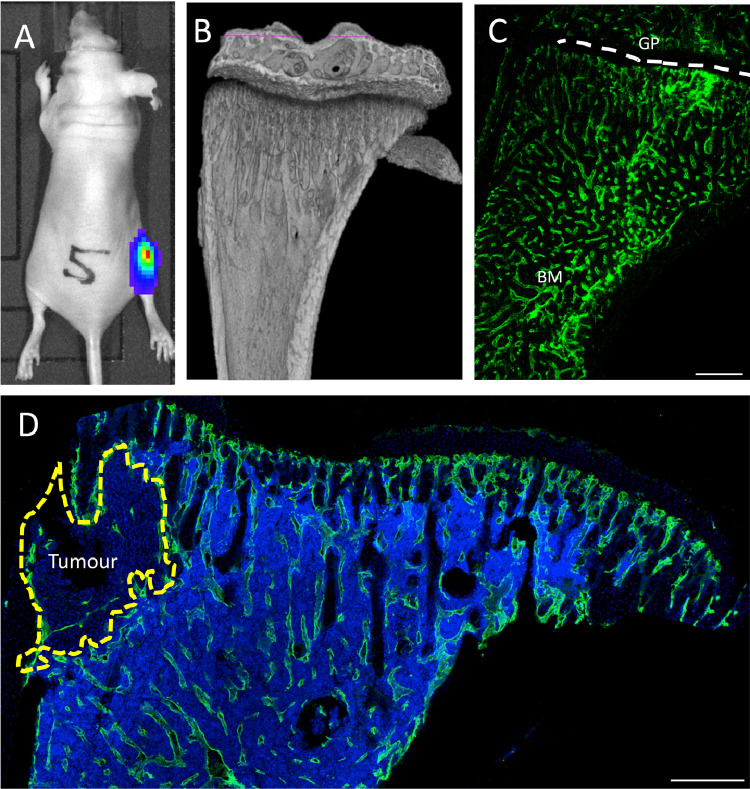
Fig. 1
In vivo tumour model and location of the bone metastatic niche. (A) Representative image of the breast cancer bone metastasis animal model. Female BALB/c nude mice were injected i.c. MDA-MB-231-GFP ce...
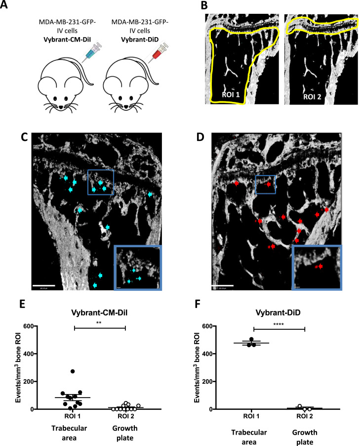
Fig. 2
Homing of the bone-seeking MDA-MB-231-GFP-IV cell-line. (A) Experimental outline of the in vivo study. 12-week old female BALB/c nude mice were injected intravenously (i.v.) with PBS on day 0 followed...
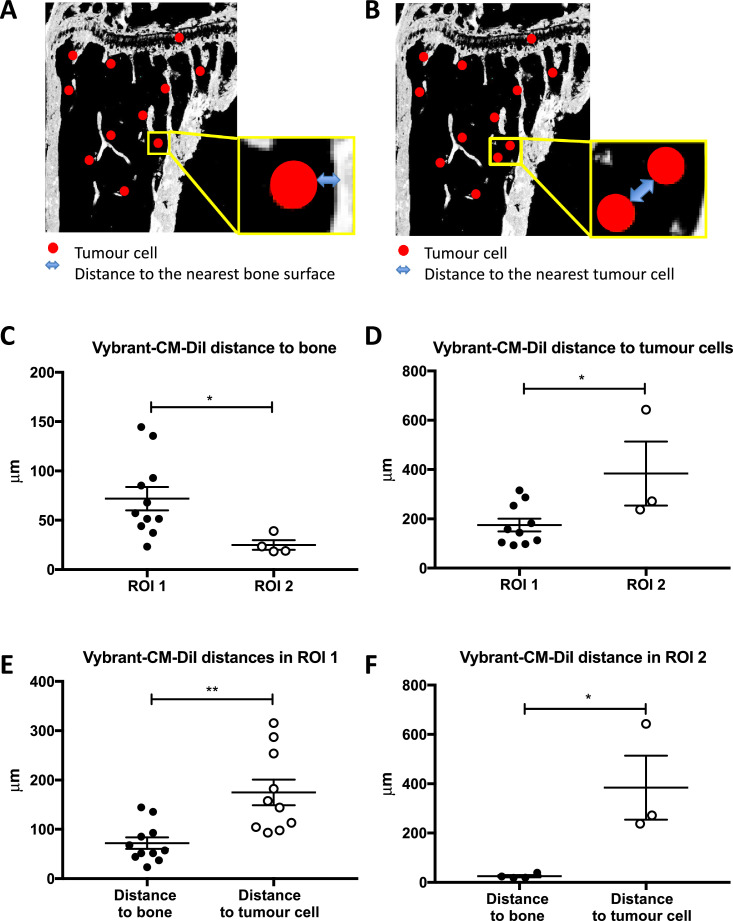
Fig. 3
Location of MDA-MB-231-GFP-IV cells in the bone microenvironment. (A) Schematic illustration of the distances measured from the edge of the tumour cells to the nearest bone surface and (B) to the clos...
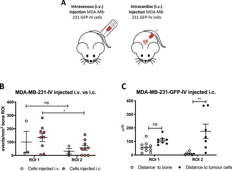
Fig. 4
Comparison between different routes of tumour cell injection. (A) Experimental outline of the in vivo study. 12-week old female BALB/c nude mice were injected either i.v. ( n =u202f1) or i.c. with 1u2...
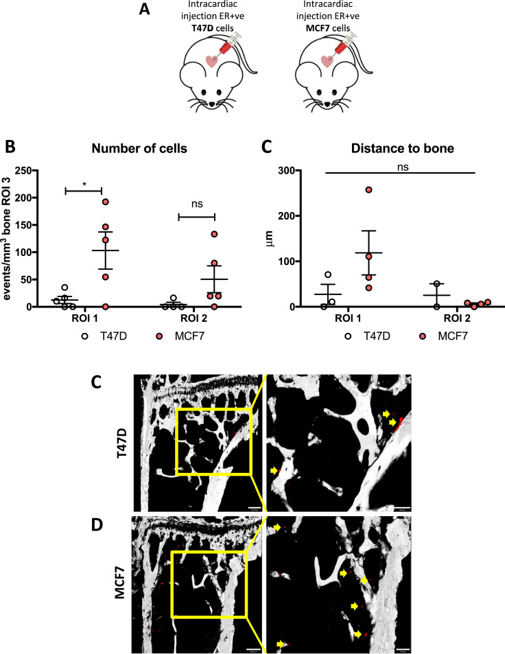
Fig. 5
Homing of ER+ve cell lines. (A) Experimental outline of the in vivo study. 12-week old female BALB/c nude mice were injected on day 1 with 1u202fu00d7u202f10 5 T47D or MCF7 Vybrant-DiD labelled cells ...
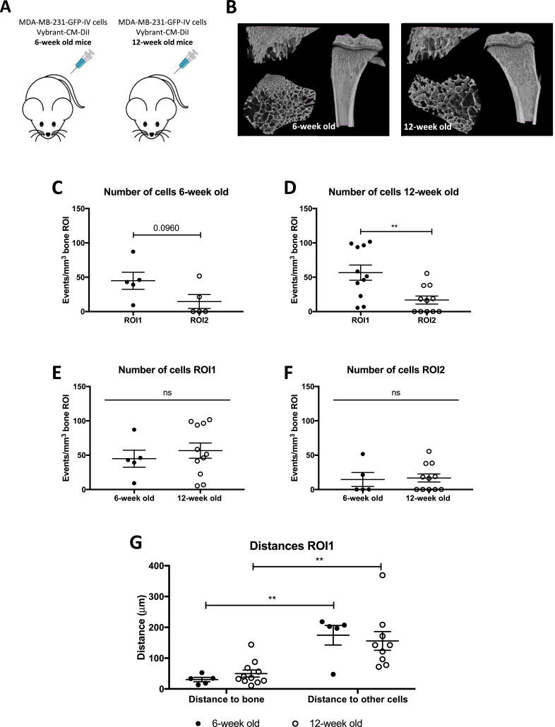
Fig. 6
Homing of MDA-MB-231-GFP-IV cells in young and mature mice. (A) Experimental outline of the in vivo study. 6- and 12-week old female BALB/c nude mice ( n =u202f8/group) were injected on day 1 with 1u2...
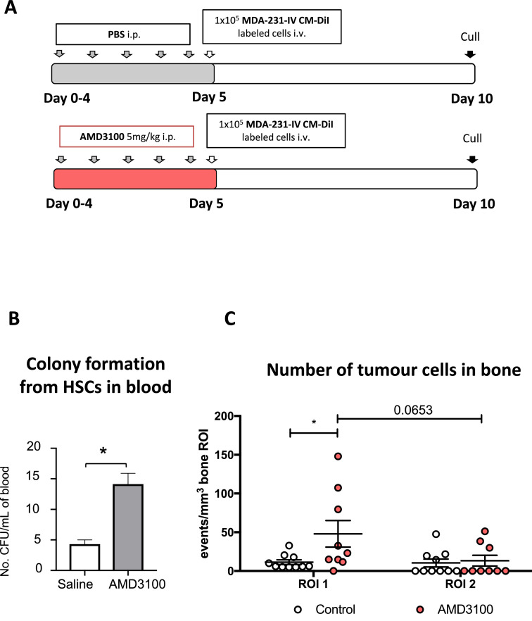
Fig. 7
Modification of the HSCs niche u2013 effect on tumour cell homing. (A) Experimental outline of the in vivo study. 12-week old female BALB/c nude mice were injected daily for 5 days with PBS or AMD3100...
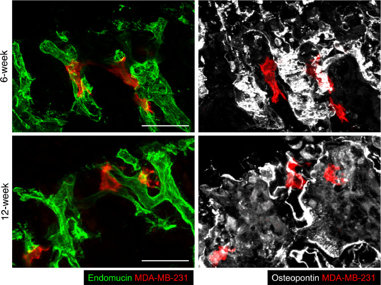
Fig. 8
Disseminated breast cancer cells reside in perivascular locations within mouse long bones. Immunofluorescent labelling of tibial cryosections from young and mature mice containing human MDA-MB-231 bre...
Figure images are served from the NIH/NLM PubMed Central Open Access Subset or Europe PMC; copyright remains with the publishers and authors.
💬 Discussion
0 commentsNo comments yet. Be the first to start a discussion!
Leave a Comment