Abstract
Mitochondrial diseases produce profound neurological dysfunction via mutations affecting mitochondrial energy production, including the relatively common Leigh syndrome (LS). We recently described an LS case caused by a pathogenic mutation in USMG5, encoding a small supernumerary subunit of mitochondrial ATP synthase. This protein is integral for ATP synthase dimerization, and patient fibroblasts revealed an almost total loss of ATP synthase dimers. Here, we utilize in situ cryoelectron tomography (cryo-ET) in a clinical case-control study of mitochondrial disease to directly study mitochondria within cultured fibroblasts from a patient with LS and a healthy human control subject. Through tomographic analysis of patient and control mitochondria, we find that loss of ATP synthase dimerization due to the pathogenic mutation causes profound disturbances of mitochondrial crista ultrastructure. Overall, this work supports the crucial role of ATP synthase in regulating crista architecture in the context of human disease.
🔬 Techniques
🧬 Organisms
✨ Fluorophores
💾 Data Repositories
🏛️ Research Organizations (ROR)
Affiliated research institutions:
📊 Figures
Figureu00a01
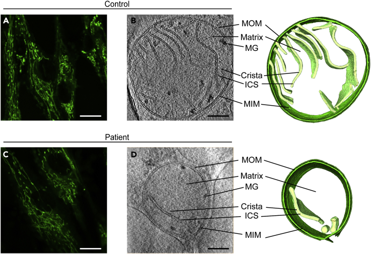
Analysis of Patient Mitochondria Reveals Abnormal Crista Membrane Architecture, Related to Figures S1 and S2 (A and C) Representative fibroblasts from control and patient were stained with MitoTracker...
Figureu00a02
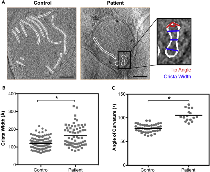
Two-Dimensional Measurements of Patient Crista Architecture Reveal Increased Width and Blunted Apex Curvature, Related to Figures S2u2013S4 and Videos S1 , S2 , S3 , and S4 (A) Representative two-dime...
Figureu00a03
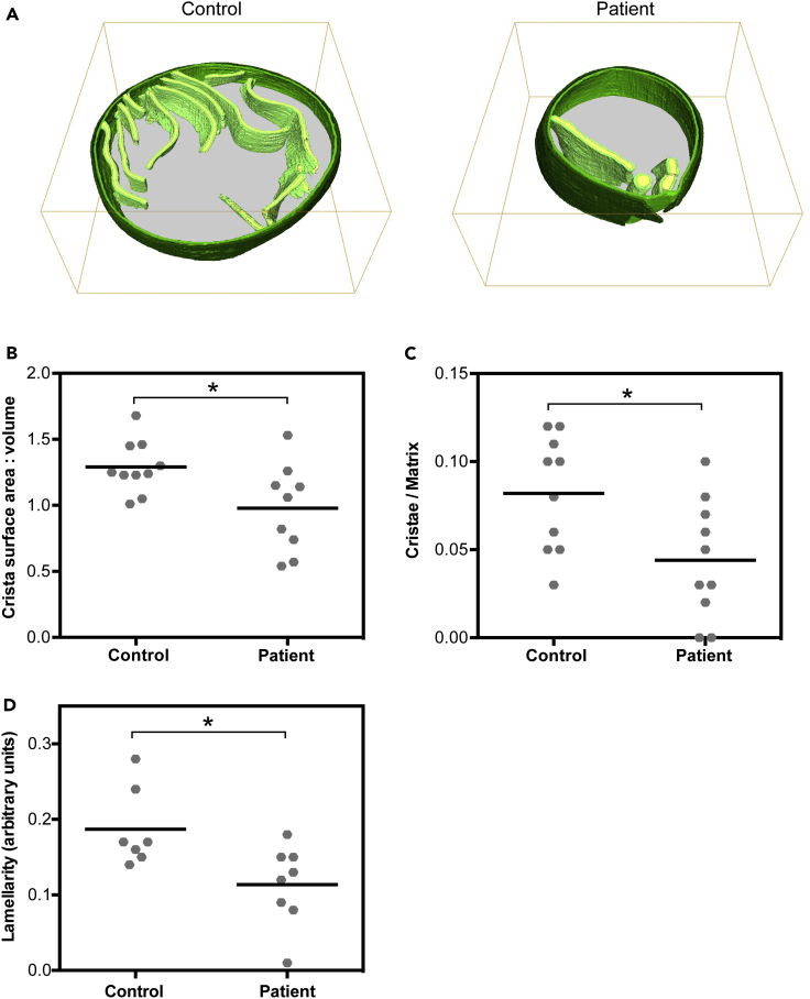
Three-Dimensional Analysis of Reconstructed Patient Mitochondria Reveals Abnormal Morphology and Distribution of Cristae, Related to Videos S3 and S4 (A) Representative three-dimensional reconstructio...
Figureu00a04
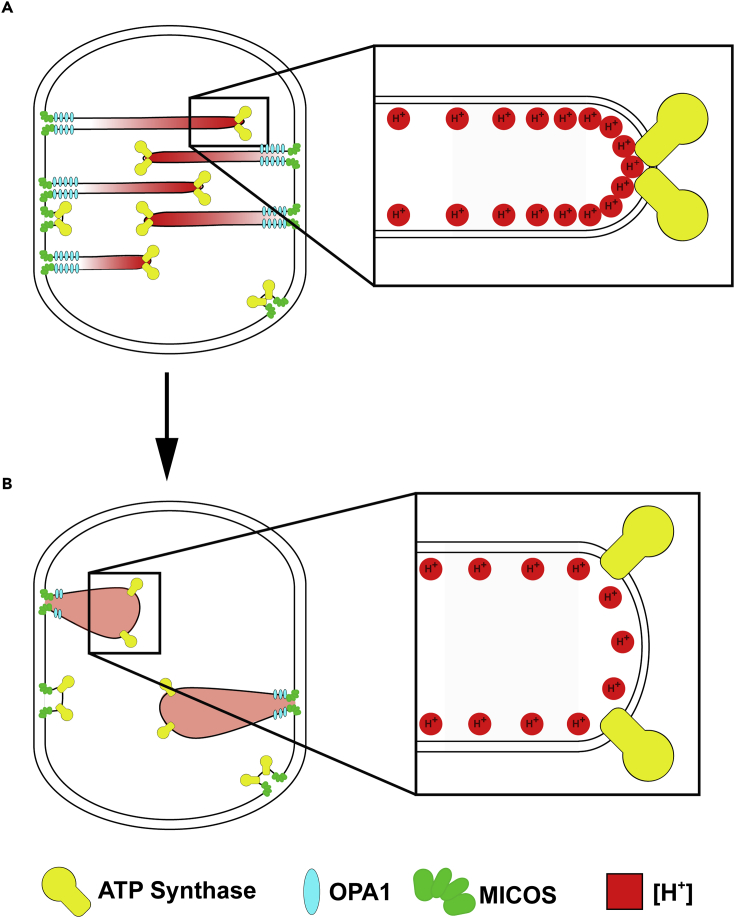
Model of Patient Pathogenic Mutation (A and B) Model representing crista architecture of mitochondria in fibroblasts from the (A) healthy control and (B) patient. In mitochondria from the healthy huma...
Figure images are served from the NIH/NLM PubMed Central Open Access Subset or Europe PMC; copyright remains with the publishers and authors.
💬 Discussion
0 commentsNo comments yet. Be the first to start a discussion!
Leave a Comment